Full Question & Answer Text (for Search Engines)
Question 1:
A 13-year-old obese boy presents with a 3-month history of vague left knee pain and a limp. Physical examination demonstrates an obligate external rotation of the left hip during flexion. Radiographs demonstrate a posterior and inferior displacement of the proximal femoral epiphysis. Through which physeal zone does the primary failure occur in this condition?
Options:
- Zone of resting cartilage
- Zone of proliferation
- Zone of hypertrophy
- Zone of provisional calcification
- Primary spongiosa
Correct Answer: Zone of hypertrophy
Explanation:
Slipped Capital Femoral Epiphysis (SCFE) characteristically involves a structural failure through the zone of hypertrophy of the physis. This zone is the weakest biomechanically because the chondrocytes are enlarged, lacking substantial extracellular matrix. This is in contrast to Salter-Harris fractures, which typically propagate through the zone of provisional calcification or the zone of hypertrophy depending on the vector, but SCFE specifically involves pathologic widening and slipping through the hypertrophic zone.
Question 2:
A 32-year-old man sustains a closed fracture of the distal third of his humeral shaft, presenting with an inability to actively extend his wrist and digits. He undergoes open reduction and internal fixation using a posterior triceps-splitting approach. Which of the following accurately describes the relationship of the radial nerve to the humerus and intermuscular septum at the level of the distal third of the humeral shaft?
Options:
- It runs distally in the posterior compartment down to the lateral epicondyle
- It pierces the lateral intermuscular septum from posterior to anterior approximately 10 cm proximal to the radiocapitellar joint
- It crosses posterior to the medial epicondyle before piercing the medial intermuscular septum
- It enters the anterior compartment 2 cm proximal to the lateral epicondyle
- It travels superficially over the brachialis muscle throughout its entire course in the arm
Correct Answer: It pierces the lateral intermuscular septum from posterior to anterior approximately 10 cm proximal to the radiocapitellar joint
Explanation:
The radial nerve spirals around the posterior humerus in the spiral groove and pieces the lateral intermuscular septum from posterior to anterior approximately 10 cm (range 7.5 to 12 cm) proximal to the radiocapitellar joint to enter the anterior compartment. This anatomical landmark is critical when performing a posterior approach to the humerus or when treating distal third humeral shaft fractures (Holstein-Lewis fractures).
Question 3:
A 15-year-old boy presents with progressive, severe night pain in his distal thigh over 2 months. Radiographs reveal a mixed lytic and sclerotic lesion in the distal femoral metaphysis with a robust periosteal reaction demonstrating a 'sunburst' pattern. Biopsy confirms high-grade intramedullary osteosarcoma. Which of the following genetic mutations is most frequently implicated in the pathogenesis of this tumor?
Options:
- t(11;22) translocation
- t(X;18) translocation
- Mutations in the RB1 and TP53 tumor suppressor genes
- t(9;22) translocation
- t(2;13) translocation
Correct Answer: Mutations in the RB1 and TP53 tumor suppressor genes
Explanation:
Osteosarcoma is strongly associated with mutations in the tumor suppressor genes RB1 (retinoblastoma gene) and TP53 (Li-Fraumeni syndrome). t(11;22) is characteristic of Ewing sarcoma. t(X;18) is seen in synovial sarcoma. t(2;13) is characteristic of alveolar rhabdomyosarcoma.
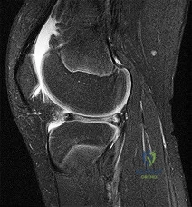
Question 4:
A 22-year-old female soccer player undergoes primary anterior cruciate ligament (ACL) reconstruction using a bone-patellar tendon-bone autograft. Postoperatively, she experiences loss of terminal extension and anterior knee pain. An MRI indicates graft impingement against the intercondylar roof. Which of the following technical errors during tunnel preparation is the most common cause of this specific impingement pattern?
Options:
- Femoral tunnel placed too anteriorly
- Tibial tunnel placed too anteriorly
- Femoral tunnel placed too vertically
- Tibial tunnel placed too medially
- Femoral tunnel placed too posteriorly
Correct Answer: Tibial tunnel placed too anteriorly
Explanation:
Placing the tibial tunnel too anteriorly is the most common cause of intercondylar roof impingement. The tibial tunnel should be placed posterior to the intersection of Blumensaat's line and the tibial plateau when the knee is in full extension. If placed anterior to this line, the graft will impinge on the notch roof during terminal extension, leading to a loss of extension and potential graft failure.
Question 5:
A 60-year-old man with poorly controlled diabetes mellitus presents with worsening back pain and low-grade fevers for 1 week. Over the past 24 hours, he developed progressive bilateral leg weakness and urinary retention. MRI reveals a massive ventral fluid collection from L2 to L4 causing severe compression of the cauda equina, consistent with a spinal epidural abscess. What is the most common causative organism isolated in this condition?
Options:
- Streptococcus pneumoniae
- Escherichia coli
- Mycobacterium tuberculosis
- Staphylococcus aureus
- Pseudomonas aeruginosa
Correct Answer: Staphylococcus aureus
Explanation:
Staphylococcus aureus is the most common organism responsible for spinal epidural abscesses, accounting for approximately 60-90% of cases. Immediate surgical decompression and targeted intravenous antibiotics are the standard of care for patients presenting with progressive neurologic deficits.
Question 6:
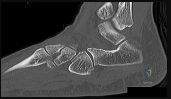
A 28-year-old professional football player sustains an axial load to his plantarflexed foot. Weight-bearing radiographs demonstrate a 4 mm diastasis between the base of the first and second metatarsals. He is diagnosed with a Lisfranc injury. Which of the following ligaments is primarily disrupted in this classic injury pattern?
Options:
- Plantar ligament extending from the medial cuneiform to the base of the first metatarsal
- Dorsal ligament extending from the middle cuneiform to the base of the second metatarsal
- Plantar ligament extending from the medial cuneiform to the base of the second metatarsal
- Dorsal ligament extending from the medial cuneiform to the base of the second metatarsal
- Interosseous ligament between the bases of the first and second metatarsals
Correct Answer: Plantar ligament extending from the medial cuneiform to the base of the second metatarsal
Explanation:
The Lisfranc ligament is an oblique, stout ligament extending from the plantar-lateral aspect of the medial cuneiform to the plantar-medial base of the second metatarsal. It is the critical stabilizer of the tarsometatarsal articulation. There is no transverse ligament directly connecting the bases of the first and second metatarsals.
Question 7:
A 24-year-old carpenter sustains a deep laceration to the volar aspect of his right index finger exactly at the level of the proximal interphalangeal (PIP) joint flexion crease. Examination shows absent active flexion at both the PIP and DIP joints. During surgical exploration, complete lacerations of the FDP and FDS tendons are identified. In which flexor tendon zone is this injury located?
Options:
- Zone I
- Zone II
- Zone III
- Zone IV
- Zone V
Correct Answer: Zone II
Explanation:
Zone II (also known historically as 'no man's land') extends from the proximal edge of the A1 pulley to the insertion of the flexor digitorum superficialis (FDS) on the middle phalanx. A laceration at the level of the PIP joint falls squarely within Zone II, where both the FDS and FDP tendons are enclosed within the tight fibro-osseous sheath.
Question 8:
A 35-year-old woman undergoes open reduction and internal fixation of a diaphyseal radial fracture using a dynamically compressed plate, achieving absolute fracture stability and anatomical reduction. Which of the following best describes the primary mechanism of bone healing expected in this specific environment?
Options:
- Endochondral ossification with massive cartilaginous callus formation
- Intramembranous ossification with robust periosteal bridging callus
- Cutting cone remodeling by osteoclasts followed directly by osteoblastic bone deposition
- Formation of woven bone directly from mesenchymal stem cells without any remodeling
- Chondrocyte hypertrophy followed by vascular invasion and subsequent osteoid deposition
Correct Answer: Cutting cone remodeling by osteoclasts followed directly by osteoblastic bone deposition
Explanation:
Absolute stability achieved through dynamic compression plating results in primary (direct) bone healing. This process bypasses callus formation. It occurs via Haversian remodeling, where 'cutting cones' of osteoclasts cross the fracture line, followed immediately by osteoblasts laying down lamellar bone. Secondary bone healing (callus formation) occurs in settings of relative stability.
Question 9:
A 55-year-old highly active man is scheduled for a total hip arthroplasty (THA). The surgeon considers utilizing a ceramic-on-ceramic bearing surface to maximize longevity. Which of the following is the most notable disadvantage or complication specific to a ceramic-on-ceramic bearing compared to ceramic-on-highly cross-linked polyethylene?
Options:
- A significantly higher rate of volumetric wear debris generation
- An increased risk of profound macrophage-mediated osteolysis
- Audible squeaking during hip articulation
- Severe adverse local tissue reactions (ALTR) mediated by a Type IV hypersensitivity
- Accelerated corrosion at the femoral head-neck trunnion
Correct Answer: Audible squeaking during hip articulation
Explanation:
Ceramic-on-ceramic (CoC) bearings offer the lowest wear rates of all THA bearing couples and do not cause significant osteolysis or metal-related hypersensitivity. However, a well-documented unique complication of CoC bearings is audible squeaking during motion, occurring in up to 10-15% of patients in some series, often associated with micro-separation or impingement.
Question 10:
A hemodynamically unstable 42-year-old male is brought to the trauma bay after a motorcycle crash. Pelvic radiographs demonstrate an APC-III pelvic ring injury ('open book' pelvis). A pelvic binder is immediately applied by the trauma team. Which of the following statements regarding the application and function of a pelvic binder is most accurate?
Options:
- It must be centered exactly over the greater trochanters to effectively close the pelvic ring
- It primarily achieves hemostasis by directly compressing bleeding from the superior gluteal artery
- It is absolutely contraindicated if a concurrent extraperitoneal bladder rupture is identified
- It should be applied tightly over the iliac crests to maximize anterior compression
- It works primarily by tamponading arterial bleeding in the presacral space
Correct Answer: It must be centered exactly over the greater trochanters to effectively close the pelvic ring
Explanation:
To be effective in reducing pelvic volume and closing an open-book pelvic injury, a pelvic binder (or sheet) must be centered low, directly over the greater trochanters. Positioning it too high over the iliac crests can actually worsen the deformity by acting as a fulcrum. Its primary mechanism of action is reducing pelvic volume to encourage tamponade of the massive venous plexus bleeding, not arterial bleeding.
Question 11:
A 6-year-old boy presents with a painless limp of 3 months' duration. His mother notes restricted hip motion, particularly in abduction and internal rotation. Radiographs reveal sclerosis, flattening, and early fragmentation of the proximal femoral epiphysis. In the natural history of Legg-Calvé-Perthes disease, which of the following represents the correct sequence of radiographic stages?
Options:
- Fragmentation, Initial (necrosis), Reossification, Healed
- Initial (necrosis), Fragmentation, Reossification, Healed
- Reossification, Fragmentation, Initial (necrosis), Healed
- Initial (necrosis), Reossification, Fragmentation, Healed
- Fragmentation, Reossification, Initial (necrosis), Healed
Correct Answer: Initial (necrosis), Fragmentation, Reossification, Healed
Explanation:
The classic radiographic progression of Legg-Calvé-Perthes disease (Waldenström stages) follows four distinct phases: 1) Initial stage (avascular necrosis, sclerosis, and growth arrest), 2) Fragmentation stage (subchondral radiolucent crescent sign, epiphysis fragmenting), 3) Reossification stage (new woven bone formation), and 4) Healed or residual stage (final shape of the femoral head).
Question 12:
A 20-year-old collegiate quarterback sustains a traumatic anterior shoulder dislocation. Post-reduction MRI demonstrates an anterior-inferior labral tear that is displaced medially and rolled down the glenoid neck, still attached to an intact but stripped anterior periosteal sleeve. Which of the following eponymous terms best describes this specific soft-tissue lesion?
Options:
- Bankart lesion
- ALPSA lesion
- Perthes lesion
- GLAD lesion
- HAGL lesion
Correct Answer: ALPSA lesion
Explanation:
An ALPSA (Anterior Labroligamentous Periosteal Sleeve Avulsion) lesion occurs when the anterior labrum is torn and displaced medially/inferiorly along the glenoid neck, remaining tethered by an intact stripped periosteum. A Bankart lesion involves a complete detachment of the labrum and periosteum. A Perthes lesion is a non-displaced tear with an intact periosteum. GLAD is a Glenolabral Articular Disruption. HAGL is Humeral Avulsion of the Glenohumeral Ligament.
Question 13:
A 32-year-old woman complains of left knee pain. Radiographs show an eccentric, purely lytic lesion in the distal femoral epiphysis extending to the subchondral bone, without a sclerotic margin. Biopsy reveals multinucleated giant cells interspersed among mononuclear stromal cells. For recurrent or surgically unsalvageable cases of this specific tumor, which of the following is the most appropriate targeted medical therapy?
Options:
- Imatinib
- Denosumab
- Methotrexate
- Doxorubicin
- Rituximab
Correct Answer: Denosumab
Explanation:
This is a Giant Cell Tumor (GCT) of bone. The neoplastic cells in GCT are actually the mononuclear stromal cells, which heavily express RANKL (Receptor Activator of Nuclear factor Kappa-B Ligand). This expression recruits and activates normal osteoclasts (the giant cells), causing massive osteolysis. Denosumab, a monoclonal antibody against RANKL, is the medical treatment of choice for unresectable or highly recurrent GCTs.
Question 14:
A 45-year-old woman presents with severe numbness and tingling in her thumb, index, and middle fingers, confirming a diagnosis of severe carpal tunnel syndrome. She is scheduled for an open carpal tunnel release. Which of the following anatomical structures is NOT contained within the true carpal tunnel?
Options:
- Flexor pollicis longus tendon
- Median nerve
- Flexor carpi radialis tendon
- Flexor digitorum superficialis tendons
- Flexor digitorum profundus tendons
Correct Answer: Flexor carpi radialis tendon
Explanation:
The carpal tunnel contains exactly 10 structures: the median nerve, 4 flexor digitorum superficialis (FDS) tendons, 4 flexor digitorum profundus (FDP) tendons, and the flexor pollicis longus (FPL) tendon. The flexor carpi radialis (FCR) tendon runs within its own separate fibro-osseous sheath within the fibers of the transverse carpal ligament, outside the main carpal tunnel.
Question 15:
A 58-year-old man with long-standing diabetic peripheral neuropathy presents with a swollen, erythematous, and warm right foot without ulcerations. He reports minimal pain but his midfoot arch has collapsed over the past month. Radiographs demonstrate fragmentation, periarticular debris, and subluxation at the tarsometatarsal joints. Which of the following is the most appropriate initial management for the acute phase of this condition?
Options:
- Immediate open reduction and midfoot arthrodesis
- Intravenous antibiotics for 6 weeks followed by bracing
- Total contact casting with strict non-weight-bearing
- Custom orthotic inserts and modified deep-toe box shoes
- Below-knee amputation
Correct Answer: Total contact casting with strict non-weight-bearing
Explanation:
This patient is presenting with acute Eichenholtz Stage I Charcot arthropathy (fragmentation stage). The hallmark of treatment for acute Charcot neuroarthropathy is strict offloading and immobilization to prevent further deformity until the active inflammatory process resolves (Stage III - consolidation). Total contact casting is the gold standard for achieving this.
Question 16:
A 52-year-old man presents with sharp neck pain radiating down his right arm. Physical examination reveals weakness in active elbow extension and wrist flexion. He also reports decreased sensation isolated to the dorsum of his middle finger. Which cervical nerve root is most likely compressed?
Options:
Correct Answer: C7
Explanation:
Compression of the C7 nerve root causes a classic radiculopathy characterized by weakness in elbow extension (triceps) and wrist flexion (flexor carpi radialis), an absent or diminished triceps reflex, and sensory changes involving the middle finger. C6 radiculopathy typically involves wrist extension weakness and numbness in the thumb and index finger. C8 involves finger flexion and numbness in the small finger.
Question 17:
In total joint arthroplasty, ultra-high-molecular-weight polyethylene (UHMWPE) is subjected to gamma irradiation to induce extensive cross-linking. While this process significantly improves wear resistance, what is the primary mechanical disadvantage introduced by highly cross-linking the polyethylene?
Options:
- Increased susceptibility to oxidative degradation in vivo
- Decreased fatigue strength and fracture toughness
- Accelerated rate of volumetric wear compared to conventional polyethylene
- Increased generation of smaller, highly biologically active wear debris particles
- A drastic increase in the modulus of elasticity leading to stress shielding
Correct Answer: Decreased fatigue strength and fracture toughness
Explanation:
Highly cross-linking UHMWPE dramatically improves its resistance to adhesive and abrasive wear. However, the process of cross-linking alters the polymer's internal structure, resulting in decreased mechanical properties, specifically diminished fatigue strength, yield strength, and fracture toughness. This makes the material more susceptible to catastrophic failure, such as rim fracture in thin acetabular liners.
Question 18:
During a primary total knee arthroplasty (TKA), the surgeon evaluates the flexion and extension gaps. Utilizing spacer blocks, the surgeon notes that the extension gap is symmetric and perfectly balanced, but the flexion gap is excessively tight. Assuming an anterior referencing sizing guide was used, which of the following surgical adjustments is the most appropriate next step to balance the knee?
Options:
- Increase the thickness of the distal femoral bone cut
- Perform a release of the posterior capsule
- Downsize the femoral component
- Decrease the posterior tibial slope
- Release the deep medial collateral ligament
Correct Answer: Downsize the femoral component
Explanation:
When using an anterior referencing system, the anterior flange of all femoral components rests on the same cut surface. Downsizing the femoral component reduces its anteroposterior dimension by removing more bone from the posterior condyles. This specifically enlarges the flexion gap without altering the extension gap, thereby resolving an isolated tight flexion gap.
Question 19:
A 27-year-old man sustains a closed midshaft tibia fracture treated with intramedullary nailing. Six hours postoperatively, he develops severe, out-of-proportion leg pain and severe pain with passive stretch of his toes. Compartment pressure monitoring reveals an anterior compartment pressure of 45 mmHg. The decision is made to perform a standard four-compartment fasciotomy of the leg via a dual-incision technique. During the lateral incision, which anatomical structure is utilized as the key landmark to identify and separate the anterior and lateral compartments?
Options:
- Anterior intermuscular septum
- Posterior intermuscular septum
- Transverse intermuscular septum
- Interosseous membrane
- Superficial peroneal nerve
Correct Answer: Anterior intermuscular septum
Explanation:
In the standard two-incision four-compartment fasciotomy of the lower leg, the lateral (or anterolateral) incision is made halfway between the tibial crest and the fibular shaft. The key fascial landmark identified through this incision is the anterior intermuscular septum. Fasciotomies are made anterior to this septum to release the anterior compartment, and posterior to it to release the lateral compartment.
Question 20:
A 22-year-old man falls onto an outstretched hand and presents with marked anatomic snuffbox tenderness. Initial radiographs are negative, but an MRI obtained 3 days later confirms a nondisplaced fracture traversing the proximal pole of the scaphoid. What is the primary anatomical reason this specific fracture pattern is highly predisposed to avascular necrosis and nonunion?
Options:
- The proximal pole is supplied exclusively by the volar carpal branch of the radial artery
- The primary blood supply enters the distal pole via the dorsal carpal branch of the radial artery and flows retrograde
- The proximal pole relies entirely on diffusion from synovial fluid without any direct intraosseous vessels
- The blood supply originates from the deep palmar arch, which is easily disrupted by capsular swelling
- The scaphoid relies entirely on the superficial palmar arch which enters through the scapholunate ligament
Correct Answer: The primary blood supply enters the distal pole via the dorsal carpal branch of the radial artery and flows retrograde
Explanation:
The major blood supply to the scaphoid (providing 70-80% of its vascularity) comes from the dorsal carpal branch of the radial artery. These vessels enter the scaphoid along its dorsal ridge at the level of the waist and distal pole, and then perfuse the proximal pole in a retrograde fashion. A fracture at the waist or proximal pole interrupts this retrograde flow, putting the proximal fragment at extremely high risk for avascular necrosis.
Question 21:
What primary manufacturing process decreases the wear rate of highly cross-linked polyethylene (HXLPE) compared to conventional polyethylene in total hip arthroplasty?
Options:
- Addition of Vitamin E as an antioxidant only
- Irradiation followed by remelting or annealing
- Ethylene oxide gas sterilization
- Forging and hot isostatic pressing
- Cold working and subsequent rapid cooling
Correct Answer: Irradiation followed by remelting or annealing
Explanation:
Highly cross-linked polyethylene (HXLPE) is created by subjecting the ultra-high-molecular-weight polyethylene (UHMWPE) to gamma or electron beam irradiation, which creates cross-links between the polymer chains, significantly increasing wear resistance. This process also generates free radicals that can cause oxidative degradation over time. To eliminate these free radicals, the material is subsequently heated through either remelting (above the melting point) or annealing (below the melting point).
Question 22:
A 4-month-old infant is treated with a Pavlik harness for developmental dysplasia of the hip. At a follow-up visit, the infant is noted to have decreased active extension of the knee on the treated side. Which of the following positioning errors is the most likely cause of this complication?
Options:
- Excessive flexion of the hip
- Excessive extension of the hip
- Excessive abduction of the hip
- Excessive adduction of the hip
- Inadequate stabilization of the chest strap
Correct Answer: Excessive flexion of the hip
Explanation:
Decreased active extension of the knee in an infant treated with a Pavlik harness is indicative of a femoral nerve palsy. The femoral nerve can become compressed against the pelvis if the hip is positioned in excessive hyperflexion (typically >120 degrees). If this occurs, the anterior straps should be loosened or the harness temporarily discontinued to allow neurological recovery. Excessive abduction is associated with avascular necrosis of the femoral head.
Question 23:
A 35-year-old male presents after a high-speed motorcycle crash. Pelvic radiographs reveal symphysis pubis widening of 3.5 cm and widening of the anterior sacroiliac joints bilaterally, with intact posterior SI ligaments. What is the most common anatomic source of life-threatening hemorrhage in this specific injury pattern?
Options:
- Superior gluteal artery
- Internal pudendal artery
- Presacral venous plexus
- Cancellous bone of the fracture surfaces
- Obturator artery
Correct Answer: Presacral venous plexus
Explanation:
The patient has an Anterior-Posterior Compression (APC) Type II pelvic ring injury. In pelvic trauma, roughly 80-90% of life-threatening hemorrhage is venous in origin, most commonly from the presacral and prevesical venous plexuses. While arterial bleeding (e.g., superior gluteal, internal pudendal) can occur and is often more rapidly fatal if present (particularly in severe APC III or highly displaced vertical shear/lateral compression patterns), the overall most common source of severe bleeding across pelvic ring disruptions is the venous plexus.
Question 24:
A 22-year-old presents with radial-sided wrist pain after a fall. A scaphoid waist fracture is diagnosed. The proximal pole of the scaphoid is at high risk for avascular necrosis due to its tenuous blood supply. Which of the following best describes the predominant arterial supply to the scaphoid?
Options:
- Palmar carpal branch of the radial artery
- Dorsal carpal branch of the radial artery
- Anterior interosseous artery
- Ulnar artery via the deep palmar arch
- Superficial palmar branch of the radial artery
Correct Answer: Dorsal carpal branch of the radial artery
Explanation:
The scaphoid receives 70-80% of its blood supply from the dorsal carpal branch of the radial artery. These vessels enter the scaphoid via a dorsal ridge at the waist and distal pole, flowing in a retrograde fashion to supply the proximal pole. Because of this retrograde flow, fractures at the waist or proximal pole highly compromise the blood supply to the proximal fragment, leading to a high rate of avascular necrosis and nonunion.
Question 25:
During an anterior cruciate ligament (ACL) reconstruction, the surgeon places the femoral tunnel excessively anterior to the anatomic footprint. What is the most likely biomechanical consequence of this malpositioning?
Options:
- The graft will be excessively tight in flexion and loose in extension
- The graft will be excessively tight in extension and loose in flexion
- The graft will be excessively tight in both flexion and extension
- The graft will be loose in both flexion and extension
- The graft will only exhibit laxity in internal rotation
Correct Answer: The graft will be excessively tight in flexion and loose in extension
Explanation:
Femoral tunnel position is critical in ACL reconstruction. If the femoral tunnel is placed too far anteriorly (shallow), the distance between the femoral and tibial tunnels increases as the knee flexes. This results in the graft being tight in flexion (potentially limiting knee flexion or causing graft failure/stretching) and loose in extension. Conversely, a tunnel placed too posterior will be tight in extension and loose in flexion.
Question 26:
A 14-year-old boy presents with severe, unrelenting thigh pain and low-grade fevers. Radiographs show a permeative diaphyseal lesion with an 'onion-skin' periosteal reaction. Biopsy reveals sheets of uniform small round blue cells. Which chromosomal translocation is most characteristic of this condition?
Options:
- t(X;18)(p11;q11)
- t(11;22)(q24;q12)
- t(12;16)(q13;p11)
- t(2;13)(q35;q14)
- t(9;22)(q34;q11)
Correct Answer: t(11;22)(q24;q12)
Explanation:
The clinical, radiographic, and histologic presentation is classic for Ewing sarcoma. The diagnostic hallmark of Ewing sarcoma is the t(11;22)(q24;q12) translocation, which results in the EWS-FLI1 fusion protein. Option A (t(X;18)) is found in synovial sarcoma. Option C (t(12;16)) is associated with myxoid liposarcoma. Option D (t(2;13)) is seen in alveolar rhabdomyosarcoma. Option E (t(9;22)) is the Philadelphia chromosome seen in CML.
Question 27:
A 65-year-old man with cervical spondylotic myelopathy exhibits a positive Hoffmann sign on physical examination. The presence of this physical sign indicates abnormal function primarily involving which of the following descending pathways?
Options:
- Spinothalamic tract
- Dorsal column medial lemniscus
- Corticospinal tract
- Vestibulospinal tract
- Rubrospinal tract
Correct Answer: Corticospinal tract
Explanation:
A positive Hoffmann sign (flicking the distal phalanx of the middle finger results in reflexive flexion of the thumb and index finger) is a clinical indicator of an upper motor neuron lesion, specifically indicating compression or dysfunction of the corticospinal tract in the cervical spinal cord. The spinothalamic tract and dorsal columns are sensory tracts.
Question 28:
A 55-year-old poorly controlled diabetic patient presents with a warm, swollen, and erythematous foot. Initial radiographs reveal fragmentation of the midfoot bones, periarticular debris, and early joint subluxation, but no signs of consolidation. According to the Eichenholtz classification of Charcot arthropathy, which stage does this represent?
Options:
- Stage 0 (Inflammation)
- Stage 1 (Fragmentation)
- Stage 2 (Coalescence)
- Stage 3 (Reconstruction)
- Stage 4 (Consolidation)
Correct Answer: Stage 1 (Fragmentation)
Explanation:
The Eichenholtz classification outlines the natural history of Charcot arthropathy. Stage 0 is the inflammatory phase (normal x-rays, localized erythema/swelling). Stage 1 is the Fragmentation/Development phase, characterized by joint debris, subluxation, dislocation, and osteochondral fragmentation. Stage 2 is Coalescence, involving absorption of debris and early fusion of fragments. Stage 3 is Reconstruction, characterized by rounding of bone ends, remodeling, and decreased sclerosis.
Question 29:
Distraction osteogenesis, as pioneered by Ilizarov, is a powerful technique for limb lengthening and deformity correction. The formation of new bone across the distraction gap primarily relies on which biological process?
Options:
- Endochondral ossification
- Intramembranous ossification
- Appositional ossification
- Creeping substitution
- Chondrolysis
Correct Answer: Intramembranous ossification
Explanation:
Distraction osteogenesis relies primarily on intramembranous ossification. Under conditions of stable fixation and controlled, gradual distraction (typically 1 mm per day), osteoblasts directly deposit osteoid along the collagen bundles in the distraction gap without a cartilaginous intermediate. Endochondral ossification (bone formation via a cartilage model) is typical of secondary fracture healing with relative stability.
Question 30:
A 13-year-old boy with a BMI of 32 presents with right knee pain and an antalgic limp. Examination reveals obligatory external rotation with hip flexion. He is diagnosed with a slipped capital femoral epiphysis (SCFE). Which of the following is considered an indication for prophylactic pinning of the contralateral asymptomatic hip?
Options:
- Patient age greater than 15 years
- Severe degree of slip on the affected side
- Presence of an underlying endocrine disorder (e.g., hypothyroidism)
- Male sex
- Associated knee pain
Correct Answer: Presence of an underlying endocrine disorder (e.g., hypothyroidism)
Explanation:
Prophylactic pinning of the contralateral hip in SCFE is controversial but is generally indicated in patients with underlying endocrine disorders (e.g., hypothyroidism, renal osteodystrophy, growth hormone deficiency), previous pelvic radiation, or highly skeletal immature patients (e.g., chronological age < 10 for boys, modified Oxford bone age < 16). These populations have a very high risk of bilateral involvement. Age > 15, male sex, and slip severity alone are not strict indications for prophylactic pinning.
Question 31:
A 28-year-old male sustains a high-energy closed tibial shaft fracture. Two hours post-admission, he complains of worsening, unrelenting pain out of proportion to his injury. His diastolic blood pressure is 80 mmHg and mean arterial pressure is 95 mmHg. Compartment pressures are measured. What measurement threshold is the most widely accepted absolute indication for a four-compartment fasciotomy?
Options:
- Absolute compartment pressure > 20 mmHg
- Absolute compartment pressure > 45 mmHg
- Delta P (Diastolic BP - Compartment Pressure) < 30 mmHg
- Delta P (Mean Arterial Pressure - Compartment Pressure) < 40 mmHg
- Delta P (Systolic BP - Compartment Pressure) < 20 mmHg
Correct Answer: Delta P (Diastolic BP - Compartment Pressure) < 30 mmHg
Explanation:
The diagnosis of acute compartment syndrome is primarily clinical, but when measurements are used, a Delta P (Diastolic Blood Pressure minus absolute Compartment Pressure) of less than 30 mmHg is the most reliable threshold for indicating fasciotomy. Absolute pressure thresholds are less reliable because tissue perfusion depends on the driving gradient between the diastolic pressure and the local tissue pressure.
Question 32:
In the surgical anatomy of the hand, Flexor Tendon Zone II (historically referred to as 'no man's land') is defined by specific anatomical landmarks. Which of the following best describes the boundaries of Zone II?
Options:
- From the carpal tunnel to the proximal edge of the A1 pulley
- From the proximal edge of the A1 pulley to the insertion of the flexor digitorum superficialis (FDS)
- From the insertion of the FDS to the insertion of the flexor digitorum profundus (FDP)
- Proximal to the transverse carpal ligament
- Specifically within the camper's chiasm only
Correct Answer: From the proximal edge of the A1 pulley to the insertion of the flexor digitorum superficialis (FDS)
Explanation:
Flexor Tendon Zone II extends from the proximal edge of the A1 pulley (at the level of the metacarpal neck/distal palmar crease) to the insertion of the flexor digitorum superficialis (FDS) on the middle phalanx. It is termed 'no man's land' because historically, primary repair of both the FDS and FDP in this tight fibro-osseous sheath yielded poor results due to adhesion formation.
Question 33:
During arthroscopic management of anterior shoulder instability, a 'remplissage' procedure is occasionally indicated for a patient with an engaging Hill-Sachs lesion. This technique involves tenodesis of the posterior joint capsule and which of the following structures into the humeral defect?
Options:
- Supraspinatus tendon
- Infraspinatus tendon
- Teres minor tendon
- Subscapularis tendon
- Long head of the biceps tendon
Correct Answer: Infraspinatus tendon
Explanation:
A remplissage (French for 'filling') procedure involves tying the posterior capsule and the infraspinatus tendon into a large, engaging Hill-Sachs defect. This converts an intra-articular defect into an extra-articular one, thereby preventing the defect from engaging the anterior glenoid rim during abduction and external rotation.
Question 34:
To avoid galvanic corrosion in orthopedic implants, dissimilar metals should generally not be in physical contact. However, combining a cobalt-chromium femoral head with a titanium alloy femoral stem is clinically common and highly successful. What is the primary electrochemical reason this specific modular combination is acceptable?
Options:
- Titanium acts as a sacrificial anode, corroding preferentially to protect the cobalt-chromium.
- Both metals spontaneously form stable passivating oxide layers that drastically limit electron transfer.
- Cobalt-chromium is highly electronegative, chemically repelling ionic degradation.
- The taper junction becomes quickly biologically sealed by an ingrowth of a fibrous tissue capsule.
- Galvanic currents are naturally neutralized by the buffering capacity of the body's physiological saline environment.
Correct Answer: Both metals spontaneously form stable passivating oxide layers that drastically limit electron transfer.
Explanation:
Both titanium alloys and cobalt-chromium alloys naturally form dense, highly stable, protective oxide layers (TiO2 and Cr2O3, respectively) on their surfaces through a process called passivation. These oxide layers are excellent insulators and substantially prevent the transfer of electrons between the dissimilar metals, virtually eliminating clinically significant galvanic corrosion as long as the passivating layers remain intact.
Question 35:
During a primary total knee arthroplasty utilizing a measured resection technique, trial components are placed. Examination of the joint reveals that the knee is well-balanced and symmetric in 90 degrees of flexion, but it is excessively tight in full extension. Which of the following is the most appropriate next step to balance the knee?
Options:
- Release the posterior cruciate ligament (PCL)
- Resect additional proximal tibia
- Downsize the femoral component
- Resect additional distal femur
- Upsize the polyethylene insert
Correct Answer: Resect additional distal femur
Explanation:
Gap balancing in TKA requires independent adjustment of the flexion and extension gaps. If the knee is balanced in flexion but tight in extension, the extension gap is too small. Resecting additional distal femur specifically opens the extension gap without affecting the flexion gap. Resecting more proximal tibia would open both gaps. Downsizing the femur increases the flexion gap. Upsizing the poly would make both gaps tighter.
Question 36:
A 68-year-old woman complains of bilateral buttock, thigh, and calf pain that occurs after walking three blocks. The pain is rapidly relieved when she sits down or leans forward over a shopping cart. Physical examination reveals normal pedal pulses. What is the primary anatomical explanation for the relief she experiences when leaning forward?
Options:
- Flexion mechanically decompresses the peripheral popliteal vascular supply.
- Flexion increases the cross-sectional area of the spinal canal and neural foramina.
- Flexion shifts the nucleus pulposus anteriorly, reducing a posterior disc herniation.
- Flexion directly inhibits the sympathetic autonomic tone to the lower extremity vasculature.
- Flexion increases tension on the ligamentum flavum, buckling it anteriorly.
Correct Answer: Flexion increases the cross-sectional area of the spinal canal and neural foramina.
Explanation:
The patient's presentation is classic for neurogenic claudication due to lumbar spinal stenosis. Patients typically adopt a flexed posture (e.g., 'shopping cart sign') because lumbar flexion increases the cross-sectional area of both the central spinal canal and the neural foramina, stretching the ligamentum flavum tighter (reducing its buckling) and temporarily relieving ischemic compression of the cauda equina nerve roots.
Question 37:
A 45-year-old woman requires open reduction and internal fixation for a volar Barton's fracture of the distal radius. The surgeon utilizes the standard volar Henry approach. Distally, this approach develops an internervous plane between which two structures?
Options:
- Flexor carpi ulnaris (FCU) and flexor digitorum superficialis (FDS)
- Flexor carpi radialis (FCR) and the radial artery
- Flexor carpi radialis (FCR) and brachioradialis (BR)
- Brachioradialis (BR) and extensor carpi radialis longus (ECRL)
- Abductor pollicis longus (APL) and extensor pollicis brevis (EPB)
Correct Answer: Flexor carpi radialis (FCR) and brachioradialis (BR)
Explanation:
The classic volar Henry approach to the distal radius utilizes the internervous plane between the flexor carpi radialis (FCR, innervated by the median nerve) and the brachioradialis (BR, innervated by the radial nerve). The radial artery is mobilized radially along with the brachioradialis to protect it while exposing the pronator quadratus and the underlying radius.
Question 38:
A 50-year-old male undergoes biopsy of a painful, enlarging proximal humerus mass, confirming the diagnosis of a conventional Grade II chondrosarcoma. Staging reveals no evidence of metastasis. What is the standard primary treatment modality for this lesion?
Options:
- Neoadjuvant chemotherapy followed by wide surgical excision
- Wide surgical excision alone
- Primary fractionated radiation therapy
- Intralesional curettage, adjuvant argon beam, and bone grafting
- Neoadjuvant radiation followed by wide excision
Correct Answer: Wide surgical excision alone
Explanation:
Conventional intermediate and high-grade chondrosarcomas are notably resistant to both chemotherapy and radiation therapy due to their low mitotic rate, poor vascularity, and abundant extracellular matrix. Therefore, the mainstay of treatment for a Grade II chondrosarcoma is wide surgical excision with negative margins. Intralesional curettage is reserved for benign cartilage lesions (e.g., enchondromas) or very select low-grade (Grade I) chondrosarcomas in the appendicular skeleton.
Question 39:
A 6-year-old boy sustains a completely displaced (Gartland Type III) supracondylar humerus fracture. Radiographs reveal that the distal fracture fragment is displaced posterolaterally. Given this specific displacement pattern, which nerve is at the highest risk of injury?
Options:
- Radial nerve
- Ulnar nerve
- Anterior interosseous nerve (AIN)
- Musculocutaneous nerve
- Axillary nerve
Correct Answer: Anterior interosseous nerve (AIN)
Explanation:
In a posterolateral displaced supracondylar humerus fracture, the proximal fragment is driven anteromedially. This specific vector threatens the median nerve (and its anterior interosseous nerve branch) as well as the brachial artery. Conversely, in a posteromedial displacement pattern, the proximal fragment displaces anterolaterally, putting the radial nerve at the greatest risk.
Question 40:
When comparing operative versus nonoperative management for acute Achilles tendon ruptures using modern functional rehabilitation protocols (early weight-bearing and motion), which of the following statements best reflects the consensus of recent high-level literature?
Options:
- Operative management has a significantly lower re-rupture rate with functional rehab protocols.
- Nonoperative management has a significantly higher re-rupture rate regardless of rehab protocol.
- There is no significant difference in re-rupture rates, but operative management has a higher risk of soft-tissue complications.
- Operative management results in measurably superior final plantarflexion strength and return to play rates.
- Nonoperative management is associated with a significantly higher rate of deep vein thrombosis (DVT).
Correct Answer: There is no significant difference in re-rupture rates, but operative management has a higher risk of soft-tissue complications.
Explanation:
Recent meta-analyses and randomized controlled trials demonstrate that when modern functional rehabilitation protocols (early motion and early weight-bearing) are utilized, there is no statistically significant difference in the re-rupture rates between operative and nonoperative management of acute Achilles tendon ruptures. However, operative management does carry a significantly higher risk of soft-tissue complications, including infection and wound breakdown.