Full Question & Answer Text (for Search Engines)
Question 1:
A 14-year-old boy sustains a hip dislocation in a motor vehicle accident. Recommended treatment is:
Options:
- Closed reduction using skeletal traction
- C losed reduction using fluoroscopy
- Open reduction through a posterior approach
- Open reduction through an open approach
- Percutaneous physeal stabilization followed by closed reduction
Correct Answer: Closed reduction using skeletal traction
Explanation:
Hip dislocations in young adolescents require a high-energy trauma, which may result in occult injury to the physis. Numerous reports of physeal separation during the reduction are found in the literature. The authors of the largest series recommend gentle closed reduction under fluoroscopy followed by prophylactic stabilization if there is evidence of physeal injury.
Question 2:
Which of the following has not shown a decrease in the success rate of flexible intramedullary nails for femur fractures in children:
Options:
- Age older than 10 years
- Weight more than 120 lbs
- Comminution
- Long oblique fracture pattern
- Transverse fracture pattern
Correct Answer: Transverse fracture pattern
Explanation:
All of the factors, except for a transverse fracture pattern, have been shown to decrease the chances of success in treating children with femur fractures using flexible intramedullary nails.
Question 3:
Which of the following is the best starting point for inserting a rigid femoral intramedullary nail in a 13-year-old boy:
Options:
- Piriformis fossa
- Medial to the tip of the greater trochanter
- Apex of the greater trochanter
- Between the tip and the growth plate of the greater trochanter
- Below the growth plate of the greater trochanter
Correct Answer: Between the tip and the growth plate of the greater trochanter
Explanation:
Avascular necrosis is a risk if a nail is inserted near the piriformis fossa in a patient younger than 15 years old with open physes. The best way to avoid this risk is to insert the intramedullary nail just lateral to the tip of the greater trochanter.
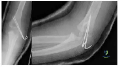
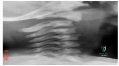
Question 4:
A patient had an elbow fracture that was openly reduced and internally fixed. The radiographs (Slide) from his first postoperative checkup are presented. Which of the following is a correct assessment:
Options:
- The fixation is adequate.
- The second pin should have been placed from the medial side.
- The two pins should have diverged in the proximal fragment.
- The pins should have been larger in diameter.
- The pins should have been buried under the skin.
Correct Answer: The two pins should have diverged in the proximal fragment.
Explanation:
This patient has a lateral condyle fracture. This type of fracture has a tendency to spread unless adequate compression is maintained. Diverging pins in the proximal fragment is the most widely advocated strategy.
Question 5:
Which of the following femur fractures is best treated with flexible intramedullary nails:
Options:
- A 12-year-old child, 45 kg, transverse fracture of the midshaft
- An 8-year-old child, 28 kg, midshaft fracture with grade 3 butterfly fragment
- An 8-year-old child, 55 kg, transverse fracture of the midshaft
- An 8-year-old child, 35 kg, oblique fracture 8 cm below the lesser trochanter
- A 4-year-old child, 20 kg, transverse midshaft fracture with 2 cm shortening
Correct Answer: An 8-year-old child, 28 kg, midshaft fracture with grade 3 butterfly fragment
Explanation:
Age older than 10 years, weight greater than 50 kg, and length-unstable fractures are associated with poor results after insertion of flexible intramedullary nails. The subtrochanteric region is defined in children as 10% femur length below the lesser trochanter. Children age 5 and younger are best treated with immediate spica cast.
Question 6:
A mutation in the gene for peripheral myelin protein 22 causes which of the following disorders:
Options:
- Friedreich ataxia
- Charcot-Marie-Tooth disease
- Cerebral palsy
- Adrenoleukodystrophy
- Fragile X syndrome
Correct Answer: Charcot-Marie-Tooth disease
Explanation:
Type I, or the hypertrophic demyelinating form, is the most common form of Charcot-Marie-Tooth disease. It is due to a mutation in the gene on chromosome 22, which encodes for peripheral myelin protein 22. Type I C harcot-Marie-Tooth disease has a prevalence of approximately 1 in 5,000 individuals.
Question 7:
Which of the following statements is true of scoliosis in patients with C harcot-Marie-Tooth disease:
Options:
- Scoliosis responds well to bracing.
- Spinal cord monitoring is usually normal.
- Scoliosis is rarely progressive.
- Thoracic curves more commonly have an apex convex to the left than the right.
- Thoracic kyphosis is less than in idiopathic scoliosis.
Correct Answer: Thoracic curves more commonly have an apex convex to the left than the right.
Explanation:
Scoliosis is more common in patients with C harcot-Marie-Tooth disease than in the general population (between 10% to 33%). Only 15% of curves are halted by bracing. Scoliosis is commonly associated with thoracic kyphosis and with a left thoracic curve pattern. Intraoperative spinal cord monitoring is often difficult to elicit even at baseline due to the underlying neurologic disorder.
Question 8:
Risk factors for developmental dysplasia of the hip (DDH) include all of the following except:
Options:
- Oligohydramnios
- Firstborn child
- C ongenital muscular torticollis
- Identical twin with DDH
- Male gender
Correct Answer: Male gender
Explanation:
Developmental dysplasia of the hip is caused by both genetic and mechanical factors. C rowding and laxity are common factors. Females, pregnancies with oligohydramnios, firstborn children, infants with congenital muscular torticollis, and those with an affected identical twin are at increased risk. Also parental hip dysplasia, congenital dislocation of the knee, and breech position increase the risk of DDH.
Question 9:
In ultrasound of the infant hip, the alpha angle is defined as:
Options:
- The angle between the acetabular roof and the midline of the pelvis
- The acute angle between the lateral wall of the ilium and the extension of the acetabular roof
- The angle between the center of the femoral head and the lateral wall of the ilium
- The angle of the thigh required to produce subluxation of the hip on the sonogram
- The angle between the acetabular roof and the transverse plane
Correct Answer: The acute angle between the lateral wall of the ilium and the extension of the acetabular roof
Explanation:
The alpha angle is the acute angle between the lateral wall of the ilium and the extension of the acetabular roof. This angle varies inversely with the acetabular index as seen on plain radiographs.
Question 10:
In a sonogram, the normal alpha angle of the neonatal hip measures:
Options:
Correct Answer: <50°
Explanation:
The alpha angle is the acute angle formed by the lateral wall of the ilium and the extension of the acetabular roof. This angle varies inversely with the acetabular index as seen on plain radiographs. An angle >50° is normal.
Question 11:
Diagnosis of Duchenne muscular dystrophy in a female patient could be explained if the patient had which of the following:
Options:
- Addison disease
- Turner syndrome
- Trisomy 21
- Klinefelter syndrome
- Fragile X syndrome
Correct Answer: Turner syndrome
Explanation:
Duchenne muscular dystrophy is the most common form of muscular dystrophy, affecting one in 3,300 males. Duchenne muscular dystrophy is an X-linked recessive defect that results from the absence of dystrophin. It may be expressed in females with Turner syndrome, which is the presence of a single X chromosome. Such patients would otherwise be carriers of Duchenne muscular dystrophy but express this recessive disorder because they only have one X chromosome. Duchenne muscular dystrophy would not occur in Klinefelter syndrome, which involves the presence of an extra X chromosome, or in trisomy 21. Steroids can help mitigate the phenotype; therefore Addison disease (hypercortisonism) would not produce the disorder. Fragile X is the most common form of inherited mental retardation but affects only males.
Question 12:
Spinal muscular atrophy is due to a mutation in the gene for which of the following proteins:
Options:
- Dystrophin
- Survival motor neuron
- Peripheral myelin protein 22
- Frataxin
- Type I collagen
Correct Answer: Survival motor neuron
Explanation:
Spinal muscular atrophy is due to a mutation in the survival motor neuron gene, which results in loss of many anterior horn cells of the spinal cord. Dystrophin abnormalities cause Duchenne and Becker dystrophies. Peripheral myelin protein 22 is disordered in Charcot-Marie-Tooth disease and frataxin in Friedreich ataxia. Type I collagen disorders cause structural skeletal dysplasias.
Question 13:
Which of the following agents is contraindicated in children with open physes:
Options:
- Pamidronate
- Alendronate
- Etidronate
- Teriparatide
- C alcium carbonate
Correct Answer: Teriparatide
Explanation:
Teriparatide, or Forteo (Eli Lilly and C ompany, Indianapolis, Ind), is contraindicated in children with open physes because of a concern for the risk of osteosarcoma.
Question 14:
A 15-month-old toddler, who is neurologically intact, presents with a fracture (pic). Which of the following is the recommended treatment:
Options:
- Posterior C1-C 2 fusion
- Posterior occiput-C2 fusion
- Open reduction and screw fixation
- C 2 corpectomy and C1-C3 fusion anteriorly
- Reduction and halo vest immobilization
Correct Answer: Posterior C1-C 2 fusion
Explanation:
This odontoid physeal fracture should be treated by postural reduction and external immobilization. The reduction maneuver is posterior translation with slight axial traction, which may be accomplished by a halter or a halo vest. Immobilization must include a device such as a Minerva cast or a halo vest that can control the head well.C orrect Answer: Reduction and halo vest immobilization
Question 15:
A 17-month-old toddler sustained a femur fracture (Slide) in a fall from a height. Which of the following is the best treatment method:
Options:
- Pavlik harness
- Spica cast
- Femoral skeletal traction
- External fixation
- Flexible intramedullary nails
Correct Answer: Spica cast
Explanation:
This toddlerâ s fracture shows minimal shortening. Spica cast treatment is ideal for fractures of the femur in children younger than school age because of their portability, overgrowth, remodeling, and lack of implant to remove. A Pavlik harness does not control a child beyond the age of a few months. Femoral skeletal traction, external fixation, and flexible intramedullary nails are more invasive than is warranted.
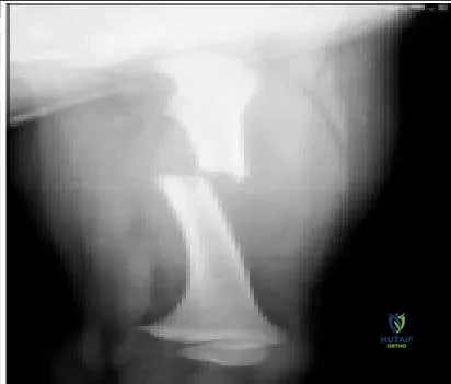
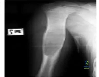
Question 16:
The lesion shown in the radiograph (pic) most likely represents which of the following processes:
Options:
- Aneurysmal bone cyst
- Fibrous dysplasia
- Unicameral bone cyst
- Osteomyelitis
- Fibrous cortical defect
Correct Answer: Unicameral bone cyst
Explanation:
The lesion shown in the radiograph is a unicameral bone cyst. The diagnosis was confirmed by aspiration and the subsequent response, filling in after autogenous marrow (pic).
An aneurysmal bone cyst is typically more septated and expansile in width, fibrous dysplasia has a more blurred zone of transition and ground-glass appearance, and a fibrous cortical defect is more eccentrically placed.
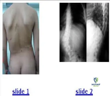
Question 17:
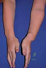
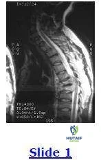
A clinical photograph (Slide 1) and radiographs (Slide 2) of a 13-year-old girl, who is neurologically normal, are presented. She does not report any pain. The most likely diagnosis is:
Options:
- Fibrous dysplasia
- Blue-rubber bleb nevus syndrome
- Idiopathic scoliosis
- Neurofibromatosis-1
- Klippel-Trenaunay syndrome
Correct Answer: Neurofibromatosis-1
Explanation:
This patient has neurofibromatosis-1. She has a dystrophic scoliosis that is sharply angulated and involves only four vertebrae (Cobb levels T12-L3). The pedicles are thinned, and the endplates are scalloped. She also has subcutaneous neurofibromas.
Question 18:
An 11-year-old girl presents with a limp. She has no history of trauma, infection, or neurologic disorder. She does not report any pain. Recommended treatment includes:
Options:
- Hip arthrodesis
- Uncemented total hip arthroplasty
- Cemented total hip arthroplasty
- Resurfacing hip arthroplasty
- No treatment
Correct Answer: No treatment
Explanation:
This patient has untreated developmental dysplasia of the hip. Her limp cannot be eliminated. If she is not experiencing pain, then no treatment is indicated.
Question 19:
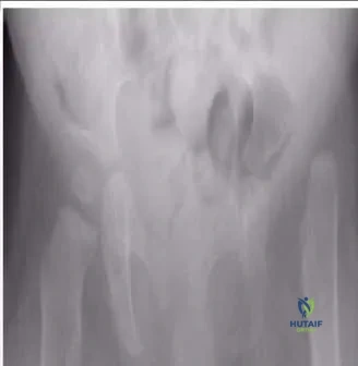
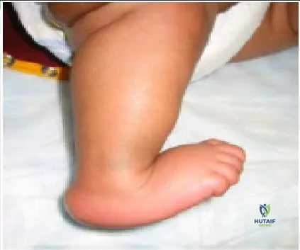
The foot pictured in this clinical photograph (pic) represents:
Options:
- Calcaneovalgus foot
- Clubfoot
- Calcaneus foot
- Congenital vertical talus
- Skewfoot
Correct Answer: Congenital vertical talus
Explanation:
The foot presented in the clinical photograph represents congenital vertical talus. Note the equinus of the hindfoot, calcaneus of the forefoot, and the crease in the sinus tarsi. In patients with a calcaneovalgus foot, the hindfoot is in calcaneus, not equinus.
Question 20:
Scapulothoracic fusion is most commonly indicated to help improve function in which of the following conditions:
Options:
- Duchenne muscular dystrophy
- Beckers muscular dystrophy
- Facioscapulohumeral dystrophy
- Charcot-Marie-Tooth disorder
- Congenital muscular dystrophy
Correct Answer: Facioscapulohumeral dystrophy
Explanation:
Facioscapulohumeral dystrophy is a rare disorder inherited in an autosomal dominant fashion. Thirty percent of affected individuals have a new mutation. The genetic abnormality is found on chromosome 4, with a decreased number of D4Z4 tandem repeats, but this does not appear to code for a protein product. In this condition, selective weakness of the serratus anterior, trapezius, and rhomboid muscles is present. Therefore, the scapula is not effectively stabilized against the trunk during use. Although the deltoid is relatively spared, it cannot work well due to a hypermobile scapula. Fusion of the scapula to the thorax improves range of abduction in this condition.
 An aneurysmal bone cyst is typically more septated and expansile in width, fibrous dysplasia has a more blurred zone of transition and ground-glass appearance, and a fibrous cortical defect is more eccentrically placed.
 This patient has neurofibromatosis-1. She has a dystrophic scoliosis that is sharply angulated and involves only four vertebrae (Cobb levels T12-L3). The pedicles are thinned, and the endplates are scalloped. She also has subcutaneous neurofibromas.
 This patient has untreated developmental dysplasia of the hip. Her limp cannot be eliminated. If she is not experiencing pain, then no treatment is indicated.
 The foot presented in the clinical photograph represents congenital vertical talus. Note the equinus of the hindfoot, calcaneus of the forefoot, and the crease in the sinus tarsi. In patients with a calcaneovalgus foot, the hindfoot is in calcaneus, not equinus.