Full Question & Answer Text (for Search Engines)
Question 1:
The best way to diagnose dysplasia epiphysealis hemimelica, in addition to history and physical, is:
Options:
- Bone scan
- Ultrasound
- Biopsy
- Plain radiograph
- Bacterial culture
Correct Answer: Plain radiograph
Explanation:
Dysplasia epiphysealis hemimelica, or Trevor disease, is an epiphyseal osteochondroma. The characteristic location, continuity with the epiphysis and alteration in growth of the epiphysis, is the most specific confirmation of the disease. Histology is less specific, as are bone scan and ultrasound.
Question 2:
Which of the following commonly used statements regarding the pelvis in patients with bladder exstrophy is true:
Options:
- The forces across the hip joints are increased in patients with bladder exstrophy.
- Athletic ability is significantly compromised in patients with bladder exstrophy.
- Significant limp persists throughout life in patients with bladder exstrophy.
- The pelvis is more vulnerable to fracture in patients with bladder exstrophy.
- Urinary continence is not possible in patients with bladder exstrophy.
Correct Answer: The forces across the hip joints are increased in patients with bladder exstrophy.
Explanation:
In patients with bladder exstrophy, computerized modeling shows that the forces across the hip are increased by approximately 30%. However, limp is not significant and the pelvis is not more prone to fracture. Most patients can be made continent through surgery.
Question 3:
A 1-year-old child requires evaluation of limb length inequality. The surgeon notices a disproportional increase in length, as well as width, of the ipsilateral upper and lower extremity. No other physical abnormalities are evident. Recommended treatment includes which of the following:
Options:
- Ultrasound periodically throughout childhood
- Brain magnetic resonance imaging (MRI)
- MRI of the affected extremities
- Spinal ultrasound
- Angiogram of the affected limbs
Correct Answer: Ultrasound periodically throughout childhood
Explanation:
Idiopathic hemihypertrophy is associated with an increased incidence of intra-abdominal tumors such as Wilmsâ tumor and hepatoblastoma. Periodic screening by ultrasound throughout childhood is recommended.
Question 4:
Which of the following statements describes the growth plate biomechanics of the distal femur:
Options:
- The collateral ligaments protect the distal femur.
- The posterior cruciate ligament protects the distal femur.
- The anterior cruciate ligaments protect the distal femur.
- The patellar ligament protects the distal femur.
- The distal femur is not protected by any ligaments.
Correct Answer: The distal femur is not protected by any ligaments.
Explanation:
Whereas the the proximal tibial physis is protected by the collateral ligaments and tibial tubercle epiphysis, the distal femoral physis is vulnerable to injury because it is not protected by any ligaments.
Question 5:
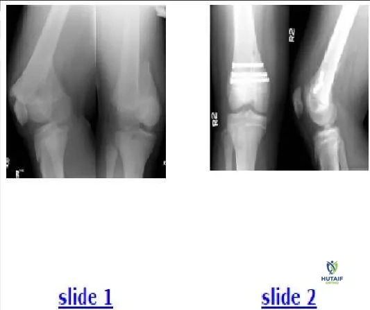
A 200-lb 13-year-old boy sustained a fracture while playing football. His radiographs are presented (Slide 1). Which of the following treatments should be attempted first:
Options:
- Closed reduction and internal fixation
- Open reduction and screw fixation
- Open reduction and plate fixation
- External fixation
- Arthroscopically assisted reduction and internal fixation
Correct Answer: Closed reduction and internal fixation
Explanation:
The radiographs show a Salter II fracture with a large metaphyseal fragment. A high likelihood exists for successful closed reduction, and the metaphyseal fragment should allow rigid fixation. This patient received closed reduction and was internally fixed with three percutaneous 7.3-mm screws (Slide 2). He did not require a cast.
Question 6:
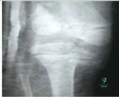
An 8-year-old boy was injured by a lawnmower. The resulting proximal tibial injury presented in the radiograph (Slide) is classified as a:
Options:
- Salter I injury
- Salter II injury
- Salter III injury
- Salter IV injury
- Salter V injury
Correct Answer: Salter IV injury
Explanation:
A Salter IV injury to the proximal tibia is apparent in the patientâ s radiograph. The fracture traverses the epiphysis, physis, and metaphysis medial to the tibial spine. No injury to the lateral portion of the plateau is present. Incidentally, the distal femoral injury is also classified as Salter IV.
Question 7:
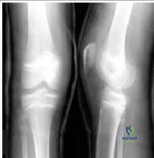
A 12-year-old boy twisted his knee while riding a bicycle. Based on his radiographs (Slide), which of the following is the most appropriate diagnosis:
Options:
- Patellar dislocation
- Traumatic osteochondritis dissecans
- Tibial spine avulsion
- Anterior cruciate ligament tear
- Tibial tubercle fracture
Correct Answer: Tibial spine avulsion
Explanation:
This patient has a tibial spine avulsion, classified as type III by McKeever and Meyers. Treatment options include closed immobilization or arthroscopic or open reduction and internal fixation.
Question 8:
What is the most common cause of intoeing in children with bilateral cerebral palsy:
Options:
- Internal pelvic rotation
- Internal hip rotation
- Increased tibial torsion
- Hindfoot rotation
- Forefoot adduction
Correct Answer: Internal hip rotation
Explanation:
The most common cause of intoeing in children with bilateral spasticity is internal hip rotation. For children with hemiplegia, the most common cause of intoeing is tibial torsion. In some patients, several causes may coexist to cause the condition.
Question 9:
Risk factors for superior mesenteric artery syndrome after adolescent idiopathic scoliosis surgery include all of the following except:
Options:
- Decreased correction of thoracic curve with preoperative bending
- Body mass index (BMI) below the 25th percentile
- Lenke lumbar modifier B or C
- Staged surgical correction
- Use of iliac crest bone graft
Correct Answer: Use of iliac crest bone graft
Explanation:
Superior mesenteric artery syndrome occurs more often in patients with decreased BMI, larger and stiffer thoracic curves, lumbar apical translation of Lenke B or C , and two staged procedures.
Question 10:
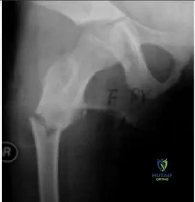
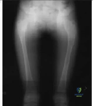
An 8-year-old girl fractures her right femur when starting a sprint. Based on her radiograph (Slide), which of the following is the most likely diagnosis:
Options:
- Unicameral bone cyst
- Aneurysmal bone cyst
- Osteogenic sarcoma
- Fibrous dysplasia
- Normal pre-existing bone
Correct Answer: Fibrous dysplasia
Explanation:
This patient has signs of a pre-existing fibrous dysplasia lesion, including a central expansion of the subtrochanteric region with a ground-glass (not lucent) appearance. She was diagnosed with fibrous dysplasia.
Question 11:
An 8-year old girl with fibrous dysplasia fractures her femur (Slide). Which of the following procedures is the best treatment option:
Options:
- Femoral traction followed by spica cast
- C urettage, bone graft, and spica cast
- Allograft strut and spica cast
- External fixation
- Hip screw with long side plate
Correct Answer: Hip screw with long side plate
Explanation:
This lesion requires mechanical support because it is vulnerable to malunion and further deformity. Bone graft would be rapidly reabsorbed and has no lasting value in this patient. A screw with a long side plate that extends well into normal bone is the best treatment option.
Question 12:
The patient presented in the radiograph (Slide) has a slight, painless limp. Which of the following is the most likely diagnosis:
Options:
- Ollier disease
- Osteogenesis imperfecta
- Fibrous dysplasia
- Neurofibromatosis
- Fibrous cortical defects
Correct Answer: Fibrous dysplasia
Explanation:
This patient has multiple â ground-glassâ lesions and one cystic lesion, as well as bowing in the subtrochanteric region. The patient was diagnosed with fibrous dysplasia.
Question 13:
A 14-year-old boy sustains an avulsion of the tibial tubercle while playing basketball. The fracture extends vertically into the joint, crossing the proximal tibial articular surface. He is at greatest risk for which of the following complications:
Options:
- Peroneal nerve injury
- Tibial nerve injury
- Popliteal artery disruption
- Anterior leg compartment syndrome
- Femoral nerve palsy
Correct Answer: Anterior leg compartment syndrome
Explanation:
Fracture of the tibial tubercle has been reported to lead to compartment syndrome, presumably due to bleeding from the geniculate vessels and the exposed bone. This would not be expected from such a proximal fracture, but orthopedic surgeons should be aware of the possible association.
Question 14:
An infant presents with idiopathic scoliosis. Which of the following factors indicates observation as the treatment of choice:
Options:
- Age younger than 1 year
- Rib-vertebral angle difference >20°
- Rib-vertebra phase 2
- C urve of 47°
- Left thoracic curve
Correct Answer: Age younger than 1 year
Explanation:
Rib-vertebral angle difference >20° rib-vertebra phase 2, and curves >45° indicate a likely progression. The majority of infantile curves are left thoracic, so this is not a factor. A patient younger than 1 year of age has a higher chance of spontaneous resolution than an older patient. Therefore, absent other risk factors, observation is the initial recommend treatment.
Question 15:
Which of the following factors predicts a lesser degree of impairment of pulmonary function in patients with adolescent idiopathic scoliosis:
Options:
- Increased C obb angle
- Decreased number of vertebrae in the curve
- Increased cephalad apex of the curve
- Decreased thoracic kyphosis
- Pectus excavatum in addition to the scoliosis
Correct Answer: Decreased number of vertebrae in the curve
Explanation:
An increased Cobb angle, increased cephalad apex of the curve, decreased thoracic kyphosis, and pectus excavatum in addition to the scoliosis are factors that predict a greater degree of pulmonary impairment. A lower number of vertebrae in the curve signals less impairment.
Question 16:
All except which of the following neurologic/muscular disorders can present undiagnosed in a patient with scoliosis:
Options:
- Friedreichâ s ataxia
- C harcot-Marie-Tooth disease
- Syringomyelia
- Duchenne muscular dystrophy
- Spinal cord tumor
Correct Answer: Duchenne muscular dystrophy
Explanation:
Scoliosis can be caused by any neurologic disorder that affects trunk balance. Scoliosis is seen in most patients with Friedreichâ s ataxia, a disorder in which patients present with an ataxic gait in preadolescence or adolescence. The curve becomes significant at about the same time as the ataxia. Scoliosis can also be seen in 10% of patients with C harcot-Marie- Tooth disease. This condition is characterized by cavus feet, intrinsic atrophy, and occasional hip dysplasia; no significant pain or clumsiness is present. Scoliosis is seen in many patients with syringomyelia. The syrinx and scoliosis both develop silently with no noticeable weakness until both the syrinx and the curve are significant. Spinal cord tumor may present with scoliosis in an ambulatory patient. Duchenne muscular dystrophy leads to scoliosis in most patients, but the patients become nonambulatory several years before the curve develops.
Question 17:
All except which of the following structural disorders often causes scoliosis and presents undiagnosed in patients:
Options:
- Marfan syndrome
- Ehlers-Danlos syndrome
- Osteogenesis imperfecta
- Loeys-Dietz syndrome
- Achondroplasia
Correct Answer: Achondroplasia
Explanation:
Although achondroplasia causes kyphosis, it is not associated with scoliosis to a significant degree. Marfan syndrome, Ehlers- Danlos syndrome, osteogenesis imperfecta, and Loeys-Dietz syndrome (a defect in TGF-beta receptor protein) are frequently associated with scoliosis.
Question 18:
Which of the following levels of evidence should be assigned to a prospective, randomized therapeutic study with 80% follow-up:
Options:
- Level I
- Level II
- Level III
- Level IV
- Level V
Correct Answer: Level I
Explanation:
A study may be level I as long as it has at least 80% follow-up.
Question 19:
A prospective comparative study should be assigned which level of evidence:
Options:
- Level I
- Level II
- Level III
- Level IV
- Level V
Correct Answer: Level I
Explanation:
A comparative study is one in which patients treated one way are compared with patients treated in another manner at the same institution. As long as the study is prospective, it can be assigned level II.
Question 20:
A case-control study should be assigned which level of evidence:
Options:
- Level I
- Level II
- Level III
- Level IV
- Level V
Correct Answer: Level I
Explanation:
A case-control study is considered level III evidence, as is a retrospective comparison study or a meta-analysis in which the lowest level of primary study is level III.