Full Question & Answer Text (for Search Engines)
Question 1:
A 12-year-old patient with fibrous dysplasia has an increasing limp and progressive bowing in the intertrochanteric and subtrochanteric regions of his femur. Recommended treatment includes:
Options:
- Allograft strut graft in situ
- Plate fixation in situ
- Rod fixation in situ
- Valgus osteotomy with plate and/or rod
- Hip arthrodesis in straight position
Correct Answer: Valgus osteotomy with plate and/or rod
Explanation:
The most important step in treating patients with fibrous dysplasia is to decrease the bending force on the patientâ s femur. A valgus osteotomy with a plate or a rod decreases the bending force on the patientâ s femur by decreasing the offset from the center of the patientâ s body mass. Allografting and plate or rod fixation in situ causes progressive bowing in the patient.
Question 2:
Which of the following bones is the most common site for involvement with fibrous dysplasia:
Options:
- Phalanges of the hand
- Radius
- Ulna
- Humerus
- Femur
Correct Answer: Femur
Explanation:
The femur and the tibia are the most common sites of fibrous dysplasia in the appendicular skeleton. Involvement of the humerus is also common. Fibrous dysplasia may also occur in the radius, ulna, and phalanges of the hand; it occurs less frequently in the femur, humerus, and pelvis.
Question 3:
What is the risk of malignant transformation over the course of a lifetime in a person with fibrous dysplasia:
Options:
- 50%
- 25%
- 15%
- 5%
- Less than 1%
Correct Answer: Less than 1%
Explanation:
Fibrous dysplasia is a relatively common disorder. The risk of malignant transformation is less than 1%. Many historic cases of malignant transformation with fibrous dysplasia occurred in patients who also received external beam irradiation.
Question 4:
The most common malignancy arising from transformation of fibrous dysplasia is:
Options:
- C hondrosarcoma
- Adamantinoma
- Giant cell tumor
- Osteosarcoma
- Fibrosarcoma
Correct Answer: Osteosarcoma
Explanation:
The most common malignancies arising from transformation of fibrous dysplasia are osteosarcoma and malignant fibrous histiocytoma. However, chondrosarcoma, giant cell tumor, adamantinoma, and fibrosarcoma have been reported to result from fibrous dysplasia. Radiation increases the risk of malignant transformation of fibrous dysplasia.
Question 5:
The pattern of genetic transmission of polyostotic fibrous dysplasia is best described as:
Options:
- Autosomal dominant with high penetrance
- Autosomal dominant with low penetrance
- Autosomal recessive
- Sex-linked dominant
- No genetic transmission (sporadic)
Correct Answer: No genetic transmission (sporadic)
Explanation:
Fibrous dysplasia is a sporadic condition. Although there are a few reports of parents and children having fibrous dysplasia, such reports are rare.
Question 6:
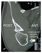
Which of the following conditions does this computerizd tomography scan (Slide) most likely represent:
Options:
- Brodies abscess
- Osteosarcoma
- Fibrous dysplasia
- Osteoid osteoma
- Enchondromas
Correct Answer: Osteoid osteoma
Explanation:
This is an osteoid osteoma of the fourth lumbar vertebra in a patient who experienced night pain and relieved the pain with nonsteroidal anti- inflammatory drugs. The location of a sclerotic nidus in the posterior elements of the vertebrae is typical for this disorder. Excision of the osteoid osteoma resulted in prompt pain relief for this patient.
Question 7:
This radiograph (Slide) shows a 9-year-old boy with scoliosis. From which of the following conditions is the boyâ s scoliosis likely to have resulted:
Options:
- Marfans syndrome
- Osteogenesis imperfecta
- Neurofibromatosis
- Fibrous dysplasia
- Juvenile idiopathic scoliosis
Correct Answer: Neurofibromatosis
Explanation:
The patientâ s scoliosis is the result of neurofibromatosis. A sharp focal curve over few vertebrae, thinning of apical pedicles, and spindling of the ribs are symptoms of neurofibromatosis that are indicated in the radiograph.
Question 8:
A 5-year-old boy presents for examination. He is diagnosed with developmental dysplasia of the hip. Recommended treatment includes:
Options:
- Closed reduction and cast application
- Traction, closed reduction, and cast
- Femoral osteotomy and closed reduction
- Open reduction and iliac osteotomy
- Open reduction, femoral osteotomy, and iliac osteotomy
Correct Answer: Femoral osteotomy and closed reduction
Explanation:
At age 5, traction or closed reduction is not likely to produce a stable joint. Femoral shortening is indicated to reduce pressure, reducing the likelihood of avascular necrosis or redislocation. The most likely option to produce a stable joint is open reduction with femoral and iliac osteotomy.
Question 9:
The radiograph (Slide) of a 16-month-old toddler is presented. Which diagnosis is most appropriate:
Options:
- Proximal focal femoral deficiency
- C ongenital coxa valga
- Traumatic hip dislocation
- Developmental dysplasia of the hip
- Femoral retroversion
Correct Answer: Developmental dysplasia of the hip
Explanation:
This patient has developmental dysplasia of the hip. The femoral head is delayed in ossifying because of lower contact pressure and the femur is anteverted, not retroverted.
Question 10:
A 4-year-old girl sustains an injury in a motor vehicle accident. She sustained a femoral artery injury, which was repaired. Her pubic diastasis is 4.5 cm. A radiograph (Slide 1) and clinical photograph (Slide 2) are presented. Which of the following treatment options is recommended:
Options:
- Accept deformity; no weight bearing for 6 to 8 weeks
- Spica cast application
- Long leg cast application
- Pelvic sling
- Open reduction internal fixation
Correct Answer: Open reduction internal fixation
Explanation:
This is an open-book injury due to direct frontal impact, which presumably also injured the femoral artery directly. A pubic diastasis .2.5 cm should be reduced. Open reduction and internal fixation is the preferred method to accomplish this, using either a wire or a plate. External fixation is also an acceptable option. Casts and sling are more likely to cause pressure sores or to be ineffective.
Question 11:
Loeys-Dietz syndrome is caused by a mutation in:
Options:
- Fibrillin -1
- Fibrillin-2
- TGF-beta receptor
- Collagen type III
- Decorin
Correct Answer: TGF-beta receptor
Explanation:
Loeys-Dietz syndrome is an autosomal dominant syndrome characterized by arterial tortuosity, aneurysms, hypertelorism, and bifid uvula or cleft palate. Scoliosis, foot deformities, ligamentous laxity, and other findings are often present. The aneurysms have particular risk for rupture at small diameters. This disorder is caused by mutations in genes encoding TGF-beta receptor 1 and 2.
Question 12:
An 11-year-old female patient with bilateral cavus feet presents with foot pain and callosities on the plantar surface of the foot. She is diagnosed with C harcot-Marie-Tooth disease and may require surgical intervention. During a standing C olemanâ s lateral block test, the patientâ s hindfoot varus corrects bilaterally when standing on a 1-inch wooden block. Which of the following surgical options is the most appropriate:
Options:
- Plantar fasciotomy
- Plantar-based opening wedge osteotomy of the medial cuneiform
- Osteotomy of the first metatarsal
- Lateral closing wedge osteotomy of the calcaneus
- Triple arthrodesis
Correct Answer: Plantar-based opening wedge osteotomy of the medial cuneiform
Explanation:
This patient has a typical cavovarus foot. The key in evaluating the treatment options is the standing block test. Because her hindfoot varus is corrected during this test, her hindfoot is flexible and a calcaneal osteotomy is not necessary. Osteotomy of the base of the first metatarsal is also not preferred in such a case because the growth plate becomes at risk for arrest, the procedure requires internal fixation, the second metatarsal head becomes at risk for a stress transfer lesion, and is not located at the side of the deformity. In C harcot-Marie-Tooth disease, the deformity apex generally lies in the tarsometatarsal articulations and the medial cuneiform.
Question 13:
Which of the following disorders is not a cause of cavus foot:
Options:
- Charcot-Marie-Tooth disease
- Friedreichâ s ataxia
- Myelomeningocele
- Poliomyelitis
- Tarsal coalition
Correct Answer: Tarsal coalition
Explanation:
All of the disorders mentioned may cause cavus foot except for tarsal coalition.
Question 14:
Based on the clinical photograph (Slide 1) and radiographs (Slide 2) of this 11-year-old boy, which of the following conditions is demonstrated:
Options:
- Idiopathic scoliosis
- Fibrous dysplasia
- Tuberous sclerosis
- Neurofibromatosis
- Diastematomyelia
Correct Answer: Neurofibromatosis
Explanation:
This patient has many typical features of neurofibromatosis 1: subcutaneous neurofibromas, large â coast of Californiaâ cafe-aulait spot with several others, and a dystrophic curve at a young age. Surgery is recommended.
Question 15:
Which of the following factors predicts an increased risk that a child sustaining a pelvis fracture will incur an unstable fracture:
Options:
- Whether the triradiate cartilages are open or closed
- C hilds body weight
- C hilds body mass index
- C hilds age
- Presence of associated injuries
Correct Answer: Whether the triradiate cartilages are open or closed
Explanation:
The closure of the triradiate cartilage of the acetabulum is associated with a significant increase in the risk of an unstable pelvis fracture, as well as the need for surgical treatment. This seems to signal the change in bone plasticity from pediatric type to adult type.
Question 16:
The mean age of triradiate cartilage closure in girls and boys is:
Options:
- 10 years for girls and 12 years for boys
- 11 years for girls and 12 years for boys
- 12 years for girls and 13 years for boys
- 12.5 years for girls and 13.5 years for boys
- 13.5 years for girls and 14.5 years for boys
Correct Answer: 12.5 years for girls and 13.5 years for boys
Explanation:
The triradiate cartilage closes at a mean of 12.5 years in girls and 13.5 years in boys. The closure of the triradiate cartilage signals the end of the peak height velocity (growth spurt). This is important for timing of scoliosis treatment and for signaling a change in pelvic fracture patterns from pediatric to adult.
Question 17:
C hildren with unstable pelvis fractures have an increased risk of late pain and dysfunction if which of the following is present:
Options:
- Triradiate cartilages are closed
- Pelvic asymmetry >0.6 cm after reduction
- Pelvic asymmetry >1.1 cm after reduction
- Aspica cast is used
- Age is older than 8 years
Correct Answer: Pelvic asymmetry >0.6 cm after reduction
Explanation:
Pediatric pelvis fractures are associated with an increased risk of late pain and dysfunction if the asymmetry after reduction is .1.1 cm.
Question 18:
Which of the following is true regarding ability to remodel after a displaced pediatric pelvic fracture:
Options:
- The pelvis will remodel if the patient is younger than 11 years old.
- The pelvis will remodel if the patient is younger than 8 years old.
- The pelvis will remodel if triradiate cartilages are open.
- The pelvis will remodel if the patient is premenarchal.
- No significant remodeling is seen at any age.
Correct Answer: No significant remodeling is seen at any age.
Explanation:
In pediatric pelvic fractures healing with asymmetry, no significant remodeling is seen at any age.
Question 19:
According to the Delbet classification, a transphyseal fracture of the pediatric proximal femur is considered type:
Options:
Correct Answer: I
Explanation:
According to the Delbet classification: Type I: Transphyseal fracture Type II: Transcervical fracture Type III: Basicervical fracture Type IV: Intertrochanteric fracture
Question 20:
According to the Delbet classification, the risk of avascular necrosis is least with which of the following pediatric hip fractures:
Options:
- Type I
- Type II
- Type III
- Type IV
- Type V
Correct Answer: Type I
Explanation:
The risk of avascular necrosis is highest with type I, followed by about equal rates for types II and III (approximately 15 %). The risk of avascular necrosis is low for type IV, intertrochanteric fractures. Type I Transphyseal Type II Transcervical Type III Basicervical Type IV Intertrochanteric

 This is an osteoid osteoma of the fourth lumbar vertebra in a patient who experienced night pain and relieved the pain with nonsteroidal anti- inflammatory drugs. The location of a sclerotic nidus in the posterior elements of the vertebrae is typical for this disorder. Excision of the osteoid osteoma resulted in prompt pain relief for this patient.
 At age 5, traction or closed reduction is not likely to produce a stable joint. Femoral shortening is indicated to reduce pressure, reducing the likelihood of avascular necrosis or redislocation. The most likely option to produce a stable joint is open reduction with femoral and iliac osteotomy.
 This patient has developmental dysplasia of the hip. The femoral head is delayed in ossifying because of lower contact pressure and the femur is anteverted, not retroverted.