Full Question & Answer Text (for Search Engines)
Question 1:
Which of the following regions of the spine is normally straight:
Options:
- T1 to T6
- T7 to T12
- T10 to L2
- L1 to L4
- T12 to S1
Correct Answer: T10 to L2
Explanation:
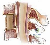
The normal range of thoracic kyphosis is 20° to 50°. The mean in normal adults is 35°. The normal range of lumbar lordosis is 40° to 80°. The mean in normal adults is approximately 60°. The spine is usually straight in the sagittal plane between T10 and L2. The majority of lumbar lordosis occurs between L4 and S1.
Question 2:
The endplates and pedicles of which of the following vertebra are normally parallel to the ground in a standing individual:
Options:
Correct Answer: L3
Explanation:
The alignment of the spine is important in normal upright posture. There is a normal degree of lordosis in the cervical and lumbar spines and a moderate degree of kyphosis in the thoracic spine. The head, spine, and pelvis are connected and balanced. If the spine is out of balance, then a deformity can develop causing fatigue of the paraspinal muscles. The normal sagittal alignment in the upright patient is as follows: Plumb line The sagittal plumb line falls from the odontoid process through the C 7-T1 intervertebral disk and then anterior to the thoracic spine. The plumb line then crosses the spine at the T12-L1 intervertebral disk, and then travels posterior to the spine. The plumb line crosses at the posterior corner of the S1 vertebra. The endplates and pedicles of the L3 vertebra are normally parallel to the ground.
Question 3:
Which of the following is true regarding the alignment of the spine with aging:
Options:
- Thoracic kyphosis decreases; lumbar lordosis increases
- Thoracic kyphosis decreases; lumbar lordosis decreases
- Thoracic kyphosis increases; lumbar lordosis decreases
- Thoracic kyphosis increases; lumbar lordosis increases
- The alignment of the spine undergoes no significant changes with aging.
Correct Answer: Thoracic kyphosis decreases; lumbar lordosis increases
Explanation:
The normal range of thoracic kyphosis is 20° to 50°. The mean in normal adults is 35°. The normal range of lumbar lordosis is 40° to 80°. The mean in normal adults is approximately 60°. The spine is usually straight in the sagittal plane between T10 and L2. The majority of lumbar lordosis occurs between L4 and S1. With aging, due to changes in the intervertebral disks, thoracic kyphosis increases and lumbar lordosis increases. There is loss of height of the intervertebral disks.
Question 4:
In reference to the normal sagittal vertical axis (sagittal plumb line), the axis normally falls from the odontoid process through the C 7-T1 intervertebral disk and anterior to the thoracic vertebra. This normal axis crosses the spinal column at which of the following levels before crossing the spinal column at the posterior superior border of the S1 vertebral body:
Options:
- T3-T4 intervertebral disk
- T6-T7 intervertebral disk
- T8-T10 intervertebral disk
- T12-L1 intervertebral disk
- L3-L4 intervertebral disk
Correct Answer: T12-L1 intervertebral disk
Explanation:
The alignment of the spine is important in normal upright posture. There is a normal degree of lordosis in the cervical and lumbar spines and a moderate degree of kyphosis in the thoracic spine. The head, spine, and pelvis are connected and balanced. If the spine is out of balance, then a deformity can develop causing fatigue of the paraspinal muscles. The normal sagittal alignment in an upright patient is as follows: Plumb line The sagittal plumb line falls from the odontoid process through the C 7-T1 intervertebral disk and then anterior to the thoracic spine. The plumb line then crosses the spine at the T12-L1 intervertebral disk, and then travels posterior to the spine. The plumb line crosses at the posterior corner of the S1 vertebra The endplates and pedicles of the L3 vertebra are normally parallel to the ground.
Question 5:
The vertebral artery on the right side of the body arises from the subclavian artery and enters the lateral mass foramen of which of the following cervical vertebra (the first one it enters) before ascending to the brain:
Options:
Correct Answer: C 6
Explanation:
The vertebral artery arises from the subclavian artery on the right side of the body and the aortic arch on the left side. The vertebral artery enters the lateral mass foramen of the sixth cervical vertebra before ascending to the brain.
Question 6:
To avoid damages to the vertebral arteries when exposing the posterior aspect of the first cervical vertebra, dissection should be limited to ______ mm from the midline on the superior aspect of C 1 and _____ mm from the midline on the posterior aspect of C 1.
Options:
- 8 mm; 12 mm
- 10 mm; 14 mm
- 12 mm; 16 mm
- 14 mm; 20 mm
- 16 mm; 22 mm
Correct Answer: 8 mm; 12 mm
Explanation:
One must be careful not to damage the vertebral artery when exposing the posterior and superior aspect of the C1 vertebra. It is especially important when using a Cobb elevator or an electrocautery not to dissect too far from the midline. The vertebral artery lies close to the midline. On the superior aspect, the groove for the vertebral artery lies 8 mm to12 mm from the midline. On the posterior aspect of the vertebral body, the vertebral artery lies 12 mm to 23 mm from the midline.
Question 7:
Which of the following levels most significantly contributes to the blood supply of the cervical spinal cord:
Options:
- C 2 (accompanying the left C 2 spinal nerve)
- C 4 (accompanying the right C 4 spinal nerve)
- C 6 (accompanying the left C 6 spinal nerve)
- C 7 (accompanying the right C 7 spinal nerve)
- T1 (accompanying the right T1 spinal nerve)
Correct Answer: C 6 (accompanying the left C 6 spinal nerve)
Explanation:
The major blood supply to the cervical spinal cord comes from the anterior spinal artery, which arises from the deep cervical artery. This vessel most commonly accompanies the left C 6 spinal nerve.
Question 8:
Patients with Brown-Séquard syndrome usually presents with:
Options:
- Ipsilateral paralysis, loss of contralateral vibration and touch sensation, and loss of ipsilateral pain and temperature sensation
- Ipsilateral paralysis and loss of contralateral vibration, and touch, pain, and temperature sensation
- Ipsilateral paralysis, loss of ipsilateral vibration and touch sensation, and loss of contralateral pain and temperature sensation
- C ontralateral paralysis, loss of ipsilateral vibration and touch sensation, and loss of contralateral pain and temperature sensation
- C ontralateral paralysis, loss of contralateral vibration and touch sensation, and loss of ipsilateral pain and temperature sensation
Correct Answer: Ipsilateral paralysis, loss of contralateral vibration and touch sensation, and loss of ipsilateral pain and temperature sensation
Explanation:
Brown-S aquard Syndrome usually results from hemisection of the spinal cord, which is often a result of trauma (eg, penetrating stab wounds). Clinical presentation usually consists of: Ipsilateral paralysis Loss of ipsilateral vibration and touch sensation Loss of contralateral pain and temperature sensation
Question 9:
Central cord syndrome is typically due to:
Options:
- An axial compression injury with resultant injury to the central gray matter
- A hyperextension injury with compression of the cord by herniated disk material anteriorly
- A hyperextension injury with compression of the cord by osteophytes anteriorly and infolded ligamentum flavum posteriorly
- A hyperflexion injury with compression of the cord by herniated disk material anteriorly
- A hyperflexion injury compression of the cord by the anterior longitudinal ligament anteriorly and osteophytes posteriorly
Correct Answer: A hyperextension injury with compression of the cord by herniated disk material anteriorly
Explanation:
Central cord syndrome is the most common incomplete spinal cord lesion and is usually seen in patients with preexisting cervical spondylosis who then sustain a hyperextension injury to the cervical spine. This mechanism causes compression of the cord by osteophytes anteriorly and the infolded ligamentum flavum posteriorly with resulting injury to the central gray matter. The clinical presentation is variable but usually consists of: Greater loss of motor neurons to the upper extremities than the lower extremities often resulting in profound weakness in the arms and hands, and some weakness in the legs and feet Variable sensory loss Patients with central cord syndrome have variable return of function but are usually left with some degree of residual deficit and spasticity.
Question 10:
Patients with anterior cord syndrome usually presents with:
Options:
- Preservation of motor function, preservation of pain and temperature sensation, and loss of vibration and touch sensation
- Preservation of motor function, with loss of pain, temperature, vibration, and touch sensation
- Motor paralysis, loss of pain, temperature, vibration, and touch sensation
- Motor paralysis, loss of pain and temperature sensation, and preservation of vibration and touch sensation
- Motor paralysis, loss of vibration and touch sensation, and preservation of pain and temperature sensation
Correct Answer: Preservation of motor function, preservation of pain and temperature sensation, and loss of vibration and touch sensation
Explanation:
Anterior cord syndrome is due to injury of the anterior elements of the spinal cord, which is usually due to a space-occupying lesion anterior to the cord such as vertebral body fracture fragments, a herniated disk, or a hematoma. The clinical presentation consists of: Complete motor paralysis (loss of anterior corticospinal tract) Loss of pain/temperature sensation (loss of lateral and anterior spinothalamic tracts) Preservation of vibration sensation/proprioception and light touch sensation (preservation of dorsal columns) In less severe cases, some motor function is preserved through the lateral corticospinal pathways. Prognosis is generally poor and in patients with absence of sacral sensation (pin prick/temperature) after 24 hours following injury, recovery is seen in 10% of patients.
Question 11:
A patient with cauda equina syndrome and the full spectrum of symptons presents with:
Options:
- Severe low back pain, sciatica, saddle anesthesia, and preservation of bladder vesicular control
- Severe low back pain, sciatica, urinary retention, and preservation of perianal sensation
- Severe low back pain, saddle anesthesia, loss of motor and sensation in the lower extremities, and preservation of bulbocavernosus reflex.
- Severe low back pain, loss of motor and sensation in the lower extremities, and preservation of bladder vesicular control
- Severe low back pain, sciatica, saddle anesthesia, urinary retention, and loss of bulbocavernosus reflex
Correct Answer: Severe low back pain, sciatica, saddle anesthesia, and preservation of bladder vesicular control
Explanation:
Cauda equina syndrome is a severe neurologic disorder that results from an injury to the neural elements within the thecal sac between the conus medullaris and the lumbosacral nerve roots (ie, cauda equina or "horse's tail"). Cauda equina syndrome usually occurs as a result of lumbar disk herniation with compression of the cauda equina and requires urgent surgical decompression. Clinical presentation includes: Severe low back pain Bilateral or unilateral sciatica Saddle anesthesia Motor or sensory deficit Bladder and bowel vesicular involvement (classically leading to urinary retention) With a complete lesion, a loss of bulbocavernosus reflex, anal wink, and reflexes in the lower extremities
Question 12:
Which of the following descriptions applies to the sacroiliac joint:
Options:
- The sacroiliac joint accounts for 15% of lower back pain.
- Pain is referred most commonly to the groin.
- Focal pain over the sacral sulcus is rare.
- Focal neurological deficits are common.
- Provocative tests (Patrick and Gaenslens) are useful predictors of joint pathology.
Correct Answer: The sacroiliac joint accounts for 15% of lower back pain.
Explanation:
Sacroiliac joint pathology accounts for 15% of lower back pain, and the sacroiliac joint is one of the most common sites of referred pain. Patients with sacroiliac joint pathology commonly experience pain above the posterior buttock and seldom have focal neurological deficits. Physical examination tests are poor predictors of sacroiliac joint pathology.
Question 13:
Which of the following statements is true regarding the sacroiliac joint:
Options:
- The anterior supporting structures are stronger than the posterior supporting structures.
- Sectioning of the sacrotuberous and sacrospinous ligaments results in increased motion.
- The sacroiliac joint withstands medially directed forces better than the lumbosacral spine.
- C ounter-nutation (forward rotation of the ilium on the sacrum) is the most common motion.
- The posterior interosseous ligaments are weak.
Correct Answer: The sacroiliac joint withstands medially directed forces better than the lumbosacral spine.
Explanation:
The sacroiliac joint is the largest axial joint in the body. The anterior capsule is thin and weaker than the posterior capsule. The posterior supporting structures are strong and are comprised of a tough interosseous ligament, a long posterior sacroiliac ligament, and strong sacrotuberous, sacrospinous ligaments. Joint innervation usually occurs anteriorly in the S2 ventral rami. Compared with the lumbosacral spine, the sacroiliac joint can better withstand medial forces, but is weaker in axial compression and in axial torsion. Nutation (backward rotation of less than 4° and 1.6 mm rotation of the ilium on the sacrum) is the most common motion in the sacroiliac joint. Increased motion of the sacroiliac joint occurs only with sectioning of the interosseous ligaments.
Question 14:
Osteochondromatosis is a hereditary genetic disorder that is:
Options:
- Autosomal recessive with incomplete penetrance
- Autosomal recessive with complete penetrance
- Autosomal dominant with incomplete penetrance
- Sex-linked dominant
- Sex-linked recessive
Correct Answer: Autosomal dominant with incomplete penetrance
Explanation:
Osteochondromatosis (also known as hereditary multiple exostoses) is a genetic disorder that is autosomal dominant with incomplete penetrance in women. The genetic defect occurs on the EXT1, EXT2, and EXT 3 genes located on chromosome 8q24.
Question 15:
Osteochondromatosis is a hereditary genetic disorder that is caused by:
Options:
- Mutation in the fibrillin-1 gene
- Translocation between chromosomes 9 and 22
- Mutation in the g-fos gene
- Translocation between chromosomes 11 and 22
- Mutation in the EXT1, EXT2, and/or EXT3 genes
Correct Answer: Mutation in the fibrillin-1 gene
Explanation:
Osteochondromatosis (also known as hereditary multiple exostoses) is a genetic disorder that is autosomal dominant with incomplete penetrance in women. The genetic defect occurs on the EXT1, EXT2, and EXT 3 genes located on chromosome 8q24. Mutation in the fibrillin-1 gene is seen in patients with Marfan syndrome. Translocation between chromosomes 9 and 22 is seen in myxoid chondrosarcoma. Mutation in the g-fos gene is seen in patients with Ollierâ s disease. Translocation between chromosomes 11 and 22 is present in patientâ s with Ewings tumor.
Question 16:
Osteochondromas in the spine most commonly occur in:
Options:
- Posterior elements of the cervical spine
- Posterior elements of the thoracic spine
- Posterior elements of the lumbar spine
- Posterior elements of the sacral spine
- Vertebral body of the thoracic spine
Correct Answer: Posterior elements of the cervical spine
Explanation:
Osteochondromas most commonly occur in the appendicular skeleton but can also occur in the spine (<5% of cases). When present in the spine, solitary osteochondromas have a predilection for the cervical spine. They can, however, also occur in the thoracic and lumbar spine. Sacral involvement is rare.
Question 17:
When an osteoid osteoma occurs in the spine, it can involve all of the following except:
Options:
- Facets
- Transverse processes
- Pedicles
- Rib heads adjacent to thoracic vertebrae
- Vertebral body
Correct Answer: Vertebral body
Explanation:
When an osteoid osteoma occurs in the spine, involvement of the posterior elements of the vertebra is typical and includes: Lamina Pedicles Transverse processes Facets Rib heads adjacent to thoracic vertebrae
Question 18:
Typical histologic features of an osteoid osteoma include all of the following except:
Options:
- C hondrocytes in an arrangement similar to that of a physis
- Nidus composed of haphazardly arranged network of osteoid trabeculae
- Varying degrees of mineralization with greatest mineralization in the center of the lesion
- Osteoblasts rimming the trabeculae
- Vascularized spindle cell stroma
Correct Answer: C hondrocytes in an arrangement similar to that of a physis
Explanation:
The histologic features of an osteoid osteoma include the following: Nidus composed of haphazardly arranged network of osteoid trabeculae Varying degrees of mineralization with greatest mineralization in the center of the lesion Loose fibrovascular connective tissue between trabeculae Osteoblasts rimming the trabeculae Vascularized spindle cell stroma
Question 19:
Treatment of a vertebral osteoid osteoma includes all of the following except:
Options:
- Surgical excision/curettage of the nidus
- En-bloc resection
- Observation if symptoms are mild
- Aspirin/salicylates/nonsteroidal anti-inflammatory drugs (NSAIDs)
- Radiofrequency ablation
Correct Answer: En-bloc resection
Explanation:
Treatment of osteoid osteomas in the spine include the following: Aspirin/salicylates/NSAIDs Administered for up to 2 years Successful in up to 50% cases Radiofrequency ablation (RFA) Usually computed tomography-guided Clinical success rates as high as 97% have been reported with 1 to 2 treatments Surgical excision of the nidus/curettage Necessary when aspirin/salicylates/NSAIDs cannot be tolerated for long periods of time and RFA is not possible or unsuccessful Can usually be accomplished through a posterior approach En-bloc resection or a more radical procedure play no role in management
Question 20:
Typical symptoms of a spinal osteoblastoma include all of the following except:
Options:
- Torticollis
- Painful scoliosis
- Stiffness
- Diskogenic pain
- Radicular symptoms
Correct Answer: Diskogenic pain
Explanation:
The most common symptoms of spinal osteoblastomas include: Pain Usually the first and most common presenting symptom Night pain is not as common as it is with osteoid osteomas Night pain is not as common as it is with osteoid osteomas Painful scoliosis Torticollis Stiffness Radicular symptoms usually due to mass effect