Full Question & Answer Text (for Search Engines)
Question 1:
Advantages of minimally invasive lumbar interbody fusion over traditional open interbody fusion include:
Options:
- Minimal muscle dissection and trauma
- Wider surgical exposure
- Better fusion rates
- Lowered risk of nerve root injury
- Better decompression
Correct Answer: Minimal muscle dissection and trauma
Explanation:
Minimally invasive lumbar interbody fusion involves less muscle dissection and trauma than traditional open approaches. The surgical exposure is more limited, though, and there is no evidence to date of minimally invasive techniques providing better fusion rates or lowered risk of nerve root injury.
Question 2:
Which of the following instruments are of value to a surgeon when performing minimally invasive lumbar fusions:
Options:
- Surgical loupes and headlight
- Operating microscope
- Intraoperative fluoroscopy
- Surgical loupes and headlight and operating microscope
- Surgical loupes and headlight, operating microscope, and intraoperative fluoroscopy
Correct Answer: Surgical loupes and headlight
Explanation:
All of the above instruments are of value to a surgeon when performing minimally invasive lumbar fusion.
Question 3:
Which of the following statements is true regarding minimally invasive posterior lumbar interbody fusion:
Options:
- Minimally invasive fusion may be safely performed only with the assistance of endoscopy.
- Minimally invasive fusion increases the risk of nerve root injury.
- Internal fixation with pedicle screws is not possible via the minimally invasive approach.
- Intraoperative fluoroscopy is of great value in minimally invasive fusion.
- Minimally invasive surgery has improved fusion rates.
Correct Answer: Minimally invasive fusion may be safely performed only with the assistance of endoscopy.
Explanation:
Intraoperative fluoroscopy or radiography is vital for the proper identification of lumbar level and vertebral structures in minimally invasive posterior lumbar interbody fusions. While endoscopic assistance has been well described as a method of minimally invasive fusion, it is not vital to this technique. There is no evidence to date of increased risk of nerve root injury with minimally invasive techniques, and it is possible to internally fixate the lumbar segment with pedicle screws through minimally invasive techniques.
Question 4:
Which of the following is not a described technique of minimally invasive anterior lumbar interbody fusion:
Options:
- Laparoscopic transperitoneal
- Endoscopic retroperitoneal
- Mini-open retroperitoneal
- All of the above are described techniques
- None of the above are described techniques
Correct Answer: All of the above are described techniques
Explanation:
All of the above are well-described techniques of minimally invasive anterior lumbar interbody fusion.
Question 5:
Which of the following statements is false regarding minimally invasive transperitoneal anterior lumbar interbody fusion:
Options:
- This technique may be safely performed at all lumbar levels.
- This technique allows direct access to pathology in the vertebral body.
- Laparoscopy is of great value in the transperitoneal approach to the anterior lumbar spine.
- There is a potential risk of injuring the aorta and its bifurcation with this technique.
- None of the above
Correct Answer: This technique may be safely performed at all lumbar levels.
Explanation:
Due to the potential risk of injury to the aorta and its bifurcation, which occurs at the L4 level, this procedure is difficult and may be impossible to perform above the L4 level. Retroperitoneal approaches allow access to more superior lumbar levels due to the more lateral trajectory taken to avoid the aorta and its bifurcation.
Question 6:
All of the following are elements of the lateral mass of cervical spinal segments except:
Options:
- Inferior articulating process
- Superior articulating process
- Spinous process
- Transverse process
- Transverse foramen
Correct Answer: Spinous process
Explanation:
The lateral mass of the cervical spinal segments includes the inferior and superior articulating processes, the transverse foramen, and the transverse process. The spinous process is not an element of the lateral mass.
Question 7:
To avoid vertebral artery injury during cervical lateral mass screw placement, it is best to:
Options:
- Start at the midpoint and aim the screw laterally
- Start at the midpoint and aim the screw medially
- Start medially and aim the screw perpendicular
- Start medially and aim the screw medially
- Start laterally and aim the screw medially.
Correct Answer: Start at the midpoint and aim the screw laterally
Explanation:
To avoid injury to the vertebral artery when placing lateral mass screws, it is best to avoid placing the screw in the medial portion of the lateral mass, where the vertebral body is most likely to be found.
Question 8:
Which of the following is/are potential complications associated with posterior cervical decompression and placement of lateral mass screws:
Options:
- Injury to the vertebral artery
- Compression injury to the spinal cord
- Traction injury to the cervical nerve roots
- Injury to the vertebral artery and traction injury to the cervical nerve roots only
- Injury to the vertebral artery, traction injury to the cervical nerve, and traction injury to the cervical nerve roots
Correct Answer: Injury to the vertebral artery
Explanation:
All of the above are potential complications associated with posterior cervical decompression and placement of lateral mass screws.
Question 9:
Which of the following statements is true regarding the C 2 lateral mass:
Options:
- The vertebral artery assumes a more lateral position at this level.
- The vertebral artery assumes a more medial position at this level.
- The vertebral artery is found outside of the transverse foramen at this level.
- The vertebral artery precludes placement of lateral mass screws at this level.
- None of the above
Correct Answer: The vertebral artery assumes a more lateral position at this level.
Explanation:
The vertebral artery assumes a more lateral position at the C 2 level; therefore, screw placement at this level should follow a medial trajectory to avoid injury to the vertebral artery.
Question 10:
The technique for C1-C 2 lateral mass fixation may involve:
Options:
- Removal of the posterior arch of C1
- Placing the C2 screws through the pedicle
- Following a medial trajectory with the C 1 screws
- Removal of the posterior arch of C 1, and placing the C 2 screws through the pedicle only
- Removal of the posterior arch of C1, placing the C2 screws through the pedicle, and following a medial trajectory with the C1 screws
Correct Answer: Removal of the posterior arch of C1
Explanation:
The C 1 and C 2 levels have unique anatomies that require variation in lateral mass screw fixation technique. Removing the C1 arch assists in proper placement of the C 1 screws via a lateral trajectory. The C 2 pedicle is large, and pedicle screws arecommonly placed at this level to avoid vertebral artery injury in the small lateral masses. C 1 lateral mass screws follow the long axis of the C 1 lateral mass as visualized on pre-operative C T scanning.
Question 11:
Which of the following conditions is not associated with cervical fractures:
Options:
- Rheumatoid arthritis
- Ossiculum terminale
- Ankylosing spondylitis
- Os odontoideum
- None of the above
Correct Answer: Ossiculum terminale
Explanation:
Rheumatoid arthritis, ankylosing spondylitis, and os odontoideum have been associated with fractures as part of their presentation or etiology. Os odontoideum is most likely an old nonunion fracture or injury to vascular supply of the developing odontoid process. However, one has to differentiate true os odontoideum from the more common ossiculum terminale, which describes the nonunion of the apex at the secondary ossification center and is not a fracture.
Question 12:
Which of the following pathogens is not typically implicated in diskitis:
Options:
- Staphylococcus aureus
- Staphylococcus albus
- Pseudomonas aeruginosa
- Staphylococcus epidermidis
- Gram-positive cocci
Correct Answer: Pseudomonas aeruginosa
Explanation:
The gram-positive cocci are typical opportunistic pathogens that are capable of causing infection in the vertebral disk space. Most commonly they seed via the hematogenous route but local translocation has also been implicated. Unless a patient has been hospitalized for a while and iatrogenesis is ruled out, Pseudomonas species usually do not cause diskitis.
Question 13:
Which imaging modality is usually the least sensitive in diagnosing discitis:
Options:
- Plain radiograph
- C omputed tomography (C T) scan
- Magnetic resonance image (MRI)
- Technetium bone scan
- Tomograms
Correct Answer: Plain radiograph
Explanation:
The least helpful modality in diagnosing early discitis is the plain radiograph. Fluoroscopy does not give insight into the state of the intervertebral disk. It can suggest loss of disk height or involvement of the vertebral bone but will not reveal infection limited to the disk. The CT scan is useful because of its excellent resolution of bony structures and associated changes secondary to disk infection. MRI is the best modality to characterize the soft tissues in the cervical spine.
Question 14:
Potts disease is most commonly treated by:
Options:
- Decompression
- Antibiotic therapy and immobilization
- Antibiotic therapy only
- Spinal orthosis
- Decompression and fusion
Correct Answer: Antibiotic therapy and immobilization
Explanation:
The treatment of tuberculous involvement of the spine is rarely surgical. Most commonly, the spine remains stable and fusion is not necessary. However, orthosis in combination with long-term antibiotic therapy is the key for successful treatment. A collar is sufficient to provide enough stability and comfort for the lesion to heal.
Question 15:
Which of the following is characteristic of patients with Klippel-Feil syndrome:
Options:
- Absence of the vertebral pedicles
- Absence of intervertebral joints
- Shortened pedicles
- A narrow spinal canal
- Increased interpediculate distance
Correct Answer: Absence of intervertebral joints
Explanation:
Klippel-Feil syndrome is a rare disorder characterized by the congenital fusion of any two of the seven cervical vertebrae. The cause is a failure in the early segmentation during fetal development. The fused segments show absence of intervertebral joints. Associated abnormalities may include scoliosis; spina bifida; anomalies of the kidneys and ribs; and other midline anomalies.
Question 16:
A burst fracture results in failure of the:
Options:
- Anterior column
- Middle column
- Posterior column
- Anterior and middle columns
- Middle and posterior columns
Correct Answer: Middle column
Explanation:
A burst fracture by definition is failure of the anterior and middle columns due to axial loading, which often leads to instability and neurologic impairment.
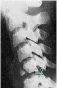
Question 17:
What type of fracture is presented in the radiograph (Slide):
Options:
- Teardrop fracture
- Burst fracture
- Compression fracture
- Hangmans fracture
- Clay-shovelers fracture
Correct Answer: Teardrop fracture
Explanation:
Clearly seen in this radiograph is a fracture along the anterior/inferior vertebral body, which is a characteristic of a teardrop fracture.
Question 18:
What type of fracture is presented in the radiograph (Slide):
Options:
- Teardrop fracture
- Burst fracture
- Compression fracture
- Hangmans fracture
- Clay-shovelerâ s fracture
Correct Answer: Teardrop fracture
Explanation:
Clearly seen in this radiograph is a fracture along the anterior/inferior vertebral body, which is a characteristic of a teardrop fracture.
Question 19:
Which of the following may be used as treatment options for bilateral facet dislocations:
Options:
- Traction reduction of dislocations
- Halo fixation
- Open reduction
- Open fixation
- All of the above
Correct Answer: All of the above
Explanation:
All of the choices are used in the treatment of bilateral jumped facets, often in combination or sequence.
Question 20:
Which of the following fracture types is the most stable fracture:
Options:
- Teardrop fracture
- Burst fracture
- Unilateral facet dislocation
- Hangmans fracture
- C lay-shovelers fracture
Correct Answer: C lay-shovelers fracture
Explanation:
The avulsion of part or all of the spinous process that occurs after a violent flexion motion is a one-column injury. The injury is a stable fracture treated by external orthosis, which rarely results in neurologic impairment. The other answer choices may be considered stable in some instances, but none of them are stable all of the time.