Full Question & Answer Text (for Search Engines)
Question 1:
Clinical symptoms of lumbar spinal stenosis usually correlate with a canal anteroposterior diameter of less than:
Options:
- 20 mm
- 15 mm
- 10 mm
- 5 mm
- There is no correlation.
Correct Answer: 10 mm
Explanation:
The clinical syndrome of lumbar stenosis correlates with a measured anteroposterior diameter of the dural sac of less than 10 mm.
Question 2:
Which of the following is the most commonly fractured location along the thoracolumbar axis:
Options:
- The cervicothoracic junction
- The mid-thoracic region
- The thoracolumbar region
- The lumbar region
- The lumbosacral junction
Correct Answer: The thoracolumbar region
Explanation:
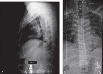
Up to 60% of spinal injuries occur between the T11 and L1 segments. The rigid thoracic rib cage and coronal orientation of the facets permit lateral bending and rotation but little flexion and extension. The facet joints then transition caudally to a more sagittal orientation in the lumbar spine, allowing increased flexion/extension but limiting lateral motion. These factors create a stress concentration at the thoracolumbar junction, which is demonstrated by the high incidence of injury at the T11 to L1 segments.
Question 3:
A 26-year-old man who was involved in a motor vehicle accident is found to have a T12 compression fracture on plain radiography without evidence of posterior extrusion. The likelihood of finding another fracture in the spinal axis with further evaluation is:
Options:
- Highly remote, these fractures are usually isolated
- 10% to 15%
- 50% to 75%
- A low thoracic fracture is almost always associated with another fracture in the spine.
- The incidence of noncontiguous-associated fractures in the spine is not known.
Correct Answer: 10% to 15%
Explanation:
A thorough workup in these patients is essential; approximately 10% to 15% of patients will have noncontiguous injuries located elsewhere in the spine.
Question 4:
Based on the three-column model of spinal stability, an unstable spinal injury is defined as:
Options:
- An injury that disrupts no less than all three columns
- Disruption of any of the three columns is considered unstable.
- Disruption of more than one column
- Disruption of all three columns plus neurological injury
- The three-column model of injury is not a reliable marker of instability.
Correct Answer: Disruption of any of the three columns is considered unstable.
Explanation:
The three-column spine consists of the anterior, middle, and posterior columns. In this widely used classification system, the middle column is the key to instability. If the middle column is disrupted, in addition to either the anterior or posterior columns, then instability results.
Question 5:
Burst fractures of the vertebral body require prompt evaluation because:
Options:
- Although burst fractures of the vertebral body are stable injuries, neurologic deterioration is likely.
- Burst fractures of the vertebral body involve two-column injury and are unstable.
- Burst fractures of the vertebral body are extremely painful to the patient.
- Burst fractures of the vertebral body are commonly associated with other noncontiguous fractures.
- Burst fractures of the vertebral body often result in spinal shock.
Correct Answer: Burst fractures of the vertebral body involve two-column injury and are unstable.
Explanation:
When the middle column is involved in a compression injury, it is classified as a burst fracture. This involves axial load on the spine, with or without a flexion component, and retropulsion of the posterosuperior vertebral body into the spinal canal, thus requiring prompt medical attention. Neurologic deficit is variable and is related to the severity of the initial injury and location of the fracture, and only loosely related to the percent of canal compromise.
Question 6:
Compression fractures of the spine, although typically considered a one- column injury, can be unstable. Findings at time of presentation suggestive of an unstable fracture include:
Options:
- Pain out of proportion to the physical examination
- Radiographic findings of more than one compression fracture
- Initial kyphosis greater than 20° to 30°
- Loss of less than 50% of anterior vertebral body height
Correct Answer: Initial kyphosis greater than 20° to 30°
Explanation:
Compression fractures are inherently stable and may be treated with extension bracing or casting. If, however, the flexion injury is severe enough, damage to the posterior ligaments can result and the injury becomes unstable. Criteria for this instability were developed by McAfee and include more than 20° to 30° of initial kyphosis or more than 50% loss of anterior vertebral height, applicable to both compression and burst fractures.
Question 7:
Which of the following is the most important factor responsible for a decreasing proportion of patients with complete paraplegia after sustaining a spinal cord injury today compared with four decades ago:
Options:
- Improvements in rehabilitative measures
- Advances in operative techniques and instrumentation
- New and novel medication therapy
- Higher patient motivation and participation in therapy and rehabilitation
- Better initial triage, resuscitation, and clinical management of patients
Correct Answer: Better initial triage, resuscitation, and clinical management of patients
Explanation:
Improvements in the initial triage, resuscitation, and clinical management of spinal cordâ injured patients are likely responsible for a decreasing proportion of patients with complete paraplegia. C urrently, approximately 45% of spinal cordâ injured patients have a complete injury, as opposed to two-thirds four decades ago.
Question 8:
Based on the current consensus on treatment of acute spinal cord injury, intravenous steroid treatment is considered to have potential benefit if begun within how many hours of original injury:
Options:
Correct Answer: 8
Explanation:
The results of the National Acute Spinal Cord Injury Study II (NASC IS II) demonstrated significant motor and sensory improvement in patients who were treated within 8 hours of injury with a methylprednisolone bolus of 30 mg/kg, followed by an infusion of 5.4 mg/kg per hour for 24 hours.
Question 9:
A 73-year-old woman with a history of cervical stenosis who sustained a fall at home yesterday is now complaining of â clums fingers and weakness in her hands. She denies any difficulty with ambulation or bowel and bladder dysfunction. She most likely has:
Options:
- Bilateral cervical radiculopathy
- Exacerbation of cervical stenosis
- Anterior cord syndrome
- Posterior cord syndrome
- C entral cord syndrome
Correct Answer: C entral cord syndrome
Explanation:
The most common incomplete spinal cord injury syndrome is most likely central cord syndrome. Central cord syndrome often occurs as a result of a pinching of the spinal cord in elderly patients who have a narrowed spinal canal as the result of degenerative spondylosis. It is a pattern of disproportionately severe upper extremity motor and sensory changes as compared to lower extremity findings.
Question 10:
A 27-year-old man was involved in a motor vehicle accident. He was resuscitated at the scene but was noted to have a prolonged hypotensive period. Upon arrival at the medical center, he is noted to be paraplegic but radiographic evaluation does not demonstrate any fracture or soft tissue abnormality. Which of the following is the most likely diagnosis:
Options:
- Occult fracture with retropulsion into the cord
- Contusion of the cord at a high thoracic level
- Spinal shock
- Spinal cord ischemic injury at the low thoracic watershed zone
- Conversion disorder
Correct Answer: Spinal cord ischemic injury at the low thoracic watershed zone
Explanation:
A watershed zone refers to an area that is supplied purely by end arteries. Therefore, during periods of hypoperfusion, it is the most likely region to sustain an ischemic injury. In the spinal cord, this region lies in the T7-T9 region as it is a watershed zone between the rostral anterior spinal artery distribution and the caudal dominant lumbar segmental artery.
Question 11:
The watershed zone of the spinal cord most closely correlates with which region of the spinal cord:
Options:
- C 5-C 7
- T4-T6
- T7-T9
- T11-L1
- L3-L5
Correct Answer: T7-T9
Explanation:
A watershed zone refers to an area that is supplied purely by end arteries. Therefore, during periods of hypoperfusion, it is the most likely region to sustain an ischemic injury. In the spinal cord, this region lies in the T7-T9 region as it is a watershed zone between the rostral anterior spinal artery distribution and the caudal dominant lumbar segmental artery.
Question 12:
Which of the following is the most common source of infection in vertebral osteomyelitis:
Options:
- Trauma
- Iatrogenic
- Hematogenous spread
- Spontaneous
- Unknown mechanism
Correct Answer: Hematogenous spread
Explanation:
Hematogenous seeding from another primary source is the most common causative agent. Hematogenous spread of infections is believed to affect the spine via septic emboli in the endarteriolar circulation of segmental spinal arteries at the vertebral endplates. The majority of cases of pyogenic spondylitis begin in the subchondral, metaphyseal region of the anterior subligamentous portion of the vertebral body â the portion with the greatest arterial supply and the most anastomoses.
Question 13:
Which of the following is the most common location of vertebral osteomyelitis along the spinal axis:
Options:
- C raniocervical junction
- Thoracic spine
- Lumbar spine
- Sacral spine
- C ervical spine
Correct Answer: Lumbar spine
Explanation:
Lumbar spine is the most common region of the spine affected by hematogenous spread of organisms leading to osteomyelitis followed by the thoracic spine.
Question 14:
Which of the following is the most common organism identified in cases of vertebral osteomyelitis:
Options:
- Staphylococcus aureus
- Streptococcus pneumoniae
- Haemophilus influenzae
- Escherichia coli
- Anaerobic gram-negative rods
Correct Answer: Staphylococcus aureus
Explanation:
Staphylococcus aureus remains the most common causative organism, but an increasing proportion of cases are due to gramnegative and anaerobic organisms such as Proteus, Escherichia coli and Pseudomonas.
Question 15:
Which of the following is the most common presentation of vertebral osteomyelitis:
Options:
- Fever of unknown origin
- Lower extremity pain and weakness
- Unrelenting back pain not relieved by rest
- Urinary incontinence
- None of the above. It is usually an incidental finding during an unrelated work-up.
Correct Answer: Unrelenting back pain not relieved by rest
Explanation:
The most common presenting sign of vertebral osteomyelitis is back pain and malaise, often of 3 monthsâ duration or greater. It is often well localized to the affected level and the nature is not unlike most degenerative spinal conditions. A high index of suspicion is essential to make a timely diagnosis. Back pain that awakens a patient at night is a hallmark of infection or tumor. Pain associated with infection tends to be relentless and not related to activity level. Most patients have percussion tenderness over the involved segments. Fevers are noted in fewer than half of patients.
Question 16:
Which of the following is the hallmark distinguishing feature of vertebral osteomyelitis when compared to a neoplastic process on imaging:
Options:
- Uniform enhancement after administration of gadolinium in the neoplasia
- Destruction of disk space and encroachment of adjacent vertebral body in vertebral osteomyelitis
- Lack of endplate involvement in the neoplastic process
- Evidence of a compression fracture in vertebral osteomyelitis
- There are no distinguishing radiographic features between vertebral osteomyelitis and a tumor.
Correct Answer: Destruction of disk space and encroachment of adjacent vertebral body in vertebral osteomyelitis
Explanation:
The crossing of the infectious process along the disk space to involve adjacent vertebrae is a hallmark feature of osteomyelitis used to differentiate it from a neoplastic process.
Question 17:
Which of the following is the imaging modality of choice with the highest relative sensitivity and specificity in patients with suspected vertebral osteomyelitis:
Options:
- Plain radiography
- C omputed tomography with contrast administration
- Magnetic resonance imaging with contrast administration
- Post myelogram computed tomography
- Vertebral osteomyelitis is primarily a clinical diagnosis
Correct Answer: Magnetic resonance imaging with contrast administration
Explanation:
Magnetic resonance imaging (MRI) is the modality of choice for spinal infections. An MRI study provides excellent visualization of the neural elements and can determine whether the inflammatory process extends beyond the margins of disk and bone. MRI also provides excellent regional anatomic information. Scans performed with and without intravenous gadolinium are diagnostic in 90% to 95% of cases.
Question 18:
Neurogenic shock is defined as:
Options:
- Decreased cardiac output due to increased parasympathetic tone
- Severe volume depletion leading to hypotension
- Widespread gram-negative septicemia with hypoperfusion
- Loss of sympathetic tone and widespread vasodilation
- Increased cardiac output due to decreased parasympathetic tone
Correct Answer: Widespread gram-negative septicemia with hypoperfusion
Explanation:
Neurogenic shock is a unique hemodynamic alteration in patients with spinal cord injuries who have their sympathetic outflow disrupted in addition to the interruption of the motor and sensory pathways. The loss of sympathetic tone to the heart and peripheral vasculature leads to bradycardia and hypotension.
Question 19:
The normal range of thoracic kyphosis is:
Options:
- 0° to 10°
- 5° to 20°
- 20° to 50°
- 35° to 50°
- 40° to 60°
Correct Answer: 20° to 50°
Explanation:
The normal range of thoracic kyphosis is 20° to 50°. The mean in normal adults is 35°. The normal range of lumbar lordosis is 40° to 80°. The mean in normal adults is approximately 60°. The spine is usually straight in the sagittal plane between T10 and L2. The majority of lumbar lordosis occurs between L4 and S1.
Question 20:
The normal range of lumbar lordosis is:
Options:
- 0° to 10°
- 10° to 20°
- 20° to 50°
- 40° to 80°
- 60° to 90°
Correct Answer: 40° to 80°
Explanation:
The normal range of thoracic kyphosis is 20° to 50°. The mean in normal adults is 35°. The normal range of lumbar lordosis is 40° to 80°. The mean in normal adults is approximately 60°. The spine is usually straight in the sagittal plane between T10 and L2. The majority of lumbar lordosis occurs between L4 and S1.