Full Question & Answer Text (for Search Engines)
Question 1:
If the C 5 cervical spine nerve root is injured during a decompression of the cervical spine, then sensation is lost over which of the following areas:
Options:
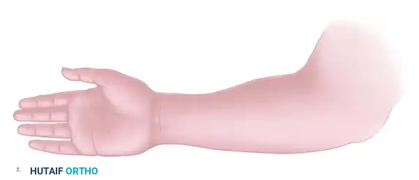
- The lateral aspect of the arm from the shoulder to the elbow
- The medial aspect of the arm from the shoulder to the elbow
- The lateral border of the forearm including the thumb
- The middle finger
- The medial border of the forearm including the little finger
Correct Answer: The lateral aspect of the arm from the shoulder to the elbow
Explanation:
The C 5 cervical spine nerve root supplies sensation from the lateral aspect of the arm from the shoulder to the elbow. C5 Lateral aspect of the arm from the shoulder to the elbow C6 Lateral border of the forearm including the thumb C7 Middle finger C8 Medial border of the forearm including the little finger T1 Medial aspect of the arm from the shoulder to the elbow
Question 2:
If the C 6 cervical spine nerve root is injured during a posterior decompression of the cervical spine, then sensation is lost in which of the following areas:
Options:
- The lateral aspect of the arm from the shoulder to the elbow
- The medial aspect of the arm from the shoulder to the elbow
- The lateral border of the forearm including the thumb
- The middle finger
- The medial border of the forearm including the little finger
Correct Answer: The lateral aspect of the arm from the shoulder to the elbow
Explanation:
If the C 6 cervical spine nerve root is injured during a posterior decompression of the cervical spine, then sensation is lost in which of the following areas: C 5 Lateral aspect of the arm from the shoulder to the elbow C 6 Lateral border of the forearm including the thumb C 7 Middle finger C 8 Medial border of the forearm including the little finger T1 Medial aspect of the arm from the shoulder to the elbow
Question 3:
If the C 7 cervical spine nerve root is injured during a posterior decompression of the cervical spine, then sensation is lost in which of the following areas:
Options:
- The lateral aspect of the arm from the shoulder to the elbow
- The medial aspect of the arm from the shoulder to the elbow
- The lateral border of the forearm including the thumb
- The middle finger
- The medial border of the forearm including the little finger
Correct Answer: The middle finger
Explanation:
The C7 cervical spine nerve root supplies sensation to the skin over the volar aspect of the middle finger. C 5 Lateral aspect of the arm from the shoulder to the elbow C 6 Lateral border of the forearm including the thumb C 7 Middle finger C 8 Medial border of the forearm including the little finger T1 Medial aspect of the arm from the shoulder to the elbow
Question 4:
If the C 8 cervical spine nerve root is injured during a posterior spinal decompression, then sensation is lost over which of the following areas:
Options:
- The lateral aspect of the arm from the shoulder to the elbow
- The medial aspect of the arm from the shoulder to the elbow
- The lateral border of the forearm including the thumb
- The middle finger
- The medial border of the forearm including the little finger
Correct Answer: The medial border of the forearm including the little finger
Explanation:
The C8 cervical spine nerve root supplies sensation to the medial border of the forearm including the little finger. C5 Lateral aspect of the arm from the shoulder to the elbow C 6 Lateral border of the forearm including the thumb C7 Middle finger C8 Medial border of the forearm including the little finger T1 Medial aspect of the arm from the shoulder to the elbow
Question 5:
If the brachioradialis reflex is diminished after a posterior spinal decompression, then which of the following nerve roots is injured:
Options:
Correct Answer: C 6
Explanation:
The brachioradialis reflex is mediated by the C 6 cervical spine nerve root. C 5 Biceps C 6 Brachioradialis C 7 Triceps
Question 6:
If the triceps muscle is weak after a spinal decompression, then which of the following nerve roots is injured:
Options:
Correct Answer: C 7
Explanation:
The triceps muscle extends the elbow and is innervated by the C 7 cervical spine nerve root. Motor innervations include: Shoulder abduction (deltoid) - - C 5 Elbow flexion - - C 5 Wrist extension - - C 6, C 7 Wrist flexion - - C 7 Finger extension - - C 7 Finger flexion - - C 8 Finger abduction/adduction - - T1
Question 7:
If the flexor carpi radialis is weak after a spinal decompression, then which of the following nerve roots is injured:
Options:
Correct Answer: C 7
Explanation:
The flexor carpi radialis is the most powerful wrist flexor and is innervated by the C 7 cervical spine nerve root. The flexor carpi ulnaris, which is weaker than the flexor carpi radialis, is innervated by the C 8 cervical spine nerve root.
Question 8:
A patient has a fracture dislocation of the cervical spine. Which of the following nerve roots must be spared to preserve intact finger extension:
Options:
Correct Answer: C 7
Explanation:
Finger extensors are innervated by the C 7 cervical spine nerve root. Motor innervations include: Shoulder abduction (deltoid) - - C 5 Elbow flexion - - C 5 Wrist extension - - C 6, C 7 Wrist flexion - - C 7 Finger extension - - C 7 Finger flexion - - C 8 Finger abduction/adduction - - T1
Question 9:
Which of the following nerve roots supplies motor innervation to the flexor digitorum superficialis (FDS):
Options:
Correct Answer: C 8
Explanation:
The FDS flexes the proximal interphalangeal joint and is innervated by the C 8 cervical spine nerve root. The FDS is innervated peripherally by the median nerve. The flexor digitorum profundus flexes the distal interphalangeal joint and is also innervated by the C 8 cervical spine nerve root. The middle and index fingers are supplied by the median nerve, and the ring and little fingers are supplied by the ulnar nerve.
Question 10:
A patient with a herniated disk has a diminished patellar tendon reflex. Which of the following lumbosacral nerve roots is affected:
Options:
Correct Answer: L4
Explanation:
The patellar tendon reflex is primarily transmitted through the L4 lumbosacral nerve root. Although the L4 lumbosacral nerve root is the primary transmitter, the L2 and L3 lumbosacral nerve roots also contribute to the fibers. A weak reflex is present if the L4 lumbosacral nerve root is completely cut and fibers of the L2 and L3 lumbosacral nerve roots are still present. The patellar tendon reflex is seldom completely absent unless a patient has primary muscle or anterior horn lesions.
Question 11:
A patient with radicular pain is experiencing skin numbness on the medial aspect of his leg and great toe. Which of the following nerve roots is effected:
Options:
Correct Answer: L4
Explanation:
When examining patients, it is important to remember the sensory dermatomes. The medial aspect of the leg, foot, and great toe are supplied by the L4 lumbosacral nerve root. The tibial crest separates the L4 and L5 dermatomes on the leg. L4 Medial aspect of leg, foot, and great toe L5 Lateral aspect of the leg and toes 2 through 4 S1 Lateral aspect of the fifth toe
Question 12:
If the extensor hallucis longus muscle is weak in a patient who has radicular pain, then which of the following lumbosacral nerve roots is compressed:
Options:
Correct Answer: L5
Explanation:
The extensor hallucis longus muscle is primarily innervated by the L5 lumbosacral nerve root. The L5 lumbosacral nerve root innervates the following muscles: Extensor hallucis longus Extensor digitorum longus and extensor digitorum brevis Gluteus medius
Question 13:
If the extensor digitorum longus and extensor digitorum brevis muscles are weak in a patient who has radicular back pain, then which of the following lumbosacral nerve roots is compressed:
Options:
Correct Answer: L5
Explanation:
The extensor hallucis longus muscle is primarily innervated by the L5 lumbosacral nerve root. The L5 lumbosacral nerve root innervates the following muscles: Extensor hallucis longus Extensor digitorum longus and extensor digitorum brevis Gluteus medius
Question 14:
Testing of the L5 lumbosacral nerve root in a patient who has radicular back pain can be accomplished through which of the following reflexes or tests:
Options:
- Patellar tendon reflex
- Achilles tendon reflex
- Tibialis posterior reflex
- Superficial anal reflex
- Beevor sign
Correct Answer: Tibialis posterior reflex
Explanation:
Although there is not a well-defined reflex arc for the L5 lumbosacral nerve root, the tibialis posterior reflex can be elicited. The tibialis posterior reflex is mediated through the L5 lumbosacral nerve root. Reflexes and associated nerve roots include: Patellar tendon --- L4 Achilles tendon --- S1 Superficial anal reflex --- S2, S3, S4 Beevor sign refers to asymmetry of the segmental innervation of the rectus abdominus muscles and when performing a situp, there is unilateral segmental nerve root loss.
Question 15:
A patient with radicular pain is experiencing skin numbness on the lateral aspect of the leg and the dorsum of the foot between the second and fourth toes. Which of the following nerve roots is being compressed:
Options:
Correct Answer: L5
Explanation:
The L5 dermatome covers the skin on the lateral leg and dorsum of the foot from the lateral border of the great toe to the medial border of the little toe. L4 Medial aspect of leg, foot, and great toe L5 Lateral aspect of the leg and toes 2 through 4 S1 Lateral aspect of the fifth toe
Question 16:
If the peroneus longus and peroneus brevis muscles are weak in a patient who has radicular back pain, then which of the following nerve roots is compressed:
Options:
Correct Answer: S1
Explanation:
The peroneus brevis and peroneus longus muscles are principally innervated by the S1 nerve root through the superficial peroneal nerve. Although the nerve is principally innervated by the S1 nerve root, the superficial peroneal nerve is derived from the L5, S1, and S2 nerve roots. The muscles principally innervated by the S1 nerve root are the: Peroneus longus and peroneus brevis Gastrocnemius-soleus complex Gluteus maximus
Question 17:
The left medial and lateral gastrocnemius muscles are weak in a patient after a lumbar spine decompression. Which of the following nerve roots is injured:
Options:
Correct Answer: S1
Explanation:
The medial and lateral gastrocnemius muscles are principally innervated by the S1 nerve root through the tibial nerve. Although the nerve is principally innervated by the S1 nerve root, the tibial nerve is derived from the L5, S1, and S2 nerve roots. The muscles principally innervated by the S1 nerve root are the: Peroneus longus and peroneus brevis Gastrocnemius-soleus complex Gluteus maximus
Question 18:
The Achilles tendon reflex (ankle reflex) is absent in a patient who has radicular back pain. Which of the following nerve roots is compressed:
Options:
Correct Answer: S1
Explanation:
The Achilles tendon reflex is based on the triceps muscle group (medial and lateral gastrocnemius muscles and soleus muscle) and is transmitted through the S1 nerve root. Reflexes and associated nerve roots include: Patellar tendon reflex L4 Posterior tibial reflex L5 Achilles tendon reflex S1
Question 19:
A patient with cauda equina syndrome has decreased perianal sensation. Which of the following groups of nerve roots is involved:
Options:
- L2, L3, and L4
- L3, L4, and L5
- L5, S1, and S2
- S1, S2, and S3
- S2, S3, S4, and S5
Correct Answer: S2, S3, S4, and S5
Explanation:
Perianal sensation is derived from the S2, S3, S4, and S5 nerve roots. The sensory distribution is as follows: S4-S5 - - Innermost perianal ring S3 - - Middle perianal ring S2 - - Outermost perianal ring
Question 20:
A patient with a fracture dislocation of the spine has a sensory level at the nipple line. Which of the following nerve root levels indicates this finding:
Options:
Correct Answer: T4
Explanation:
In addition to knowing the innervation of selected muscles and the deep tendon reflexes, the clinician should also know the sensory levels to localize pathologic processes. T4 Nipple line T7 Xiphoid process T10 Umbilicus T12 Groin