Corrective Osteotomy for Forearm Diaphyseal Malunions: An Intraoperative Masterclass
Key Takeaway
This masterclass details corrective osteotomy for forearm malunions, crucial for restoring function. We cover comprehensive anatomy, meticulous preoperative planning, and granular, step-by-step intraoperative execution of volar, dorsal, and ulnar approaches. Fellows will learn precise instrument use, critical neurovascular protection, and strategies for optimal reduction and plating, ensuring restoration of the radial bow and forearm kinematics.
Introduction and Epidemiology
Malunion of the radial or ulnar shaft represents a complex reconstructive challenge that can lead to debilitating pain, loss of motion, loss of grip strength, and profound instability at the level of the wrist or elbow. Malrotation, angulation (with subsequent narrowing of the interosseous space between the radius and ulna), shortening, and loss of the radial bow have been shown in various biomechanical and clinical studies to lead to significantly decreased functional outcomes.

Arthritis has been extensively reported at the level of the proximal radioulnar joint (PRUJ) in the setting of long-standing malunions, although the distal radioulnar joint (DRUJ) is most commonly affected by forearm malunions due to alterations in load transmission and ulnar variance. Both-bone forearm fractures occur through a variety of mechanisms, including indirect trauma (such as falls on an outstretched arm or motor vehicle collisions) and direct trauma (such as high-energy blows to the forearm). Acute diaphyseal fractures treated non-operatively with closed reduction and casting, or those managed with flexible intramedullary nailing techniques, demonstrate a statistically higher propensity to heal in a malunited position compared to those treated with rigid plate osteosynthesis.

Radius malunions assert a substantially greater deleterious effect on forearm rotation than isolated ulna malunions. A torsional deformity of greater than 30 degrees in the radius inevitably leads to a significant loss of forearm motion, primarily due to the complex kinematic relationship between the two bones. Furthermore, changes in the length-tension curve of the interosseous membrane resulting from radial shortening or angulation further restrict rotation and alter longitudinal load transfer across the radiocarpal and ulnocarpal joints. Corrective osteotomy aims to restore the precise three-dimensional anatomy of the radius, thereby re-establishing native kinematics, alleviating pain, and preventing progressive joint degeneration.
Surgical Anatomy and Biomechanics
The forearm functions biomechanically as an articulated ring, connected proximally at the PRUJ, centrally via the interosseous membrane (IOM), and distally at the DRUJ. Force transmission occurs primarily through the central band of the interosseous membrane, transferring axial loads from the radius distally to the ulna proximally.

Radius Anatomy and Kinematics
The radius lies parallel to the ulna in full supination. During pronation, the radius rotates around the ulna, while the ulna maintains a relatively static position throughout the arc of forearm rotation. The fibers of the interosseous membrane are at their maximum length with the forearm in neutral rotation and shorten in both terminal pronation and supination.
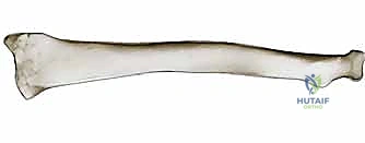
The radial shaft is triangular in cross-section, with the apex directed medially toward the attachment of the interosseous membrane. It contains three distinct surfaces: anterior, lateral, and posterior. The diaphyseal shaft possesses a gentle, complex bow, with the volar surface concave and the dorsal and lateral surfaces convex. Schemitsch and Richards devised a critical radiographic formula that locates the apex and defines the magnitude of the radial bow for each individual, which is essential for anatomical restoration.

Ulna and Radioulnar Joints
The ulna is a long bone characterized by a triangular cross-section in its proximal two-thirds, transitioning to a circular cross-section distally. It possesses three surfaces: anterior, posterior, and medial. The proximal half of the ulnar shaft is slightly concave volarly, whereas the distal half remains relatively straight.
The PRUJ consists of the radial head, the radial notch of the ulna, the annular ligament, and the quadrate ligament. Distally, the DRUJ consists of the sigmoid notch of the radius, the ulnar head, the dorsal and volar radioulnar ligaments, the extensor carpi ulnaris (ECU) subsheath, and the triangular fibrocartilage complex (TFCC).

Biomechanical studies indicate that a loss of the radial bow magnitude by more than 5% or a shift in the location of the maximum bow alters the axis of rotation, leading to impingement of the radius against the ulna. Rotational malalignment alters the tension of the DRUJ ligaments; excessive pronation deformity of the radius relative to the ulna tightens the dorsal radioulnar ligament, while supination deformity tightens the volar radioulnar ligament, leading to restricted terminal motion and chronic DRUJ pain.
Indications and Contraindications
Patient selection is paramount when considering a corrective osteotomy of the radius. The primary goal is the restoration of function and alleviation of pain caused by mechanical impingement or joint incongruity.
Operative Indications
Surgical intervention is indicated in symptomatic patients demonstrating objective functional deficits directly attributable to the osseous deformity. Prophylactic osteotomy in asymptomatic patients is generally not recommended, though impending DRUJ arthrosis in a young, high-demand patient may warrant early intervention.
| Indication Parameter | Clinical Presentation | Radiographic Findings |
|---|---|---|
| Functional Deficit | Pronation/Supination arc < 100 degrees | Impingement of radius on ulna |
| Pain | Activity-related forearm or wrist pain | DRUJ subluxation or incongruity |
| Deformity Magnitude | Visible clinical deformity, grip weakness | Angulation > 10°, Malrotation > 30° |
| Ulnar Variance | Ulnar-sided wrist pain, TFCC symptoms | Positive ulnar variance > 2-3mm |
| Radial Bow Loss | Loss of rotation, interosseous space narrowing | > 5% loss of maximum radial bow |
Contraindications
Absolute contraindications include active surgical site infection, chronic osteomyelitis, and medically unstable patients unable to tolerate general or regional anesthesia. Relative contraindications encompass advanced, irreversible osteoarthritic changes at the radiocarpal joint or DRUJ, where salvage procedures (e.g., Darrach procedure, Sauvé-Kapandji procedure, or total wrist arthrodesis) may be more appropriate. Poor soft tissue envelopes, severe osteoporosis compromising hardware fixation, and non-compliant patients also represent significant relative contraindications.
Pre Operative Planning and Patient Positioning
Successful corrective osteotomy relies heavily on meticulous preoperative planning. Standard posteroanterior (PA) and true lateral radiographs of both the affected and contralateral (normal) forearms must be obtained. The contralateral limb serves as the patient's internal template for determining the native magnitude and location of the radial bow, as well as native ulnar variance.
Advanced Imaging and Virtual Surgical Planning
While plain radiography provides baseline metrics, high-resolution computed tomography (CT) of both forearms is now considered the gold standard for complex malunions. Three-dimensional (3D) reconstructions allow for precise quantification of angular, translational, and rotational deformities.

Virtual surgical planning (VSP) software enables the surgeon to mirror the contralateral healthy radius onto the deformed radius. This facilitates the exact calculation of the osteotomy plane, the required wedge angle (opening or closing), and the degree of derotation needed.

Patient-specific instrumentation (PSI), including 3D-printed cutting guides and pre-contoured plates, has significantly enhanced the accuracy of deformity correction while reducing intraoperative fluoroscopy and tourniquet times. The guides are designed to fit precisely onto the unique topography of the malunited bone, dictating the exact trajectory of the oscillating saw and pre-drilling trajectories for the fixation construct.
Patient Positioning
The patient is placed in the supine position on the operating table. A radiolucent hand table or arm board is attached to the operative side. A pneumatic tourniquet is applied to the proximal brachium. The entire upper extremity is prepped and draped in a standard sterile fashion, allowing access to the iliac crest if autogenous structural bone grafting is anticipated. Intraoperative fluoroscopy (C-arm) must be positioned to allow unhindered orthogonal views of the entire forearm, wrist, and elbow.
Detailed Surgical Approach and Technique
The choice of surgical approach depends on the location of the malunion, the direction of the deformity apex, and the planned position of the fixation plate.
Volar Henry Approach
The volar approach is generally preferred for middle and distal third radial diaphyseal malunions. It utilizes the internervous plane between the brachioradialis (innervated by the radial nerve) and the flexor carpi radialis (innervated by the median nerve).

The incision is made along a line connecting the biceps tendon proximally to the radial styloid distally. The superficial fascia is incised, and the plane between the brachioradialis and FCR is developed. The radial artery is identified and carefully retracted ulnarly. Proximally, the supinator muscle is identified. To expose the proximal radius safely, the forearm is maximally supinated, and the supinator is detached from its ulnar insertion and reflected radially, protecting the posterior interosseous nerve (PIN) within its substance. Distally, the pronator quadratus and flexor pollicis longus are elevated to expose the volar cortex.
Dorsal Thompson Approach
The dorsal approach may be indicated for proximal third malunions or when a dorsal opening wedge is planned. It exploits the internervous plane between the extensor carpi radialis brevis (radial nerve) and the extensor digitorum communis (PIN).

The incision extends from the lateral epicondyle toward the Lister tubercle. The fascia is incised, and the interval between the ECRB and EDC is developed. The supinator is exposed, and the PIN must be meticulously identified and protected as it exits the supinator to innervate the extensor mass. The supinator is then elevated off the radius to expose the malunion site.
Osteotomy Execution
Once the malunion site is adequately exposed, the periosteum is elevated sparingly to preserve osseous vascularity. If PSI guides are utilized, they are applied to the bone footprint and secured with K-wires.

For an opening wedge osteotomy (preferred to restore length and correct positive ulnar variance), a single transverse or oblique cut is made at the apex of the deformity using a fine-toothed oscillating saw under continuous saline irrigation to prevent thermal necrosis. The distal segment is mobilized. Soft tissue contractures, particularly the interosseous membrane and brachioradialis, may require fractional lengthening or release to achieve the planned correction. The radius is derotated and angulated to match the preoperative template.
Bone Grafting and Fixation
An opening wedge defect requires structural support. Tricortical autograft harvested from the iliac crest is the gold standard, providing both osteoconductive scaffold and osteoinductive factors, alongside structural integrity. The graft is meticulously contoured to fit the exact dimensions of the defect.

Fixation is achieved using a robust, low-profile plate. A 3.5mm limited contact dynamic compression plate (LC-DCP) or locking compression plate (LCP) is typically utilized. The plate should be pre-contoured to accommodate the restored radial bow.

The plate is applied to the tension side of the bone (typically volar for the radius). At least three bicortical screws (six cortices) must be achieved both proximally and distally to the osteotomy site. Locking screws are advantageous in osteoporotic bone or when bridging a large structural graft.

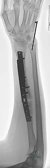
Intraoperative fluoroscopy is utilized to confirm plate position, screw length, restoration of the radial bow, and congruent reduction of the DRUJ and PRUJ. The forearm is taken through a full arc of supination and pronation to ensure absence of impingement and stability of the radioulnar joints.
Complications and Management
Corrective osteotomies of the forearm are technically demanding procedures associated with a distinct complication profile. Meticulous surgical technique and adherence to biomechanical principles mitigate these risks.
| Complication | Incidence Range | Etiology and Management Strategy |
|---|---|---|
| Nonunion / Delayed Union | 3% - 10% | Caused by inadequate fixation, thermal necrosis, or poor soft tissue envelope. Managed with revision osteosynthesis and autologous bone grafting. |
| Hardware Failure | 2% - 5% | Result of nonunion, premature weight-bearing, or insufficient plate length. Requires revision plating. |
| Nerve Injury (PIN/SRN) | 1% - 4% | Iatrogenic traction or direct laceration during exposure. Prevented by strict adherence to internervous planes. Most traction neurapraxias resolve spontaneously. |
| DRUJ Instability | 5% - 12% | Failure to restore precise length/rotation, or unrecognized TFCC tear. May require secondary soft tissue reconstruction or ulnar shortening osteotomy. |
| Infection | 1% - 3% | Superficial infections managed with oral antibiotics. Deep space infections require aggressive surgical debridement, hardware retention (if stable), and IV antibiotics. |

Hardware failure and nonunion are significant concerns, particularly in cases involving large opening wedges with inadequate structural grafting or insufficient plate length. If nonunion occurs, revision surgery with robust locking plates and fresh autogenous cancellous bone grafting is mandatory.

Persistent DRUJ instability following a technically successful radial osteotomy suggests chronic ligamentous insufficiency. In such scenarios, if the bony anatomy has been perfectly restored, secondary procedures such as TFCC repair, stabilization using the flexor carpi ulnaris (e.g., Adams-Berger procedure), or salvage operations may be indicated.
Post Operative Rehabilitation Protocols
Postoperative rehabilitation must be tailored to the rigidity of the fixation and the quality of the host bone. The primary goal is to initiate early range of motion to prevent adhesive capsulitis and soft tissue contractures while protecting the osteotomy site from excessive torsional or axial loads.
Phase 1 Immediate Postoperative (Weeks 0-2)
The patient is placed in a bulky soft dressing supported by a volar and dorsal plaster splint, immobilizing the wrist and elbow, with the forearm in neutral rotation. Elevation and active digital range of motion are encouraged immediately to reduce edema and prevent extensor tendon adhesions.
Phase 2 Early Motion (Weeks 2-6)
At the two-week mark, sutures are removed. If rigid fixation was achieved, the patient is transitioned to a removable Munster-style or long-arm thermoplastic splint. Active and active-assisted range of motion of the elbow, wrist, and forearm (pronation/supination) is initiated under the guidance of a specialized hand therapist. Passive stretching and forceful mobilization are strictly prohibited during this phase to prevent hardware failure or displacement of the structural graft.
Phase 3 Strengthening and Consolidation (Weeks 6-12)
Radiographs are obtained at 6 weeks to assess callus formation and graft incorporation. Once clinical and radiographic signs of early union are present, the splint is discontinued. Progressive resistance exercises and grip strengthening are introduced. Patients can gradually resume activities of daily living, but heavy lifting and high-impact activities remain restricted.
Phase 4 Return to Activity (Months 3-6)
Upon radiographic confirmation of complete union, all restrictions are lifted. Patients are cleared for heavy manual labor and contact sports. Maximal functional recovery of forearm rotation may take up to one year postoperatively as soft tissues remodel.
Summary of Key Literature and Guidelines
The evolution of corrective osteotomy for radial malunion is deeply rooted in biomechanical research and advancing imaging technologies.
The foundational work by Schemitsch and Richards (1992) remains the cornerstone of modern forearm reconstruction. Their study unequivocally demonstrated that restoration of the radial bow is the single most critical factor in regaining functional forearm rotation following diaphyseal fractures. They established that failure to restore the magnitude and location of the bow leads to significant loss of pronation and supination, cementing the need for precise anatomic contouring of fixation plates.

Trousdale et al. further elucidated the effects of angular deformities, noting that angulation greater than 10 degrees in any plane significantly alters the kinematics of the PRUJ and DRUJ, predisposing the patient to early-onset osteoarthritis and chronic pain. Their work highlights the necessity of multiplanar correction during osteotomy.
More recently, Jupiter and colleagues have published extensively on the utilization of 3D computer-assisted virtual surgical planning for forearm malunions. Their outcomes demonstrate that PSI significantly decreases operative time, reduces fluoroscopy exposure, and yields superior radiographic and clinical outcomes compared to traditional freehand osteotomy techniques. The integration of VSP is now widely considered the standard of care in academic orthopedic centers for the management of complex, multiplanar diaphyseal malunions of the radius.
Clinical & Radiographic Imaging
You Might Also Like