Atypical Lipomatous Tumor (ALT) & Well-Differentiated Liposarcoma: Epidemiology, Genetics, & Surgical Management
Key Takeaway
Atypical Lipomatous Tumor (ALT), also known as Well-Differentiated Liposarcoma (WDL), is an intermediate-grade, locally aggressive neoplasm characterized by adipocytic differentiation and MDM2/CDK4 amplification. While typically non-metastatic, it poses risks of local recurrence and dedifferentiation into higher-grade liposarcomas. Management differs significantly between extremity and challenging retroperitoneal locations, demanding precise surgical planning due to varying anatomical complexities.
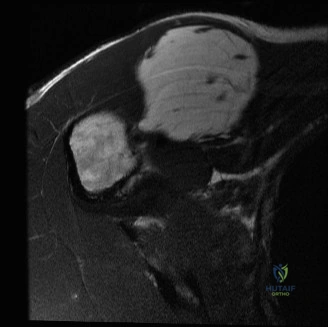
A 55-year-old patient presents with a slowly enlarging, painless, deep-seated mass in the proximal thigh. An MRI scan shows a large, predominantly lipomatous lesion with thick, irregular septations and nodular non-fatty components. What is your differential diagnosis, and what is your immediate priority?
Candidate: I would be concerned about an Atypical Lipomatous Tumor (ALT) or well-differentiated liposarcoma, given the deep location and the presence of septations/nodules on the MRI. My priority is to confirm the diagnosis via image-guided core needle biopsy and then discuss the case at a sarcoma multidisciplinary team (MDT) meeting.
A failing candidate suggests an immediate "excision biopsy" to remove the mass. This is a critical error, as it risks improper planning of the incision, lack of oncological margins, and contamination of the surgical bed, which significantly complicates definitive wide local excision.
The candidate must state: "My priority is staging and tissue diagnosis. I would request an MRI of the entire thigh (if not already done) to assess neurovascular involvement. I would perform an ultrasound-guided core needle biopsy, ensuring the tract is placed so it can be excised in the final definitive surgery. I would order MDM2/CDK4 immunohistochemistry and FISH analysis on the biopsy specimen to confirm the molecular signature of ALT/WDL, and definitively present the case to a specialist Sarcoma MDT prior to any surgical planning."
The patient is confirmed to have an ALT. During your discussion with the patient, they ask, "Since this is low-grade, can we just watch it and avoid surgery?" How do you respond?
Candidate: I would explain that while ALT is slow-growing and doesn't typically metastasize, it is locally aggressive and has a significant risk of local recurrence if not removed. Most importantly, I would highlight the risk of "dedifferentiation," where the tumor transforms into a higher-grade, more aggressive sarcoma that *can* metastasize, which makes surgical removal the standard of care.
Failing to mention the biological risk of "dedifferentiation." Candidates who focus only on the mass causing pain/functional issues miss the core oncological rationale for surgery in ALT: preventing the transformation to a lethal, high-grade liposarcoma.
The candidate should structure the answer by explaining: (1) Local progression/mass effect risk, (2) The potential for dedifferentiation into a higher-grade sarcoma, (3) The difficulty of treating a recurrence if it occurs, and (4) The importance of achieving an R0 (negative margin) resection now while the tumor is in its well-differentiated state. The "Gold Standard" mentions that non-operative management is strictly reserved for patients with absolute medical contraindications to surgery.
You are in the operating theater performing the resection. How do you ensure an oncologically sound "wide local excision"?
Candidate: I would use a longitudinal incision to allow for extensile exposure. I would aim for an en bloc resection, taking a cuff of at least 1-2 cm of normal tissue around the tumor. Crucially, I would include the biopsy tract in my incision to prevent recurrence along the track. I would also preserve major neurovascular structures unless they are macroscopically invaded.
Ignoring the biopsy tract or failing to articulate the need for a 3D margin (taking deep margins against bone/fascia). Candidates who don't mention orientation of the specimen for the pathologist (using sutures/clips) lose points on the practical "surgical safety" aspect of the exam.
A perfect answer discusses: (1) Longitudinal incision planning, (2) En bloc removal (avoiding piecemeal excision), (3) Inclusion of the biopsy scar/track, (4) Use of internal anatomical barriers (like fascia), (5) Careful dissection around critical structures, (6) Marking the specimen for the pathologist for margin orientation, and (7) Meticulous hemostasis to prevent hematoma-related complications.
