Diffuse Tenosynovial Giant Cell Tumor (TGCT) of the Knee: Pathophysiology & Surgical Anatomy
Key Takeaway
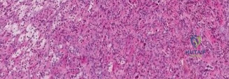
Tenosynovial Giant Cell Tumor (TGCT), diffuse type, formerly PVNS, is a rare, locally aggressive proliferative disorder primarily affecting the knee. Characterized by synovial hypertrophy, it leads to cartilage destruction and pain. Its etiology involves a CSF1 gene translocation, overexpressing CSF1, which recruits macrophages driving tumor growth and joint damage.
A 35-year-old female presents with a 2-year history of progressive swelling, pain, and a mechanical catching sensation in her left knee. She describes recurrent effusions that resolve with rest but return quickly upon activity. Physical examination reveals a diffuse synovial thickening and a moderate effusion. What is your primary differential diagnosis, and what imaging would you request?
Candidate: Given the age, chronic nature, and clinical presentation of "mechanical block" with recurrent effusions, I suspect Pigmented Villonodular Synovitis (PVNS), now referred to as diffuse-type Tenosynovial Giant Cell Tumor (TGCT). I would request an MRI of the knee to characterize the lesion, assess for hemosiderin deposition, and check for extra-articular extension.
Candidates often jump straight to suggesting an arthroscopy or biopsy. Failing to mention MRI as the gold standard for staging or failing to recognize the "blooming artifact" on GRE sequences is a major oversight. Additionally, some candidates forget to mention other differentials like chronic inflammatory arthropathies or synovial chondromatosis.
A structured response is essential: "My primary differential is diffuse-type TGCT (formerly PVNS). Other considerations include synovial chondromatosis, rheumatoid arthritis, or tubercular synovitis. I require an MRI with T1, T2, and GRE sequences. I will be looking for low-signal synovial proliferation—indicative of hemosiderin—and a 'blooming artifact' on the GRE sequence. I will also assess for extra-articular extension through capsular defects, particularly in the posterior compartments, which dictates the surgical approach."
Look at this MRI image. What are the key features demonstrated, and how does this change your surgical planning for this patient?
Candidate: The MRI shows extensive, low-signal synovial hypertrophy consistent with hemosiderin deposition, characteristic of diffuse TGCT. The involvement of the posterior recesses is critical. This necessitates a comprehensive synovectomy, and because of the posterior involvement, I would plan for a trans-septal arthroscopic approach or potentially an open posterior arthrotomy if there is extra-articular extension near the neurovascular bundle.
Ignoring the anatomy. Candidates who say "I'll do an arthroscopic synovectomy" without specifying the *posterior compartment* or the *trans-septal portal* display a lack of depth. They often forget that the popliteal artery is at risk during deep posterior dissection.
The perfect answer highlights the danger: "The scan confirms diffuse synovial disease. Crucially, I must evaluate the proximity of the tumor to the popliteal vessels. I would plan a systematic 360-degree synovectomy. I will use superolateral and superomedial portals for the suprapatellar pouch. For the posterior compartments, I will employ the Gillquist maneuver to establish a trans-septal portal. If the imaging suggests the tumor encases the neurovascular bundle, I would shift from arthroscopic to an open posterior approach to ensure safe, oncologically sound resection under direct vision."
The patient returns 18 months post-operatively with recurrent knee swelling and restricted motion. The MRI shows recurrence of the disease. What is the evidence-based management strategy for recurrent diffuse TGCT?
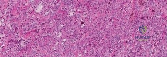
Candidate: Recurrence is common, occurring in 30-50% of cases. I would refer the patient to a multidisciplinary tumor board. Options include repeat surgical synovectomy, but if the patient has had multiple surgeries or has significant arthrofibrosis, I would consider systemic therapy with a CSF1R inhibitor like pexidartinib, or possibly adjuvant radiation therapy.
Suggesting immediate repeat surgery without considering the patient's functional state or the high risk of further arthrofibrosis. Failing to mention the multidisciplinary tumor board or newer systemic therapies like CSF1R inhibitors marks the candidate as out of date with current literature (e.g., the ENLIVEN trial).
A high-scoring answer demonstrates modern management: "Management of recurrent TGCT must be multidisciplinary. Repeat surgery has diminishing returns and increases the risk of severe stiffness. I would discuss the role of medical management using CSF1R inhibitors (e.g., pexidartinib), particularly if the tumor is unresectable or causing significant morbidity. If the patient has advanced degenerative changes, a TKA with a formal synovectomy may be necessary. Adjuvant treatments like Yttrium-90 radiosynoviorthesis or external beam radiation are also considered as part of a tailored, multi-modal strategy."
