Chondrosarcoma Diagnosis: A Detailed Clinical & Imaging Case Study
Key Takeaway
Diagnosing distal femur chondrosarcoma starts with X-rays revealing lytic lesions and 'rings and arcs' calcification. MRI details soft tissue extension; CT maps cortical destruction; PET-CT aids systemic staging. Definitive diagnosis relies on image-guided biopsy, confirming atypical chondrocytes and tumor grade, as exemplified by this Grade II case.
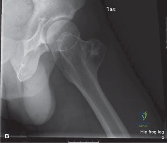
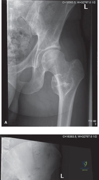
A 58-year-old male presents with 8 months of insidious, worsening distal thigh pain, now constant and disrupting sleep. Physical exam reveals a 6cm firm, fixed mass deep to the vastus lateralis. Based on the radiographs provided, what is your initial assessment and how would you characterize the lesion?
Candidate: Given the age and symptoms, I suspect a primary bone malignancy, likely a chondrosarcoma. The radiographs show an aggressive, lytic metadiaphyseal lesion with cortical destruction and "rings and arcs" calcification, suggesting a chondroid matrix. The history of night pain and the palpable mass point toward a high-grade or locally aggressive process.
Candidates often jump straight to "it's a cancer" without describing the lesion in academic terms. Failing to mention the "wide zone of transition," "endosteal scalloping," or the specific chondroid matrix morphology demonstrates a lack of radiographic fluency. Ignoring the clinical differentiation between mechanical vs. rest pain is also a major oversight.
A structured response is essential: 1. Radiographic Description: "I see an aggressive, lytic metadiaphyseal lesion of the distal femur with a wide zone of transition, cortical breakthrough, and a pathognomonic 'rings and arcs' calcified matrix." 2. Clinical Correlation: "The patient’s rest pain and the clinical finding of a fixed, palpable mass correlate with an aggressive, likely malignant, cartilaginous lesion." 3. Differential: "Primary considerations are conventional chondrosarcoma vs. chondroblastic osteosarcoma. Metastatic disease is less likely given the matrix, but must be ruled out via staging."
The biopsy confirms a Grade 2 conventional chondrosarcoma. You are now planning the surgical management. Discuss the oncologic principles for resection and the reasoning behind your reconstructive choice.
Candidate: Conventional chondrosarcomas are chemo- and radio-resistant, so the primary treatment is wide surgical excision to achieve negative margins. Given the location and the Enneking Stage IIB status, I would perform a distal femoral resection and reconstruct with a modular endoprosthesis, likely a rotating hinge knee.
Candidates often fail to explicitly state that chondrosarcoma is resistant to adjuvant therapy. They might suggest simple curettage, which is a disastrous mistake for a Grade 2 lesion, leading to inevitable local recurrence. They also often neglect the importance of the biopsy tract management.
A high-scoring answer addresses: 1. Oncologic Logic: "Because these tumors are resistant to chemotherapy and radiation, local control relies entirely on wide surgical margins." 2. Stage-Appropriate Surgery: "This is an Enneking IIB lesion; intralesional surgery is contraindicated. En bloc resection is required." 3. Reconstruction Rationale: "I would use a modular endoprosthetic replacement. A rotating hinge is mandatory here because the collateral ligaments are resected, and the hinge design reduces torsional stress at the bone-implant interface, lowering the risk of aseptic loosening."
