Grand Rounds: Giant Cell Tumor of Bone – A Detailed Diagnostic Case Study
Key Takeaway
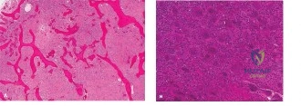
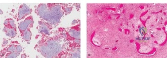
Giant Cell Tumor (GCT) diagnosis involves a comprehensive approach, typically starting with clinical presentation like pain and swelling. Imaging, including X-ray, CT, and MRI, reveals characteristic lytic, expansile lesions and assesses soft tissue extension. Definitive diagnosis relies on histopathological confirmation via biopsy, showing multinucleated osteoclast-like giant cells within mononuclear stromal cells.
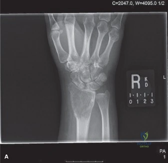
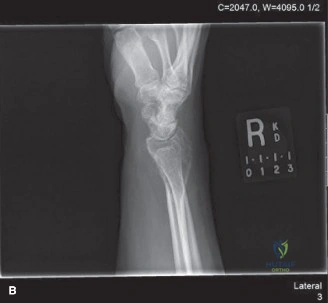
A 28-year-old male presents with 6 months of worsening lateral knee pain. Examination reveals minimal effusion but no mass. You are presented with these initial radiographs. Describe your findings and formulate a differential diagnosis.
Candidate: The radiograph shows a lytic lesion in the distal femur metadiaphysis extending into the epiphysis. It appears eccentric with a narrow zone of transition and no sclerotic rim. There is cortical thinning. My differentials would include a Giant Cell Tumor, Chondroblastoma, or potentially an Aneurysmal Bone Cyst.
Candidates often jump straight to "It's a GCT" without describing the lesion systematically (Location, Zone of Transition, Matrix, Cortical involvement). Failing to mention skeletal maturity (fused physis) is a critical error, as it narrows the differential significantly.
Systematically describe: 1. Location (epiphyseal-metaphyseal, subchondral involvement), 2. Character (purely lytic, eccentric, expansile), 3. Margins (narrow zone of transition, lack of sclerotic rim), 4. Host bone (skeletally mature). Key differential: GCTB is the primary consideration given the age and location. Must exclude Chondroblastoma (usually skeletally immature), ABC (usually metaphyseal, fluid-fluid levels), and Telangiectatic Osteosarcoma (more aggressive, wide zone of transition).
You have confirmed a Giant Cell Tumor of the distal femur via biopsy. The patient is a 28-year-old male. Based on the imaging provided, how would you classify this lesion, and what are the implications for surgical management?
Candidate: This is a Campanacci Grade III lesion because there is cortical destruction and extraosseous extension. Historically, these required wide resection, but I would now consider extended intralesional curettage with high-speed burring and adjuvants, provided the joint surface is stable.
Candidates often forget to justify the treatment. Simply saying "curettage" is insufficient. A high-scoring answer must mention addressing the "microscopic disease" in the cavity walls, which is the cause of the 50% recurrence rate in simple curettage.
Classify as Campanacci Grade III/Enneking Stage 3. Explain the surgical goal: "Local control with limb preservation." Discuss the technique: Extended curettage (burring 2-3mm of subchondral bone), use of chemical adjuvants (Phenol/Hydrogen Peroxide), and thermal adjuvants (PMMA). Justify PMMA for its three roles: thermal necrosis, structural support, and radiographic surveillance.
You have opted for extended curettage and PMMA reconstruction. Why is prophylactic internal fixation necessary in this case, and what is your follow-up protocol for local recurrence?
Candidate: Prophylactic fixation with a locking plate is used because the cortical destruction and the large lateral cortical window create a significant stress riser and structural defect that cement alone cannot reliably bridge, risking a pathological fracture. For recurrence, I would follow them with serial radiographs looking for a progressive radiolucent line at the bone-cement interface.
Candidates often fail to mention the systemic risk—ignoring the 2-4% rate of pulmonary metastasis. Any question about follow-up for a bone tumor must include both local (radiographs) and systemic (chest imaging) surveillance.
Structure the answer: 1. Biomechanics: Fixation is required because of the large cortical defect and potential for peri-prosthetic fracture. 2. Local Surveillance: Serial radiographs to monitor the cement-bone interface for a progressive >2mm radiolucent halo. 3. Systemic Surveillance: Annual chest imaging for 5 years to screen for pulmonary metastases, as GCTB is "benign" but can metastasize.
