

A 42-year-old active marathon runner presents with a 9-month history of worsening mid-substance Achilles pain. He has failed a structured 6-month Alfredson-based eccentric loading program. On examination, there is a palpable fusiform thickening 4cm proximal to the calcaneal insertion. What are the key elements of your surgical decision-making and what specific anatomical structure must be protected during your approach?
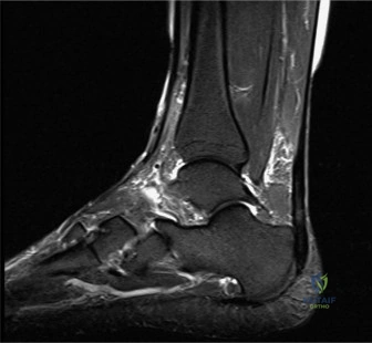
Candidate: I would consider surgical debridement given his failure of non-operative management. I would perform an open longitudinal incision, excise the degenerative tissue, and protect the sural nerve. I'd also check if the defect requires augmentation, perhaps using an FHL transfer if more than 50% of the tendon is compromised.
Failure to explicitly mention the location of the sural nerve in relation to the incision (posterolateral), or failing to quantify the threshold for augmentation (the "50% rule"). Candidates often forget to mention the importance of the paratenon preservation.
My decision-making follows a failure of 6-12 months of high-quality conservative care. I would counsel the patient on surgical risks, specifically wound complications and sural nerve injury. During the approach, I would utilize a longitudinal incision—medial is safer to avoid the sural nerve, but lateral is acceptable if careful dissection is performed to identify the nerve approximately 10-15cm proximal to the insertion. Key surgical steps include careful preservation of the paratenon, longitudinal tenotomy, excision of mucoid/degenerative tissue, and reassessment of tendon integrity. If >50% of the tendon cross-section is excised, I would plan for an FHL augmentation to ensure structural integrity and improve healing potential.
During your workup for chronic insertional Achilles pain, you obtain the following lateral radiograph. What are the key radiographic findings, and how does this change your surgical approach compared to non-insertional tendinopathy?
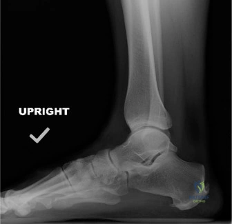
Candidate: The radiograph shows a Haglund's deformity—a prominent posterosuperior calcaneal tuberosity—and likely retrocalcaneal bursitis. Unlike NIAT, where I simply debride the mid-substance, here I must address the bone impingement by resecting the prominence and likely reattaching the tendon.
Neglecting to mention the necessity of protecting the tendon insertion or the need for specific suture anchor fixation. Failing to discuss the risk of "too much" bone resection leading to calcaneal fracture or persistent pain.
The imaging confirms a Haglund's deformity. My surgical approach requires: 1) Excision of the posterosuperior calcaneal prominence to eliminate impingement (the "bony bump"). 2) Debridement of the retrocalcaneal bursa. 3) Careful handling of the Achilles insertion; if I must detach the tendon to adequately remove the deformity, I am committed to a formal reattachment using suture anchors. The biomechanical goal is to create 2-3mm of clearance and re-establish a healthy tendon-bone footprint. I would warn the patient specifically about the higher risk of wound healing complications in the posterior heel compared to mid-substance approaches.
You have performed a successful debridement of a significant mid-substance degenerative Achilles lesion. You are now ready for closure. Walk me through your postoperative management and the key milestones for rehabilitation.
Candidate: I'd place them in a splint in equinus. Then move to a CAM boot, gradually load-bearing, and eventually start eccentric exercises at about 6 weeks.
Lack of structure. A high-scoring candidate must divide this into clear "Phases" (Protection, Controlled Motion, Strengthening, Return to Sport). Forgetting DVT prophylaxis for a prone-positioned patient is a major oversight.
My rehabilitation protocol follows a phased approach: 1. Protection (0-2/4 weeks): Immobilization in equinus with NWB to allow initial healing. 2. Controlled Motion (4-8 weeks): Transition to a CAM boot with a gradual reduction of the plantarflexion wedge. Initiate PWB as tolerated. 3. Strengthening (8-12 weeks): Full weight-bearing, initiation of formal eccentric calf strengthening (Alfredson protocol) and proprioception work. 4. Return to Activity (3-6+ months): Gradual return to impact loading and sport-specific training. Throughout, I emphasize DVT prophylaxis during the NWB phase, vigilant wound monitoring, and the critical importance of not accelerating the loading phase to avoid re-rupture.
Detailed Chapters & Topics
Dive deeper into specialized chapters regarding foot-and-ankle-cases-33