
العربية
Comprehensive Master Guide · Medically Reviewed

14 Pediatrics Cases: What's Your Next Step in Management?

CASE                               14 A 5-year-old boy has been transferred from an outside hospital to your hospital’s ER for further evaluation. Last evening…

15 Detailed Chapters
16 min read
Updated: Jun 2026
Dr. Mohammed Hutaif Clinic
Medically Reviewed by
Prof. Dr. Mohammed Hutaif Clinic
Verified Content Expert Reviewed

Quick Medical Answer

We review everything you need to understand about 14 Pediatrics Cases: What's Your Next Step in Management?. For a child with symptoms suggestive of septic arthritis, the next key step in management, beyond a physical examination, is to obtain an Erythrocyte Sedimentation Rate (ESR) and C-Reactive Protein (CRP). These laboratory tests are crucial for assessing the probability of septic arthritis, with CRP being particularly helpful for early diagnosis.

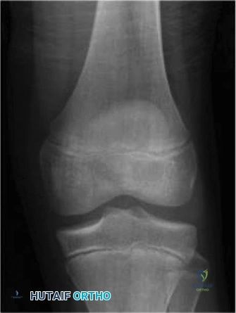
Illustration of step in management - Dr. Mohammed Hutaif
🎓

FRCS Masterclass: Clinical Viva

Interactive Examiner Scenario • Test your knowledge before revealing the answers.

👨‍⚕️ Examiner Scenario

A 5-year-old boy is transferred with a 12-hour history of severe left groin pain, fever (38.6°C), and absolute refusal to bear weight. He is holding the hip in extension and external rotation. Ultrasound shows a significant joint effusion. Discuss your immediate diagnostic priorities and how you would apply clinical prediction algorithms to this case.
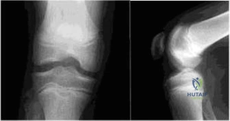
Clinical Image
Ultrasound revealing a significant joint effusion

Candidate: I would immediately suspect septic arthritis. I’d order inflammatory markers (ESR, CRP) and a blood culture. I’d use the Kocher criteria to see if he needs surgery. Since he has a fever, can't bear weight, and has a painful hip, he meets the criteria, so I would plan for an urgent arthrotomy.

❌ Common Pitfall (Poor Answer)

Failing to address the atypical "extension" resting position or waiting for an MRI to "confirm" the diagnosis. Candidates often treat the Kocher criteria as a rigid, automatic rule rather than a guide, and neglect to mention the importance of blood cultures BEFORE starting antibiotics.

⭐ The Gold Standard (Perfect Answer)

The candidate must systematically categorize the patient: 1) Clinical assessment (toxic appearance, non-weight bearing); 2) Labs (WBC, ESR, CRP—highlighting that CRP is more sensitive in the acute <24hr phase than ESR); 3) Imaging (Radiographs to rule out malignancy/trauma, Ultrasound for effusion). Crucially, they should justify the departure from "classic" FABER positioning by mentioning early-stage distension or compensatory muscle spasm. They must emphasize that if the suspicion is high (≥3 Kocher/modified criteria), delay for MRI is contraindicated, and the priority is emergent surgical lavage after blood cultures.

👨‍⚕️ Examiner Scenario

The child is now in the operating room. Describe your surgical approach, the key anatomical structures you will encounter, and the specific pitfalls to avoid during the exposure and washout.
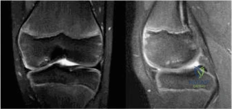
Clinical Image
Anterior surgical approach to the pediatric hip

Candidate: I would use an anterior (Smith-Petersen) approach. I’d go between the sartorius and the tensor fasciae latae, then retract the rectus femoris medially to reach the capsule. I'd perform a T-capsulotomy, irrigate, and leave a drain.

❌ Common Pitfall (Poor Answer)

Forgetting to mention the ascending branch of the lateral femoral circumflex artery. Failing to emphasize the need to wash out the dependent posterior/inferior recesses of the joint, which is the most common cause of persistent infection.

⭐ The Gold Standard (Perfect Answer)

Describe the Smith-Petersen interval (Sartorius/Femoral nerve vs. TFL/Superior Gluteal nerve). Highlight the anatomical danger zone: the ascending branch of the lateral femoral circumflex artery which must be ligated. Detail the importance of a thorough washout using a soft catheter or suction tip to reach the posterior inferior recess. Note the importance of synovectomy for tissue culture and leaving the capsule open for continuous decompression.

👨‍⚕️ Examiner Scenario

Post-operatively, the child remains irritable. How do you monitor progress, and what are the indications for re-operation or further investigation?
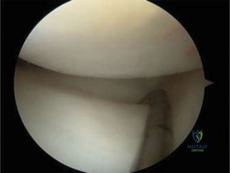
Clinical Image
Post-operative tracking of inflammatory markers

Candidate: I would watch his temperature and keep checking his blood work. If his CRP doesn't go down, I would repeat the surgery to wash it out again. I'd transition to oral antibiotics when he looks better.

❌ Common Pitfall (Poor Answer)

Being too vague. Failing to distinguish between the kinetics of CRP (rapidly responsive) vs. ESR (delayed). Ignoring the need to rule out other sites of infection or failure to consider late complications like AVN.

⭐ The Gold Standard (Perfect Answer)

Use a structured monitoring plan: Clinical (systemic signs), Laboratory (daily CRP tracking—a failure of CRP to downtrend after 48-72h mandates repeat imaging/washout), and Radiographic (serial follow-up to detect AVN/chondrolysis). Mention the transition to oral antibiotics based on clinical improvement and CRP normalization. Mention that persistent symptoms post-washout should trigger an MRI to rule out synchronous osteomyelitis or inadequately drained sub-capsular collections.

Detailed Chapters & Topics

Dive deeper into specialized chapters regarding 14-pediatrics-cases

15 Chapters
01
Chapter 1 19 min

Pediatric OCD: When to Consider Drilling for the Femoral

CASE                               15 A 12-year-old male presents to the orthopaedic clinic with several months of righ…

02
Chapter 2 15 min

Pediatric Pain & Swelling: 16 Cases Solved & Diagnoses Revealed

CASE                               16 A 12-year-old boy presents to your office 2 days after sustaining a noncontact in…

03
Chapter 3 12 min

Pediatric Anterior Knee Pain: Solve the Puzzle with Case Insights

CASE                               19 A 14-year-old female soccer player presents with a 1 year history of anterior kne…

04
Chapter 4 20 min

9 Pediatrics Cases: Essential Insights on Rotation of the Foot

CASE                                9 A 14-year-old boy is brought to the ER with complaints of right knee pain after h…

05
Chapter 5 16 min

Unraveling 10 Pediatric Cases: Surgical Reduction and Internal Fixation

CASE                               10 You are called to the emergency room to evaluate a 13-year-old male complaining o…

06
Chapter 6 18 min

8 Pediatric Cases: Critical Decisions on Exits from the Physis

Review 8 pediatric orthopedic cases detailing critical decisions on exits from the physis. Essential Viva exam prep by …

07
Chapter 7 15 min

Pediatric Cases: Optimal Closure of the Distal Femur

CASE                                8 A 7-year-old girl is transferred to the ER after suffering an injury playing tack…

08
Chapter 8 24 min

Challenging Orthopedic Case: Multiple Epiphyseal Dysplasia & Severe Hip Osteoarthritis

Explore this challenging orthopedic case study of a 28-year-old male with Multiple Epiphyseal Dysplasia (MED) and sever…

09
Chapter 9 17 min

Distal Hamstring Lengthening and Rectus Femoris Transfer for Cerebral Palsy: An Intraoperative Masterclass

Discover how to manage crouch gait in cerebral palsy with our intraoperative masterclass on distal hamstring lengthenin…

10
Chapter 10 13 min

Arthroscopic Femoroacetabular Impingement: A Masterclass in Hip Preservation

Master Femoroacetabular Impingement (FAI) with our expert guide. Explore pincer and cam mechanisms, and learn arthrosco…

11
Chapter 11 17 min

Pericapsular Acetabular Osteotomies: A Masterclass in Pemberton and Dega Techniques for Hip Dysplasia

Master pediatric hip dysplasia correction. Explore the biomechanics and key differences between Pemberton and Dega peri…

12
Chapter 12 18 min

Salter Innominate Osteotomy: A Masterclass in Acetabular Redirection for DDH

Master the Salter Innominate Osteotomy for DDH. Learn the biomechanics of the developing hip, acetabular dysplasia path…

13
Chapter 13 19 min

Congenital Knee Dislocation: Pathology & Surgical Management

Comprehensive guide on congenital knee dislocation (CDK), detailing grading, biomechanics, non-operative management, an…

14
Chapter 14 10 min

Congenital Dislocation of the Patella: Comprehensive Surgical Management

Explore the pathoanatomy and comprehensive surgical management of congenital patellar dislocation, a rare but severe pe…

15
Chapter 15 10 min

Congenital Upper Extremity and Cervical Deformities: Master Surgical Techniques

Master surgical techniques for congenital upper extremity and cervical deformities. Learn operative management of Spren…

Dr. Mohammed Hutaif Clinic
Medically Verified Content by
Prof. Dr. Mohammed Hutaif Clinic
Consultant Orthopedic & Spine Surgeon
Guide Overview