Tibial Plateau Fractures: Your Complete Guide to Diagnosis & Care
Key Takeaway
This topic focuses on Tibial Plateau Fractures: Your Complete Guide to Diagnosis & Care, Plateau fractures tibial are breaks in the upper portion of the tibia, involving the knee joint's articular surfaces. They constitute 1% of all fractures and commonly result from varus or valgus forces with axial loading. Lateral plateau fractures are most common (55-70%), while medial ones, though less frequent, are often linked to higher-energy injuries and soft tissue damage.
Tibial Plateau Fractures Your Complete Guide to Diagnosis Care
Introduction and Epidemiology
Tibial plateau fractures represent a significant challenge in orthopedic trauma, necessitating precise diagnosis and often complex surgical intervention to restore articular congruity and limb function. These intra-articular injuries to the proximal tibia carry a substantial risk of long-term morbidity, including post-traumatic osteoarthritis.
Epidemiologically, tibial plateau fractures constitute approximately 1% of all fractures seen in orthopedic practice, with a higher incidence observed in specific demographic groups. Notably, they account for up to 8% of all fractures in the elderly population, often occurring in the setting of low-energy falls due to osteopenic bone quality. In younger, more active individuals, these fractures are typically the result of high-energy mechanisms, such as motor vehicle accidents or sports-related trauma.
The distribution of fracture patterns varies anatomically. Isolated injuries to the lateral tibial plateau are the most prevalent, accounting for 55% to 70% of all tibial plateau fractures. This preponderance is attributed to the relative biomechanical weakness of the lateral plateau and the common valgus loading mechanism. In contrast, isolated medial plateau fractures are less common, comprising 10% to 25% of cases, and are often indicative of higher energy trauma due to the inherent strength of the medial condyle. Bicondylar lesions, involving both medial and lateral plateaus, represent 10% to 30% of these injuries and frequently present with severe soft tissue compromise. Furthermore, 1% to 3% of all tibial plateau fractures are open injuries, underscoring the potential for significant associated soft tissue damage and increased risk of complications such as infection.
Classification systems such as the Schatzker classification and the AO/OTA classification are invaluable tools for describing fracture morphology, guiding treatment decisions, and facilitating communication among orthopedic surgeons. The Schatzker system categorizes fractures into six types based on increasing severity and comminution, predominantly on the lateral side, with types V and VI denoting bicondylar involvement. The AO/OTA classification offers a more comprehensive alphanumeric system, detailing fracture location, articular involvement, and stability. A thorough understanding of these systems is crucial for accurate diagnosis and preoperative planning.
Surgical Anatomy and Biomechanics
A detailed appreciation of the surgical anatomy and biomechanics of the proximal tibia is fundamental to the effective management of tibial plateau fractures. The tibial plateau is formed by the articular surfaces of the medial and lateral tibial condyles, which articulate with the femoral condyles to form the tibiofemoral joint. These articular surfaces are covered by hyaline cartilage and are protected and stabilized by the fibrocartilaginous menisci.
The medial tibial plateau is typically larger and possesses a concave morphology in both the sagittal and coronal planes, providing a more conforming articulation. In contrast, the lateral tibial plateau extends more superiorly and is convex in both sagittal and coronal planes, making it inherently less stable and more susceptible to fracture. The normal posterior tibial slope, averaging 10 degrees, is a critical anatomical feature that influences knee kinematics and must be meticulously restored during surgical reconstruction.
The two plateaus are separated by the intercondylar eminence, a non-articular bony prominence that serves as the crucial tibial attachment site for the anterior cruciate ligament (ACL) and posterior cruciate ligament (PCL). Anteriorly, the ACL primarily inserts onto the intercondylar eminence, while the PCL inserts posteriorly. The menisci are firmly attached to the intercondylar area via their horns and peripherally to the joint capsule. The medial meniscus is more C-shaped and firmly adherent to the medial collateral ligament (MCL) and joint capsule, making it less mobile and more prone to tear. The lateral meniscus is more O-shaped and less firmly attached, affording it greater mobility but still susceptible to injury.
Distal to the tibial plateau, several important bony prominences and soft tissue attachments warrant attention. Approximately 2 to 3 cm distally, the tibial tubercle is located anteriorly, serving as the insertion site for the patellar ligament. Medially, the pes anserinus insertion provides attachment for the sartorius, gracilis, and semitendinosus muscles. Laterally, Gerdy's tubercle is the insertion point for the iliotibial band (ITB). These structures define the surgical corridors and potential pitfalls during operative approaches.
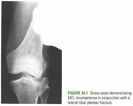
The structural integrity of the medial articular surface and its supporting medial condyle is inherently stronger than that of their lateral counterparts. This biomechanical difference, coupled with common injury mechanisms, explains the higher incidence of lateral plateau fractures. Medial plateau fractures, or those with significant medial involvement, typically result from higher-energy injuries and are more frequently associated with severe soft tissue damage. This can include disruptions of the lateral collateral ligament (LCL) complex, injuries to the common peroneal nerve, and potentially catastrophic damage to the popliteal vessels, given their close proximity to the posterior medial tibia. Therefore, comprehensive neurovascular assessment is paramount in these injury patterns. The integrity of the menisci and the collateral and cruciate ligaments plays a vital role in knee stability and load transmission, and associated injuries to these structures significantly impact treatment strategies and prognosis.
Indications and Contraindications
The decision to proceed with operative or non-operative management for tibial plateau fractures hinges on a multifactorial assessment, considering fracture morphology, displacement, articular congruity, stability, soft tissue status, and patient factors. The primary goals of treatment, regardless of method, are to restore a stable, congruent articular surface, achieve appropriate mechanical axis alignment, and preserve knee function.
Operative Indications
Operative intervention is generally indicated for fractures that compromise joint stability, articular congruity, or mechanical alignment, or in situations demanding immediate stabilization. Key indications include:
- Articular Step-Off or Gap: Greater than 2-3 mm of articular depression or step-off is widely accepted as an indication for operative reduction and internal fixation to mitigate the risk of post-traumatic osteoarthritis.
- Joint Instability: Significant instability of the knee joint demonstrated clinically or radiographically, often due to associated ligamentous injury or extensive bony comminution.
- Mechanical Axis Deviation: Varus or valgus malalignment exceeding 5-10 degrees, depending on patient age and activity level, which predisposes to abnormal loading and accelerated degenerative changes.
- Open Fractures: All open tibial plateau fractures require urgent surgical debridement, irrigation, and provisional stabilization to minimize infection risk and prepare for definitive fixation.
- Associated Vascular Injury: Fractures compromising vascular integrity necessitate immediate surgical exploration and repair, often followed by staged fracture fixation.
- Compartment Syndrome: Clinical signs of acute compartment syndrome require emergent fasciotomy, frequently performed concurrently or prior to fracture fixation.
- Bicondylar Fractures (Schatzker V/VI): These complex injuries almost universally require operative management due to their inherent instability and articular disruption.
- Medial Plateau Fractures (Schatzker IV): Often associated with high-energy mechanisms and significant soft tissue injury, making operative stabilization generally preferred.
- Floating Knee Injuries: Concomitant ipsilateral femur and tibia fractures often warrant operative fixation of the plateau fracture.
- Soft Tissue Impingement: Large incarcerated soft tissue fragments preventing closed reduction.
Non-Operative Indications
Non-operative management is reserved for a select group of stable, minimally displaced fractures with intact soft tissue envelopes. These include:
- Minimally Displaced Fractures: Articular step-off or gap less than 2 mm, with no significant articular depression.
- Stable Fractures: Fractures that demonstrate stability to varus and valgus stress testing in full extension and 30 degrees of flexion.
- Intact Mechanical Axis: No significant varus or valgus malalignment.
- Non-Ambulatory or Low-Demand Patients: In certain elderly or medically frail patients with limited functional goals, conservative management may be preferred even for slightly displaced fractures, provided the fracture pattern is stable.
Contraindications
Absolute contraindications to immediate definitive internal fixation are few and typically relate to the patient's physiological status or severe local conditions. Relative contraindications guide the timing and staging of surgical intervention.
- Absolute Contraindications:
- Severe medical comorbidities precluding safe anesthesia and surgery.
- Active local or systemic infection.
- Relative Contraindications:
- Severe soft tissue compromise (e.g., extensive blistering, crush injury, severe degloving, compromised skin viability) which may necessitate delayed definitive fixation after soft tissue healing, often with initial spanning external fixation.
- Patient non-compliance or inability to adhere to postoperative rehabilitation protocols.
- Uncontrolled diabetes or other systemic conditions that significantly impair wound healing and increase infection risk.
| Indication Category | Operative Management | Non-Operative Management |
|---|---|---|
| Articular Integrity | Articular step-off/gap > 2-3 mm, articular depression | Articular step-off/gap < 2 mm, no significant depression |
| Joint Stability | Significant instability to stress testing | Stable to stress testing |
| Mechanical Alignment | Varus/valgus malalignment > 5-10 degrees | Maintained mechanical axis |
| Fracture Pattern | Bicondylar (Schatzker V/VI), Medial (Schatzker IV) | Minimally displaced lateral (Schatzker I/II) |
| Soft Tissue Status | Open fractures, impending compartment syndrome, vascular injury | Closed fracture, stable soft tissue envelope |
| Patient Factors | Young, active, high-demand patient | Non-ambulatory, low-demand, high surgical risk |
| Special Cases | Floating knee, incarcerated soft tissue |
Pre Operative Planning and Patient Positioning
Thorough preoperative planning is paramount for successful outcomes in tibial plateau fractures, particularly given their intra-articular nature and common association with soft tissue injury. This phase encompasses detailed imaging review, soft tissue assessment, patient optimization, and meticulous surgical templating.
Diagnostic Imaging
Standard radiographic evaluation begins with AP, lateral, and oblique views of the knee, including a full-length lower extremity alignment view to assess overall limb axis. However, plain radiographs often underestimate the degree of articular depression, comminution, and displacement.

Computed Tomography (CT) scans are indispensable for nearly all tibial plateau fractures requiring surgical intervention. A fine-cut CT with 2D reconstructions (coronal and sagittal) and 3D reconstructions provides an unparalleled understanding of the fracture pattern, fragment location, articular involvement, and posterior condylar displacement. This allows for detailed surgical templating, identification of optimal screw trajectories, and determination of necessary plate length and contour.


Magnetic Resonance Imaging (MRI) is not routinely indicated for acute fracture planning but can be highly valuable for evaluating associated soft tissue injuries, particularly meniscal tears (which occur in up to 50% of cases) and ligamentous disruptions (ACL, PCL, collateral ligaments). Knowledge of these concomitant injuries influences surgical approach, fixation strategy, and rehabilitation.
Soft Tissue Assessment
The condition of the soft tissue envelope dictates the timing of surgery. A thorough examination for open wounds, blisters, abrasions, and swelling is critical. For high-energy injuries, the "wrinkle sign" (presence of skin wrinkles indicating decreased swelling) is a useful clinical indicator for allowing definitive fixation. Often, initial management involves splinting the knee, elevation, ice, and, if significant swelling or instability is present, application of a spanning external fixator. This allows the soft tissues to recover, reducing the risk of wound complications. Prophylactic antibiotics are indicated for open fractures.
Patient Optimization and Templating
Patient comorbidities, nutritional status, and coagulation profiles should be optimized. For surgical templating, identify the primary fracture fragments, determine the need for bone graft or substitutes, and select appropriate implant types and sizes (e.g., locking plates, rafting screws). Consideration of the required surgical approach(es) and potential need for dual incisions is also part of this phase.
Patient Positioning
The patient is typically positioned supine on a radiolucent operating table. A bump or bolster can be placed under the ipsilateral buttock to internally rotate the leg and improve access to the lateral plateau. A tourniquet is routinely applied to the thigh for bloodless field creation. Fluoroscopy is essential for intraoperative assessment of reduction and hardware placement, necessitating careful positioning to allow unhindered AP, lateral, and oblique views. The leg should be draped free to allow full range of motion, facilitating valgus/varus stress testing and direct visualization of articular reduction in certain approaches. For posterior approaches, the patient may need to be positioned in the prone or lateral decubitus position.
Detailed Surgical Approach and Technique
The surgical approach and technique for tibial plateau fractures are dictated by the specific fracture pattern, fragment displacement, and associated soft tissue injuries. The overarching goals are anatomical articular reduction, restoration of mechanical alignment, and stable internal fixation to permit early motion.
General Principles of Reduction and Fixation
- Reduction: Achieved through a combination of indirect (ligamentotaxis, distraction, manipulation) and direct techniques (articular elevation with bone tamps or elevators). For depressed fragments, metaphyseal windows can be created to elevate the articular surface from below.
- Bone Grafting: Autogenous cancellous bone graft (from iliac crest or proximal tibia) or allograft/synthetic bone substitutes are often used to fill metaphyseal defects created by elevated articular fragments, preventing collapse and providing structural support.
- Fixation: Typically involves buttress plating with screws. Locking plates are frequently utilized, especially in osteopenic bone, as they provide angular stability and do not rely on plate-bone compression for fixation, making them less disruptive to periosteal blood supply. Rafting screws, placed from distal to proximal underneath depressed articular fragments, are critical to support the articular surface.
Lateral Tibial Plateau Fractures (Schatzker I, II, III)
Lateral plateau fractures are often treated through an anterolateral approach.
Anterolateral Approach
- Incision: A curvilinear or straight longitudinal incision is made over the anterolateral aspect of the knee, extending from just proximal to the joint line to approximately 10-15 cm distal to it, centered over Gerdy's tubercle.
- Dissection: The incision is carried down through subcutaneous tissue. Full-thickness skin flaps are developed. The anterior compartment fascia is incised longitudinally, typically lateral to the anterior tibialis muscle, to expose the proximal tibia. The common peroneal nerve must be protected, especially in more posterolateral extensions of the incision.
- Internervous Plane: No true internervous plane is utilized; rather, direct access to the lateral aspect of the tibia is achieved by elevating the muscle belly of the tibialis anterior and potentially partially detaching the ITB from Gerdy's tubercle.
- Reduction and Fixation:
- The lateral meniscus may be elevated to visualize the articular surface.
- For split depression (Schatzker II) or pure depression (Schatzker III) fractures, a metaphyseal window (cortical fenestration) can be created in the lateral metaphysis, distal to the fracture line.
- A bone tamp or elevator is inserted through this window to gently elevate the depressed articular fragments, restoring the joint line under direct visualization or fluoroscopic guidance.
- The metaphyseal void created by fragment elevation is packed with bone graft or substitute.
- A precontoured locking buttress plate is applied to the lateral aspect of the tibia. Proximal screws, including rafting screws, are directed to support the articular surface, while distal screws secure the plate to the tibial shaft.
- The plate acts as a buttress, preventing recurrent collapse of the elevated articular segment.
Medial Tibial Plateau Fractures (Schatzker IV)
Medial plateau fractures are typically managed via an anteromedial or posteromedial approach.
Anteromedial Approach
- Incision: A longitudinal incision is made over the anteromedial aspect of the proximal tibia, parallel to the patellar tendon.
- Dissection: The sartorius muscle is identified and reflected posteriorly. The pes anserinus attachments may need to be partially released. The MCL is typically preserved but may need to be mobilized.
- Internervous Plane: Between the sartorius (femoral nerve) and vastus medialis (femoral nerve) anteriorly and the semitendinosus/gracilis (obturator/tibial nerve) posteriorly, but primarily involves direct muscle retraction.
- Reduction and Fixation: Similar principles to the lateral plateau, often using a medial buttress plate. Care must be taken to protect the saphenous nerve and vein.
Posteromedial Approach
- Incision: A posteromedial incision along the posterior border of the tibia, typically between the medial head of the gastrocnemius and the soleus, provides direct access to the posteromedial corner.
- Dissection: The saphenous nerve and vein are identified and protected. The medial head of the gastrocnemius is retracted laterally, and the neurovascular bundle (popliteal artery, vein, tibial nerve) must be meticulously protected.
- Reduction and Fixation: This approach is crucial for fractures with posterior shear components or posteromedial comminution. A posteromedial locking plate is applied, often providing excellent buttress support against posterior displacement.
Bicondylar Tibial Plateau Fractures (Schatzker V, VI)
Bicondylar fractures are complex, involving both medial and lateral plateaus, often with significant comminution and displacement.
- Approaches: Frequently require dual incisions for adequate exposure and fixation, most commonly an anterolateral approach combined with a posteromedial approach. Staged procedures are often necessary if the soft tissue envelope is severely compromised, with initial external fixation followed by definitive internal fixation once soft tissues have recovered.
- Reduction and Fixation: The strategy involves reconstructing one condyle first, typically the medial, as it often provides a more stable base. Once one condyle is reduced and provisionally fixed, attention turns to the other. Anatomical reduction of the articular surface is paramount, achieved by direct visualization or arthroscopy. Stable fixation with two independent plates (one lateral, one posteromedial) provides robust construct stability for early motion.

Open Fractures
Open tibial plateau fractures require immediate and aggressive management:
1. Emergent Debridement and Irrigation: Copious irrigation with saline and surgical debridement of all devitalized tissue is performed in the operating room.
2. Provisional Stabilization: Often achieved with spanning external fixation to allow for soft tissue swelling to subside and wound care.
3. Delayed Definitive Fixation: Once the soft tissues are clean and stable, definitive internal fixation is performed, typically within 5-7 days.
4. Antibiotics: Broad-spectrum intravenous antibiotics are initiated immediately and continued postoperatively.
Role of Arthroscopy
Arthroscopic assistance can be a valuable adjunct in certain tibial plateau fractures, particularly for visualizing articular reduction directly, assessing meniscal and ligamentous injuries, and performing associated repairs (e.g., meniscal repair). It can help confirm articular congruity achieved with indirect reduction techniques and minimizes further soft tissue stripping.
Complications and Management
Tibial plateau fractures are associated with a range of potential complications, both early and late, which can significantly impact patient outcomes. Vigilant monitoring and proactive management are essential.
Early Complications
- Compartment Syndrome: Incidence ranges from 1% to 11%, particularly in high-energy injuries, bicondylar fractures, and those with associated vascular injury. Clinical suspicion (pain out of proportion, pallor, paresthesia, pulselessness, paralysis) warrants emergent intracompartmental pressure monitoring and, if elevated, fasciotomy.
- Neurovascular Injury: The common peroneal nerve is at risk with lateral plateau fractures and posterolateral approaches, while the popliteal artery and tibial nerve are vulnerable with medial or bicondylar injuries and posterior approaches. Incidence varies but can be up to 5-10% for nerve injuries and 1% for vascular injuries. Prompt diagnosis and surgical repair or decompression are critical.
- Infection: Open fractures have a high risk (up to 20%). Closed fractures can also become infected, especially with extensive soft tissue dissection or prolonged surgical time (incidence 1-5%). Management involves surgical debridement, appropriate antibiotic therapy, and potential hardware removal if infection persists.
- Wound Complications: Blisters, skin necrosis, and dehiscence are common, especially with compromised soft tissue envelopes or premature surgical intervention. Managed with local wound care, delayed primary closure, or reconstructive plastic surgery if needed.
- Deep Vein Thrombosis (DVT) and Pulmonary Embolism (PE): High risk due to lower extremity trauma and immobilization. Prophylaxis with anticoagulants is standard practice.
Late Complications
- Post-Traumatic Arthritis: The most common long-term complication, occurring in 20-60% of cases, directly related to residual articular incongruity, malalignment, and associated meniscal/ligamentous injuries. Management includes activity modification, injections, osteotomy to correct malalignment, and ultimately total knee arthroplasty in severe cases.
- Malunion/Nonunion: Malunion (healing in an unacceptable position) can lead to mechanical axis deviation and accelerated arthritis. Nonunion (failure to heal) is less common but may necessitate revision surgery with debridement, bone grafting, and revised fixation.
- Stiffness and Arthrofibrosis: Common after intra-articular fractures and prolonged immobilization. Aggressive physical therapy, manipulation under anesthesia, or arthroscopic lysis of adhesions may be required.
- Chronic Pain: Can result from arthritis, hardware irritation, nerve injury, or soft tissue scarring.
- Hardware Irritation/Failure: Prominent hardware may require removal after fracture union. Hardware failure (plate or screw breakage) indicates nonunion or excessive stress on the construct and requires revision.
| Complication Category | Common Complication | Incidence | Salvage Strategies |
|---|---|---|---|
| Acute | Compartment Syndrome | 1-11% | Emergent fasciotomy |
| Neurovascular Injury | 1-10% (nerve), 1% (vascular) | Immediate exploration, repair/decompression | |
| Infection (Open Fractures) | Up to 20% | Debridement, irrigation, antibiotics, hardware management | |
| Wound Dehiscence/Necrosis | 5-15% | Local wound care, delayed closure, flap reconstruction | |
| DVT/PE | High risk | Anticoagulation prophylaxis, early mobilization | |
| Late | Post-Traumatic Arthritis | 20-60% | Activity modification, injections, osteotomy, total knee arthroplasty |
| Malunion | Variable | Corrective osteotomy | |
| Nonunion | < 5% | Revision fixation, bone grafting, biologic stimulation | |
| Stiffness/Arthrofibrosis | 10-30% | Aggressive PT, manipulation under anesthesia, arthroscopic lysis of adhesions | |
| Chronic Pain | Variable | Multimodal pain management, hardware removal, nerve block | |
| Hardware Irritation/Failure | Variable | Hardware removal, revision fixation |
Post Operative Rehabilitation Protocols
Postoperative rehabilitation is a critical determinant of functional outcome following surgical fixation of tibial plateau fractures. Protocols are tailored based on fracture stability, fixation strength, soft tissue integrity, and associated injuries (e.g., meniscal repair, ligament reconstruction). The overriding principle is to balance protection of the healing fracture with early, controlled mobilization to prevent stiffness and optimize cartilage health.
Immediate Postoperative Phase (Weeks 0-6)
- Pain Management: Multimodal analgesia including regional blocks, NSAIDs, and opioids.
- Wound Care: Meticulous wound care to prevent infection and monitor for complications.
- DVT Prophylaxis: Pharmacological (e.g., LMWH) and mechanical (e.g., compression devices) prophylaxis.
- Weight-Bearing (WB) Status:
- Non-Weight-Bearing (NWB): Most common initially, especially for complex or comminuted fractures, or in osteopenic patients, for 6-12 weeks. Use of crutches or a walker.
- Touch-Down Weight-Bearing (TDWB): For more stable constructs, allowing proprioceptive input without significant load, typically 10-15 lbs.
- Protected Weight-Bearing (PWB): May be initiated earlier for very stable, simple fractures.
- Range of Motion (ROM):
- Passive Range of Motion (PROM): Often initiated immediately or within the first week, either manually by a therapist or with a continuous passive motion (CPM) machine. Goal is typically 0-90 degrees flexion by 4-6 weeks, respecting pain limits.
- Active-Assisted Range of Motion (AAROM): Gradual progression as pain allows.
- Active Range of Motion (AROM): Progressed cautiously, ensuring no stress on the healing fracture.
- Muscle Activation: Isometric quadriceps sets, gluteal sets, and ankle pumps to maintain muscle tone and improve circulation.
- Splint/Brace: A hinged knee brace is commonly used, locked in extension initially for stability, then gradually unlocked to allow increasing ROM.
Intermediate Phase (Weeks 6-12)
- Weight-Bearing Progression: Gradual progression from NWB/TDWB to partial weight-bearing (PWB) (25-50% body weight) and then full weight-bearing (FWB) as determined by radiographic union and clinical stability. Fluoroscopic weight-bearing assessment may be used.
- ROM Progression: Continue to increase knee flexion and work towards full extension. Emphasis on regaining full, pain-free ROM.
- Strengthening: Initiate light resistance exercises for quadriceps, hamstrings, and calf muscles. Closed kinetic chain exercises (e.g., mini-squats, wall slides) are generally preferred initially for stability.
- Proprioception: Balance and proprioceptive exercises (e.g., single-leg stance, wobble board) are introduced.
- Gait Training: Progression from assisted ambulation to independent ambulation with a normal gait pattern.
Advanced Phase (Weeks 12-24+)
- Full Weight-Bearing and Functional Training: Patient should be FWB with good control. Focus shifts to regaining strength, endurance, and sport-specific function.
- High-Level Strengthening: Advanced resistance training, plyometrics (if appropriate), agility drills.
- Sport-Specific Training: Gradual return to activities, starting with low-impact exercises and progressing to higher-impact or sport-specific drills.
- Return to Activity: Typically not before 6-9 months, and often up to 12-18 months for high-demand activities or contact sports, depending on the severity of the injury and functional recovery. Return to sport decisions should be guided by objective functional testing and clinical assessment.
Key Considerations:
- Meniscal/Ligamentous Repairs: If concomitant meniscal repair or ligamentous reconstruction was performed, the rehabilitation protocol will be modified to protect these repairs (e.g., slower ROM progression, delayed WB).
- Bone Graft: If significant bone grafting was utilized, WB progression may be delayed to allow for graft incorporation.
- Patient Compliance: Adherence to the rehabilitation protocol is crucial. Regular communication with the physical therapist is essential.
Summary of Key Literature and Guidelines
The management of tibial plateau fractures has evolved significantly with advancements in imaging, surgical techniques, and implants. The literature consistently emphasizes several critical principles for optimal outcomes.
Anatomical Reduction and Stable Fixation: Numerous studies, including those by Schatzker and colleagues, underscore that anatomical reduction of the articular surface (less than 2 mm step-off or gap) and stable fixation are paramount to prevent post-traumatic osteoarthritis. A meta-analysis by Zong et al. (2018) highlighted that accurate articular reduction significantly correlated with improved long-term functional scores and reduced incidence of arthritis.
Role of CT Imaging: Modern guidelines strongly recommend CT scans for all displaced tibial plateau fractures to accurately characterize fracture morphology, guide surgical planning, and identify specific fragments for reduction. Luo et al.'s classification, based on CT imaging, provides a practical framework for surgical approaches, particularly for complex bicondylar injuries.
Associated Soft Tissue Injuries: The high incidence of meniscal (30-70%) and ligamentous injuries (10-40%) with tibial plateau fractures is well-documented. Delamarter et al. (1990) and others have shown that meniscal tears, particularly lateral, are common with lateral plateau fractures, emphasizing the need for thorough intraoperative assessment and repair when feasible to restore joint stability and distribute load. MRI plays a crucial role in preoperative identification of these injuries.
Surgical Approaches: The choice of surgical approach depends on the fracture pattern. The anterolateral approach is standard for lateral plateau fractures. For bicondylar and posteromedial fractures, dual incisions (e.g., anterolateral and posteromedial) or specific posterior approaches are increasingly recognized as superior for achieving direct visualization and robust fixation, as evidenced by studies on the modified posteromedial approach for specific fracture types.
Fixation Principles: Locking plate technology has become a cornerstone in the treatment of tibial plateau fractures, particularly in osteoporotic bone. The concept of "rafting screws" placed parallel to the subchondral bone, supporting the articular surface, is widely accepted and critical for stability. The use of bone graft or substitutes to support elevated articular fragments is a standard practice to prevent late collapse.
Staged Protocol for High-Energy Injuries: For severe high-energy injuries with compromised soft tissues, a staged protocol involving initial external fixation followed by delayed definitive internal fixation once the soft tissue envelope has improved is a well-established principle to reduce wound complication rates.
Rehabilitation: Early, controlled range of motion (CPM) and delayed weight-bearing based on fracture stability and fixation strength are key tenets of rehabilitation. Research consistently supports the benefit of early motion to prevent stiffness and promote cartilage healing. However, aggressive weight-bearing before adequate fracture consolidation can lead to loss of reduction or hardware failure.
In conclusion, the current paradigm for managing tibial plateau fractures integrates meticulous preoperative planning with advanced imaging, anatomical reduction, stable internal fixation, judicious management of soft tissues, and a structured rehabilitation protocol. Ongoing research continues to refine these principles, but the core tenets remain focused on restoring articular congruity and mechanical alignment to optimize long-term functional outcomes and mitigate the risk of post-traumatic arthritis.
