Part of the Master Guide

Orthopedic Surgery Board Review MCQs: Arthroplasty, Trauma & Spine Part 255

Orthopedic Board Review MCQs: Fracture, Dislocation & Nerve | Part 93

23 Apr 2026 49 min read 64 Views
Orthopedic Surgery Board Review MCQs: AAOS Master Bank Part 93

Key Takeaway

This page offers Part 93 of a comprehensive orthopedic surgery board review. Featuring 50 high-yield MCQs, it's designed for orthopedic residents and surgeons preparing for OITE and AAOS certification exams. Enhance your knowledge across key topics like fracture, hip, and elbow with detailed explanations and literature references in both study and exam modes.

Orthopedic Board Review MCQs: Fracture, Dislocation & Nerve | Part 93

Comprehensive 100-Question Exam


00:00

Start Quiz

Question 1

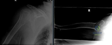
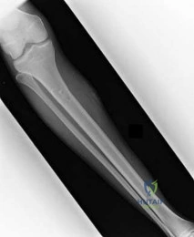
A 35-year-old man sustains a closed spiral fracture of the distal third of the humeral shaft (Holstein-Lewis type). On examination in the emergency department, he is unable to extend his wrist or fingers, but triceps function is completely intact.

What is the most appropriate initial management of this nerve injury?


Explanation

Most radial nerve palsies associated with closed humeral shaft fractures, including Holstein-Lewis types, represent neurapraxia and will resolve spontaneously. The standard of care is initial observation for 3 to 4 months with supportive splinting. Surgical exploration is indicated if there is an open fracture, if the paralysis occurs acutely after a closed reduction attempt, or if there is no clinical or EMG evidence of recovery at 3 to 4 months.

Question 2

A 24-year-old athlete sustains an anterior shoulder dislocation during a rugby tackle.

After successful closed reduction, he is noted to have decreased sensation over the lateral aspect of his deltoid. Which of the following physical examination findings is most specifically associated with this nerve injury?


Explanation

The axillary nerve is the most commonly injured nerve in anterior shoulder dislocations. It innervates the deltoid and teres minor muscles. The deltoid acts as the primary abductor of the shoulder from 15 to 90 degrees. Initiation of abduction (0-15 degrees) is primarily a function of the supraspinatus (suprascapular nerve). External rotation at the side is primarily driven by the infraspinatus (suprascapular nerve).

Question 3

A 6-year-old boy presents with a completely displaced extension-type supracondylar fracture of the humerus.

Which of the following clinical deficits represents the most common nerve injury associated with this specific fracture pattern?


Explanation

The anterior interosseous nerve (AIN) is the most commonly injured nerve in extension-type supracondylar humerus fractures. Injury to the AIN manifests as weakness or inability to flex the interphalangeal joint of the thumb (flexor pollicis longus) and the distal interphalangeal joint of the index finger (flexor digitorum profundus), resulting in an inability to form the 'OK' sign.

Question 4

A 7-year-old girl falls on an outstretched hand and sustains a Bado Type I Monteggia fracture-dislocation.

She is noted to have a nerve palsy on presentation. What is the expected clinical presentation of her neurological deficit?


Explanation

The posterior interosseous nerve (PIN) is classically injured in Monteggia fractures, particularly Bado Type I and III. The PIN is a purely motor branch of the radial nerve that innervates the extensor muscles of the digits and the extensor carpi ulnaris (ECU). Because the extensor carpi radialis longus (ECRL) is innervated by the radial nerve proper proximal to the PIN bifurcation, wrist extension is preserved but deviates radially due to the paralyzed ECU. There is no sensory loss.

Question 5

A 28-year-old male is involved in a high-speed motor vehicle collision and sustains a severe traumatic knee dislocation resulting in a complete foot drop.

Given the mechanism and neurological deficit, which adjacent vascular structure is most critically at risk and requires emergent evaluation?


Explanation

Traumatic knee dislocations are highly associated with popliteal artery injuries due to the artery's anatomic tethering at the adductor hiatus proximally and the soleus arch distally. A concomitant common peroneal nerve injury (indicated by the foot drop) occurs in 15-40% of knee dislocations and typically results from a severe stretch injury. Emergent evaluation of the popliteal artery via ABI and/or CT angiography is mandatory.

Question 6

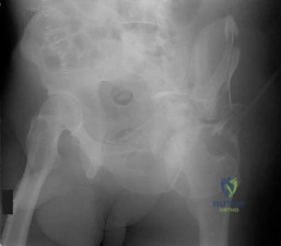
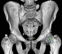
A 45-year-old man falls from a roof, sustaining a Zone 3 sacral fracture according to the Denis classification.

Which of the following neurological deficits is most commonly associated with this specific fracture zone?


Explanation

The Denis classification of sacral fractures divides the sacrum into three zones. Zone 3 involves the central sacral canal. Fractures in this zone have the highest rate of neurological injury (up to 60%), specifically affecting the lower sacral roots (S2-S4), which leads to sphincter disturbances resulting in bowel, bladder, and sexual dysfunction.

Question 7

A 33-year-old unrestrained driver sustains a posterior wall acetabular fracture with a posterior hip dislocation.

On initial evaluation, the patient has weakness in ankle dorsiflexion and great toe extension, with completely intact ankle plantar flexion. Which nerve division is most likely injured?


Explanation

The common peroneal division of the sciatic nerve is injured much more frequently than the tibial division during posterior hip dislocations or posterior wall acetabular fractures. This division is more susceptible to stretch injury because it is located laterally (closer to the displacing femoral head), is tethered at the fibular head, and possesses larger fascicles with less protective epineurial connective tissue.

Question 8

A 65-year-old female sustains a closed, displaced intra-articular distal humerus fracture (AO type 13-C3).

Preoperatively, she reports numbness and tingling in her ring and small fingers. What is the most appropriate intraoperative management of the ulnar nerve during open reduction and internal fixation (ORIF)?


Explanation

In the surgical management of distal humerus fractures, the ulnar nerve must be identified and protected. Current evidence suggests that routine transposition is not required and may increase the risk of devascularization and subsequent neuropathy. The nerve should be mobilized enough to allow safe fracture fixation and then left in situ, provided it rests comfortably without tension or direct impingement by the implants.

Question 9

A 25-year-old male sustains an anteroposterior compression (APC) type III pelvic ring injury. He undergoes anterior plating of the symphysis pubis and percutaneous posterior sacroiliac (SI) joint screw fixation. Postoperatively, he is noted to have a foot drop and weakness in great toe extension, but sensation to the plantar aspect of the foot is intact. Which of the following nerve roots is most likely injured due to its precise anatomical relationship to the sacral ala?


Explanation

The L5 nerve root courses directly over the anterior aspect of the sacral ala as it joins the lumbosacral trunk. It is highly susceptible to stretch injury during significant SI joint disruptions or iatrogenic injury during placement of iliosacral screws if they breach the anterior cortex of the sacral ala. Injury presents with weakness in ankle dorsiflexion and great toe extension (foot drop).

Question 10

A 19-year-old cyclist sustains a closed, highly displaced midshaft clavicle fracture that is treated nonoperatively. Several weeks later, he presents with new-onset progressive paresthesias in his medial forearm and hand, alongside weakness of intrinsic hand muscles.

Which of the following is the most likely cause of his neurological symptoms?


Explanation

Late-onset brachial plexus palsy after a clavicle fracture is typically caused by compression of the neurovascular bundle by hypertrophic callus from a nonunion or malunion. Due to its inferior anatomical location, the medial cord or lower trunk is most frequently compressed, leading to neurological symptoms in an ulnar nerve distribution (medial forearm/hand paresthesias and intrinsic weakness).

Question 11

A 30-year-old soccer player sustains a twisting injury to the knee. Radiographs reveal an isolated, slightly displaced fracture of the fibular head. He exhibits a complete inability to dorsiflex his foot and has numbness over the entire dorsum of the foot, involving both the superficial and deep peroneal nerve territories. What is the typical mechanism of injury for the nerve in this specific scenario?


Explanation

Common peroneal nerve palsy associated with proximal fibula fractures or multiligamentous knee injuries is almost exclusively a traction/stretch injury. The nerve is tethered as it wraps around the fibular neck and passes under the peroneus longus fascia, making it highly vulnerable to stretch from varus stress or internal rotation forces. Direct laceration is exceedingly rare.

Question 12

A 45-year-old man sustains a comminuted fracture of the scapular body that extends into the spinoglenoid notch.

If the nerve passing through this specific notch is entrapped by fracture callus, which of the following physical examination findings would be exclusively expected?


Explanation

The suprascapular nerve innervates both the supraspinatus and infraspinatus muscles. It passes first through the suprascapular notch (giving motor branches to the supraspinatus) and then continues through the spinoglenoid notch to innervate the infraspinatus. Entrapment at the spinoglenoid notch results in isolated infraspinatus weakness (weak external rotation), while supraspinatus function (initiation of abduction) remains intact.

Question 13

A 55-year-old female sustains a completely displaced, volarly angulated fracture of the distal radius (Smith fracture). In the emergency department, she complains of severe, progressively worsening burning pain in her hand and numbness in her thumb, index, and middle fingers. The pain is exacerbated by passive extension of her digits. What is the most appropriate immediate next step in management?


Explanation

Acute carpal tunnel syndrome is a known complication of displaced distal radius fractures due to fracture displacement increasing pressure within the carpal tunnel, or from a fracture hematoma. The initial and most effective treatment is prompt, anatomically aligned closed reduction of the fracture, which usually relieves the pressure on the median nerve. If severe symptoms persist unchanged after an adequate reduction, urgent surgical decompression is indicated.

Question 14

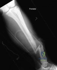
A 22-year-old man undergoes intramedullary nailing of a midshaft femur fracture on a fracture table.

The procedure takes 4 hours due to significant difficulty achieving a closed reduction. Postoperatively, he complains of numbness in his perineal region and presents with erectile dysfunction. What is the most likely cause of this complication?


Explanation

Pudendal nerve palsy is a well-documented complication of using a fracture table, resulting from prolonged compression of the nerve against the perineal post. It manifests as perineal numbness and erectile dysfunction. Preventive measures include minimizing traction time, ensuring adequate padding, and occasionally releasing traction if a delay occurs during surgery.

Question 15

A 30-year-old construction worker drops a heavy steel beam on his midfoot, sustaining a severe Lisfranc fracture-dislocation.

In the emergency department, he complains of significant numbness over the dorsal aspect of the first web space of his foot. Which nerve is most likely compromised, and what is its anatomic course relative to the injury?


Explanation

The deep peroneal nerve provides sensory innervation to the first dorsal web space of the foot. It courses dorsally over the midfoot (tarsometatarsal joints) in close proximity to the dorsalis pedis artery. In severe midfoot crush injuries or Lisfranc fracture-dislocations, this nerve is vulnerable to direct contusion, stretch, or compression from localized swelling and fracture displacement.

Question 16

A 40-year-old male presents with a chronic anterior shoulder dislocation missed for 6 weeks. During open reduction and stabilization using a Latarjet procedure, the conjoint tendon is aggressively retracted medially to gain exposure to the glenoid. Postoperatively, the patient is unable to flex his elbow against resistance when the forearm is supinated and has sensory loss over the lateral forearm. Which nerve was most likely injured?


Explanation

The musculocutaneous nerve branches from the lateral cord and enters the coracobrachialis muscle approximately 5 to 8 cm distal to the tip of the coracoid process. Aggressive or prolonged medial retraction of the conjoint tendon during anterior shoulder approaches (like the Latarjet procedure) places this nerve at high risk for a stretch injury. Deficits include weakness of the biceps and brachialis muscles (elbow flexion) and sensory loss in the lateral antebrachial cutaneous nerve distribution.

Question 17

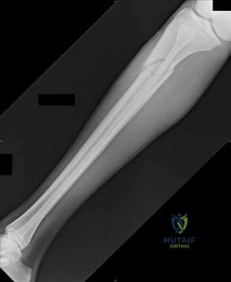
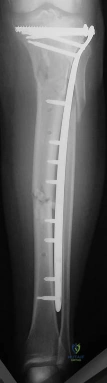
A 35-year-old man is struck by a car and sustains a Schatzker VI tibial plateau fracture.

He undergoes temporary spanning external fixation. Twelve hours later, he develops intractable leg pain out of proportion to his injury, severe pain with passive toe flexion, and decreased sensation in the first web space. His dorsalis pedis pulse remains palpable. Which of the following is the most accurate statement regarding his condition?


Explanation

The patient exhibits classic signs of acute compartment syndrome (ACS) of the anterior leg. The anterior compartment contains the tibialis anterior, EHL, and EDL muscles; passive toe flexion stretches these extensors, eliciting severe pain. The deep peroneal nerve runs within the anterior compartment and provides sensation to the first web space; its compromise is an early sign of ACS. A palpable pulse does not rule out ACS, as tissue perfusion ceases at pressures well below systolic arterial pressure.

Question 18

In a Holstein-Lewis fracture (a spiral fracture of the distal third of the humerus), the radial nerve is particularly vulnerable to entrapment or laceration.

At what specific anatomical location does the nerve typically become entrapped by the fracture fragments?


Explanation

The radial nerve is relatively fixed and tethered as it pierces the lateral intermuscular septum to pass from the posterior compartment to the anterior compartment of the arm, approximately 10 cm proximal to the lateral epicondyle. In a Holstein-Lewis fracture, the distal fracture fragment typically displaces proximally, trapping or impaling the tethered radial nerve between the bone ends.

Question 19

A 32-year-old male sustains a posterior hip dislocation. He presents with a foot drop but retains full strength in ankle plantar flexion and toe flexion. Why is the peroneal division of the sciatic nerve more frequently and severely injured than the tibial division in this clinical scenario?


Explanation

The common peroneal division of the sciatic nerve is injured more frequently in posterior hip dislocations due to its anatomical and histological properties. It lies lateral to the tibial division (closer to the posteriorly displacing femoral head), is tethered between the sciatic notch and the fibular neck, and histologically contains larger fascicles with significantly less protective epineurial connective tissue compared to the tibial division.

Question 20

A 45-year-old roofer falls from a ladder, sustaining a severely comminuted, depressed intra-articular calcaneus fracture. It is treated non-operatively due to severe patient comorbidities. Six months later, he complains of a burning, neuropathic pain in the plantar aspect of his foot and toes that worsens with weight-bearing. Tinel's sign is strongly positive just posterior to the medial malleolus. What anatomical structure forms the roof of the fibro-osseous tunnel causing the compression of the involved nerve?


Explanation

The patient is presenting with post-traumatic tarsal tunnel syndrome, a known complication of calcaneus fractures due to hindfoot varus malunion, loss of calcaneal height (settling), or scar tissue formation. The tibial nerve is compressed within the tarsal tunnel, the roof of which is formed by the flexor retinaculum (also known anatomically as the laciniate ligament), which spans between the medial malleolus and the medial calcaneus.

Question 21

An 18-year-old male sustains a closed distal third spiral humeral shaft fracture.

On physical examination, he is unable to actively extend his wrist or digits. What is the most appropriate initial management of this nerve palsy?


Explanation

This patient has a Holstein-Lewis fracture (distal third spiral humerus fracture) with an associated radial nerve palsy. The vast majority of these injuries are neuropraxias. The standard of care for a closed humeral shaft fracture with an acute radial nerve palsy is conservative management with closed reduction, splinting, and observation. Surgical exploration is indicated if the palsy occurs after a closed reduction attempt (iatrogenic), if it is an open fracture, or if there is no recovery after 3-4 months.

Question 22

A 25-year-old male presents with an anterior shoulder dislocation following a rugby tackle. Post-reduction, he reports numbness over the lateral aspect of his shoulder. If this nerve injury persists, which muscle's function will most likely demonstrate profound weakness on subsequent examination?


Explanation

The patient has an axillary nerve palsy, which is the most common nerve injury associated with anterior shoulder dislocations. The axillary nerve innervates the deltoid and the teres minor. The teres minor is an external rotator of the shoulder. The supraspinatus and infraspinatus are innervated by the suprascapular nerve, while the subscapularis is innervated by the upper and lower subscapular nerves.

Question 23

A 6-year-old boy falls on an outstretched hand and sustains a Bado Type I Monteggia fracture-dislocation. On examination, he is unable to extend his thumb and digits at the metacarpophalangeal joints, but active wrist extension is preserved, accompanied by radial deviation. Which nerve is most likely injured?


Explanation

The posterior interosseous nerve (PIN) is commonly injured in Monteggia fracture-dislocations, particularly Bado types I (anterior) and III (lateral). The PIN innervates the extensor carpi ulnaris (ECU) and the extensor digitorum communis (EDC), but spares the extensor carpi radialis longus (ECRL), which is innervated by the radial nerve proper before it bifurcates. This results in preserved wrist extension but with a radial deviation bias, along with inability to extend the digits at the MCP joints.

Question 24

A 5-year-old boy presents with a Gartland type III extension-type supracondylar humerus fracture. Which specific physical examination finding is the hallmark of the most common nerve injury associated with this fracture pattern?


Explanation

The anterior interosseous nerve (AIN) is the most commonly injured nerve in extension-type supracondylar humerus fractures. The AIN is a purely motor branch of the median nerve that innervates the flexor pollicis longus (FPL), the flexor digitorum profundus (FDP) to the index and middle fingers, and the pronator quadratus. Injury results in the inability to form an 'OK' sign, manifesting as an inability to flex the IP joint of the thumb and the DIP joint of the index finger.

Question 25

A 30-year-old male sustains a high-energy traumatic knee dislocation.

Following reduction, he presents with a foot drop and numbness over the dorsum of the foot. Which specific ligamentous injury pattern is most highly associated with this neurologic deficit?


Explanation

Common peroneal nerve injury is a frequent complication of knee dislocations, occurring in up to 25-30% of cases. It is most highly associated with injuries to the posterolateral corner (PLC) and lateral collateral ligament (LCL) because the mechanism of injury (varus stress and hyperextension) strongly stretches the nerve as it wraps around the fibular neck.

Question 26

A 40-year-old female sustains a comminuted Denis Zone III sacral fracture after a fall from a height. Based on the anatomic location of this fracture, which of the following neurologic deficits is most frequently encountered?


Explanation

Denis classified sacral fractures into three zones: Zone I (alar), Zone II (foraminal), and Zone III (central canal). Zone III fractures frequently involve the sacral nerve roots (S2-S4) within the central canal, leading to a high incidence of bowel, bladder, and sexual dysfunction (cauda equina-like syndrome), manifesting as loss of perianal sensation and decreased anal sphincter tone.

Question 27

A 28-year-old male is involved in a motor vehicle collision, sustaining a posterior wall acetabular fracture and a posterior hip dislocation.

Following a successful closed reduction, he demonstrates profound weakness in ankle dorsiflexion but preserved plantarflexion. Which division of the sciatic nerve is most commonly injured in this clinical scenario?


Explanation

Sciatic nerve injury occurs in 10-20% of posterior hip dislocations and is particularly common when associated with posterior wall acetabular fractures. The peroneal division of the sciatic nerve is laterally positioned and tightly tethered at the sciatic notch and fibular head, making it significantly more susceptible to stretch injury than the medial tibial division.

Question 28

A 62-year-old female undergoes open reduction and internal fixation of a distal radius fracture using a standard Henry (volar) approach. Post-operatively, she is unable to actively flex the interphalangeal joint of her thumb. Which structure was most likely subjected to excessive retraction during the procedure?


Explanation

In the volar Henry approach to the distal radius, deep retractors placed around the radius can inadvertently compress or stretch the anterior interosseous nerve (AIN) or the flexor pollicis longus (FPL) muscle belly. AIN palsy presents as weakness or loss of thumb interphalangeal flexion and index distal interphalangeal flexion (positive Kiloh-Nevin sign / inability to make an 'OK' sign).

Question 29

A 35-year-old male sustains a severely displaced scapular fracture with a fracture line extending deep into the spinoglenoid notch. He complains of persistent shoulder weakness. Which specific physical examination finding is most characteristic of nerve entrapment at this anatomic location?


Explanation

The suprascapular nerve innervates the supraspinatus muscle as it passes through the suprascapular notch, and then travels through the spinoglenoid notch to innervate the infraspinatus. Entrapment or injury at the spinoglenoid notch affects only the infraspinatus (causing weakness in external rotation), while sparing the supraspinatus (preserving abduction).

Question 30

A 25-year-old male undergoes intramedullary nailing of a femoral shaft fracture on a fracture table utilizing a perineal post. Post-operatively, he complains of numbness in his perineum and erectile dysfunction. What is the most appropriate initial management?


Explanation

Pudendal nerve neuropraxia is a known complication of utilizing a perineal post on a fracture table, resulting from direct compression or traction. Symptoms include perineal numbness and erectile dysfunction. The vast majority of these injuries are transient and resolve completely with observation and supportive care over weeks to months.

Question 31

A 68-year-old female sustains a 3-part proximal humerus fracture. Due to severe pain, comprehensive motor testing is difficult. What is the most sensitive and reliable physical examination maneuver to assess the integrity of the nerve most commonly injured in this setting?


Explanation

The axillary nerve is the most frequently injured nerve in proximal humerus fractures. In the acute setting, pain often precludes reliable motor testing of the deltoid. Testing the sensation over the lateral aspect of the arm (regimental badge area), innervated by the superior lateral cutaneous nerve of the arm (a branch of the axillary nerve), is the most reliable way to assess its integrity.

Question 32

A 45-year-old male sustains a pronation-external rotation (PER) ankle fracture with lateral dislocation. Post-reduction, he notes numbness on the dorsal aspect of the foot (sparing the first web space) and weakness in active foot eversion. Which nerve is involved?


Explanation

The superficial peroneal nerve innervates the muscles of the lateral compartment of the leg (peroneus longus and brevis), which are responsible for eversion. It also provides sensation to the majority of the dorsum of the foot, with the exception of the first web space (deep peroneal nerve). Stretch injuries to this nerve can occur during significant rotational ankle injuries.

Question 33

A 10-year-old boy sustains a 'floating elbow' injury consisting of an ipsilateral displaced supracondylar humerus fracture and a both-bone forearm fracture. He is splinted in the emergency department. Two hours later, he develops severe, unrelenting pain in his forearm, and severe pain with passive extension of his fingers. What is the most appropriate NEXT step?


Explanation

The patient is exhibiting classic signs of acute compartment syndrome. The most critical and immediate first step in management is the complete removal of all constrictive dressings, casts, and splints down to the skin. This simple maneuver can reduce compartmental pressures by up to 50-85%. If symptoms do not rapidly improve, emergent fasciotomy is indicated.

Question 34

A 55-year-old female presents with a non-displaced distal radius fracture treated with a well-molded short arm cast. Two days later, she reports severe, progressively worsening numbness and tingling in her thumb, index, and middle fingers, as well as worsening pain. What is the most appropriate next step in management?


Explanation

Acute carpal tunnel syndrome can occur following a distal radius fracture, exacerbated by fracture hematoma and a tight cast. The initial step should always be to bi-valve or completely split the cast and all underlying padding to relieve extrinsic pressure. If symptoms persist despite decompression of the cast, surgical carpal tunnel release is indicated.

Question 35

A 30-year-old man sustains a severe grade IIIA open midshaft humerus fracture resulting from a motorcycle crash. On initial presentation, he has an isolated radial nerve palsy. He is scheduled for urgent surgical debridement. What is the recommended management for the radial nerve during this procedure?


Explanation

While closed humeral shaft fractures with radial nerve palsy are managed non-operatively initially, an open humeral shaft fracture with a radial nerve palsy is an absolute indication for primary surgical exploration of the nerve at the time of initial irrigation, debridement, and fracture stabilization to evaluate for nerve laceration or entrapment.

Question 36

A 70-year-old female sustains a primary anterior shoulder dislocation. After a successful and atraumatic closed reduction, she demonstrates persistent, profound weakness in active external rotation and active shoulder abduction. However, sensation over the lateral deltoid remains completely intact. What is the most likely etiology of her weakness?


Explanation

In older adults (particularly those over 40, and risk increases significantly over 60), anterior shoulder dislocations have a high association with massive rotator cuff tears. These tears present with profound weakness that can mimic a nerve palsy. The intact sensation over the deltoid makes an axillary nerve injury less likely, pointing strongly to a structural cuff failure.

Question 37

A 15-year-old gymnast sustains a medial epicondyle fracture that is displaced 15 mm into the joint, necessitating open reduction and internal fixation. During the surgical approach, the ulnar nerve is identified. According to current orthopedic literature, what is the most appropriate management of the ulnar nerve?


Explanation

In the surgical treatment of pediatric medial epicondyle fractures, routine anterior transposition of the ulnar nerve is not recommended unless there is pre-existing significant nerve tension, instability, or the nerve impedes anatomical reduction. Standard practice involves identifying and protecting the nerve, leaving it in situ, and proceeding with fixation.

Question 38

A 32-year-old male sustains a severe Hawkins type III talar neck fracture following a fall from height. Given the typical direction of displacement of the talar body in this specific fracture pattern, which neurovascular structure is at greatest risk of impingement or injury?


Explanation

A Hawkins type III fracture is a talar neck fracture with dislocation of both the subtalar and tibiotalar joints. The talar body characteristically extrudes posteromedially. In this position, it places direct pressure on the posteromedial neurovascular bundle, putting the posterior tibial artery and the tibial nerve at high risk of injury.

Question 39

A collegiate football player sustains a direct blow to the lateral aspect of his knee, resulting in a fibular neck fracture and a subsequent complete foot drop due to common peroneal nerve injury. During his rehabilitation phase, return of function in which of the following muscles would serve as the earliest clinical indicator of nerve recovery?


Explanation

Following a common peroneal nerve injury at the fibular neck, the nerve regenerates proximally to distally. The tibialis anterior is the most proximally innervated muscle in the anterior compartment by the deep peroneal branch. Therefore, return of active ankle dorsiflexion (tibialis anterior function) is typically the earliest clinical sign of re-innervation.

Question 40

A 45-year-old male patient with a historically malunited, highly displaced midshaft clavicle fracture presents with progressive upper extremity weakness, numbness, and tingling. Examination reveals intrinsic hand muscle wasting and sensory deficits along the medial aspect of the forearm and hand. Hypertrophic fracture callus is identified on imaging. Which nerve roots are most likely compressed in this form of Thoracic Outlet Syndrome?


Explanation

Malunited clavicle fractures with massive hypertrophic callus can cause secondary thoracic outlet syndrome (TOS). This typically results in compression of the lower trunk of the brachial plexus (C8 and T1 nerve roots) or the medial cord, as these structures pass inferior to the clavicle and over the first rib. Symptoms include ulnar neuropathy-like findings and intrinsic hand wasting.

Question 41

A 26-year-old unrestrained driver is involved in a motor vehicle collision. He presents with his right hip flexed, adducted, and internally rotated. Following closed reduction of the hip, he is unable to dorsiflex his right foot or extend his toes, but plantar flexion is symmetric to the contralateral side. Which of the following best explains the specific pattern of this neurological deficit?


Explanation

The common peroneal division of the sciatic nerve is more susceptible to injury in posterior hip dislocations because its fascicles are larger, fewer in number, and have less protective connective tissue compared to the tibial division.

Question 42

A 7-year-old girl falls from monkey bars and sustains a Bado Type III Monteggia fracture-dislocation. She exhibits a nerve palsy characterized by weakness in thumb and finger extension, but normal wrist extension with radial deviation. Which nerve is most likely injured?


Explanation

Bado Type III Monteggia fractures are highly associated with Posterior Interosseous Nerve (PIN) injuries. PIN palsy causes weakness in finger/thumb extensors and ECU, leading to radial deviation during wrist extension.

Question 43

A 34-year-old man sustains a Grade II open midshaft humerus fracture. On initial evaluation, he is unable to actively extend his wrist or fingers, and he has decreased sensation over the dorsal first web space. What is the most appropriate management of his nerve injury?


Explanation

Immediate surgical exploration of the radial nerve is indicated in the setting of an open humerus shaft fracture associated with a radial nerve palsy. Closed fractures with primary palsies can typically be observed.

Question 44

A 5-year-old boy presents with a displaced flexion-type supracondylar humerus fracture after falling directly onto a flexed elbow. Which of the following nerve injuries is most frequently associated with this specific fracture pattern?


Explanation

While extension-type supracondylar humerus fractures most commonly injure the anterior interosseous nerve (AIN), flexion-type fractures have a higher association with ulnar nerve injuries due to posterior displacement of the proximal fragment.

Question 45

A 45-year-old man is undergoing open reduction and internal fixation of a posterior wall acetabular fracture via a Kocher-Langenbeck approach. What intraoperative leg position is most critical to minimize iatrogenic tension on the sciatic nerve?


Explanation

During the Kocher-Langenbeck approach, the sciatic nerve is at significant risk of iatrogenic stretch injury. Extending the hip and flexing the knee minimizes tension on the sciatic nerve.

Question 46

A 22-year-old football player sustains a high-energy Lisfranc injury with lateral displacement of the second through fifth metatarsals. If he develops a neurological deficit secondary to this injury, what physical examination finding is most likely?


Explanation

The deep peroneal nerve and dorsalis pedis artery run between the first and second metatarsals. A severe Lisfranc dislocation can compress or stretch this nerve, resulting in numbness in the first dorsal web space.

Question 47

A 48-year-old man presents with progressive numbness in his small and ring fingers, accompanied by intrinsic muscle wasting in his dominant hand. He reports having 'broken his elbow' as a young child. Radiographs reveal a severe cubitus valgus deformity. Nonunion of which of the following pediatric fractures is the most likely underlying cause?


Explanation

Nonunion of a pediatric lateral condyle fracture typically leads to a progressive cubitus valgus deformity. Years later, this abnormal valgus angle causes stretching of the ulnar nerve, known as a tardy ulnar nerve palsy.

Question 48

A 29-year-old man falls on an outstretched hand. Lateral radiographs of his wrist show the lunate displaced volarly and 'spilled' from the radius, while the capitate remains aligned with the radius. He reports severe pain and numbness in his index and middle fingers. What is the most appropriate management?


Explanation

This is a lunate dislocation, which frequently causes acute median nerve compression in the carpal tunnel. Urgent open reduction, ligamentous repair, and carpal tunnel release are required to prevent permanent nerve damage.

Question 49

A 31-year-old female sustains an anterior knee dislocation (KD-III) following a trampoline injury. Vascular exam is normal, but she has a complete foot drop and cannot actively extend her toes. Where is the most likely anatomic site of nerve tethering causing this injury?


Explanation

Knee dislocations have a high rate of common peroneal nerve injury (especially posterolateral corner injuries). The nerve is firmly tethered at the fibular neck as it wraps around the bone, making it highly susceptible to traction.

Question 50

A 28-year-old male sustains a posterior hip dislocation. Following successful closed reduction, he is noted to have a dense foot drop and absent sensation over the dorsal aspect of his foot. Which anatomic characteristic makes the primarily involved nerve division most susceptible to this specific injury?


Explanation

The common peroneal division of the sciatic nerve is laterally positioned and has larger fascicles with less protective connective tissue (perineurium). This makes it highly susceptible to traction injury during a posterior hip dislocation.

Question 51

A 7-year-old girl presents with a flexion-type supracondylar humerus fracture. Her hand is well-perfused, but she exhibits a specific neurologic deficit. Which nerve is most commonly injured in this specific fracture pattern?


Explanation

Unlike extension-type supracondylar fractures where the anterior interosseous nerve is most commonly injured, flexion-type supracondylar fractures place the ulnar nerve at the greatest risk of injury.

Question 52

A 22-year-old male sustains a traumatic knee dislocation (KD-III). Vascular examination is normal, but he exhibits a complete common peroneal nerve palsy. Assuming a closed injury with a grossly stable reduction, what is the most appropriate initial management of the nerve injury?


Explanation

Peroneal nerve palsies associated with knee dislocations are typically traction injuries (neuropraxia/axonotmesis). Observation is indicated initially, with EMG obtained at 6 weeks to 3 months to monitor for subclinical reinnervation.

Question 53

A 6-year-old child presents with a Bado Type III Monteggia fracture-dislocation. Following closed reduction, the patient cannot extend the fingers at the metacarpophalangeal joints but demonstrates normal wrist extension with radial deviation. What is the expected natural history of this neurologic deficit?


Explanation

The posterior interosseous nerve (PIN) is classically injured in Bado Type III (lateral) Monteggia fractures. It is almost always a neuropraxia that resolves spontaneously within 3 to 6 months without surgical intervention.

Question 54

A 29-year-old male falls from a roof and sustains a dorsal perilunate dislocation. He presents with severe wrist pain, absent 2-point discrimination in the thumb and index finger, and weakness in thumb opposition. What is the most appropriate initial surgical management?


Explanation

Acute median neuropathy following a perilunate dislocation represents an acute carpal tunnel syndrome. It is a surgical emergency requiring immediate carpal tunnel release in conjunction with open reduction of the carpus.

Question 55

A 40-year-old male undergoes intramedullary nailing of a comminuted femoral shaft fracture utilizing a fracture table. Postoperatively, he complains of perineal numbness and erectile dysfunction. What is the most likely etiology of this complication?


Explanation

Pudendal nerve neuropraxia is a known complication of utilizing a fracture table for femoral nailing. It is caused by prolonged, excessive traction against a hard or poorly padded perineal post.

Question 56

A 35-year-old female sustains a U-type sacral fracture extending through the central sacral canal (Denis Zone III). Based on this specific fracture classification, she is most at risk for which of the following neurologic deficits?


Explanation

Denis Zone III sacral fractures involve the central sacral canal. These injuries carry a high risk (up to 60%) of cauda equina syndrome, presenting as bowel, bladder, and sexual dysfunction.

Question 57

A 45-year-old man is involved in a motor vehicle collision, sustaining a posterior hip dislocation and a posterior wall acetabular fracture. Following closed reduction, he is unable to dorsiflex his ankle or extend his great toe, though plantar flexion remains intact. Which division of the affected nerve is most likely injured, and what is its typical relative prognosis?


Explanation

Posterior hip dislocations most commonly injure the sciatic nerve, specifically the peroneal division due to its lateral position and secure tethering at the sciatic notch. The peroneal division has a significantly poorer prognosis for spontaneous recovery compared to the tibial division.

Question 58

A 22-year-old male sustains a KD-III knee dislocation. Post-reduction, he exhibits completely absent ankle dorsiflexion and eversion. At 3 months follow-up, there is zero return of clinical function, and an EMG shows absent motor unit action potentials in the anterior compartment musculature. What is the most appropriate next step in management?


Explanation

After a high-energy stretch injury like a knee dislocation, a common peroneal nerve palsy showing no clinical or electrodiagnostic recovery by 3 months warrants surgical exploration. Tendon transfers are typically reserved as salvage procedures if nerve reconstruction fails or presents late.

Question 59

A 7-year-old girl falls and sustains a Bado Type III Monteggia fracture-dislocation. On examination, she is unable to actively extend the MCP joints of her fingers, but wrist extension is preserved with radial deviation. Which nerve structure is injured?


Explanation

Bado Type III injuries (lateral dislocation of the radial head) carry the highest risk of posterior interosseous nerve (PIN) palsy. PIN injury causes loss of finger and thumb extension, as well as loss of ulnar wrist extension (ECU), leading to radial deviation during active wrist extension.

Question 60

A 55-year-old female presents with a completely displaced fracture of the distal radius. After a closed reduction and splinting, she complains of rapidly escalating pain and progressive loss of two-point discrimination in her thumb, index, and long fingers within 2 hours. What is the definitive management?


Explanation

Progressive, severe median nerve neuropathy following reduction of a distal radius fracture signifies acute carpal tunnel syndrome. This is a surgical emergency requiring immediate carpal tunnel release and simultaneous rigid fracture stabilization.

Question 61

A 30-year-old man sustains a closed mid-distal humeral shaft fracture. His initial neurologic examination in the trauma bay is entirely normal. Following an attempt at closed reduction and application of a coaptation splint, he is noted to have a complete loss of wrist and finger extension. What is the most appropriate management?


Explanation

A secondary radial nerve palsy that develops strictly after a closed reduction attempt of a humeral shaft fracture is an absolute indication for immediate surgical exploration. This presentation suggests the nerve may have become entrapped within the fracture site during manipulation.

Question 62

An 8-year-old boy presents to the emergency department with a flexion-type supracondylar humerus fracture. Which of the following nerve injuries is most frequently associated with this specific fracture configuration?


Explanation

While extension-type supracondylar humerus fractures are most commonly associated with anterior interosseous nerve injuries, flexion-type fractures are classically and most frequently associated with ulnar nerve palsy.

Question 63

A 25-year-old male is involved in a severe crush injury resulting in a Denis Zone III sacral fracture. Which of the following neurologic deficits is most specifically characteristic of this injury pattern?


Explanation

Denis Zone III sacral fractures involve the central sacral canal and carry a very high risk (>50%) of cauda equina syndrome. Injury to the lower sacral nerve roots classically results in saddle anesthesia, as well as bowel, bladder, and sexual dysfunction.

Question 64

A 40-year-old motorcyclist presents after a high-speed crash with massive soft tissue swelling over the left shoulder. Radiographs demonstrate lateral displacement of the scapula, an intact but widened acromioclavicular joint, and a severely displaced clavicle fracture. Radial pulses are diminished. Which nerve injury is most common in this scenario?


Explanation

This clinical and radiographic picture defines a scapulothoracic dissociation, which acts as a closed forequarter amputation. It is highly associated with catastrophic subclavian vascular disruption and complete brachial plexus avulsions, carrying a dismal functional prognosis.

Question 65

A 35-year-old man sustains a lateral compression (LC-1) pelvic ring injury involving a significantly displaced fracture through the sacral ala. On physical examination, he demonstrates weakness in ankle dorsiflexion and great toe extension. Which nerve root is most likely compromised by this specific fracture?


Explanation

The L5 nerve root courses directly over the anterior aspect of the sacral ala before joining the sacral plexus. Displaced fractures of the sacral ala can stretch or impale the L5 root, causing profound weakness in ankle and hallux dorsiflexion.

Question 66

A 65-year-old woman sustains a 3-part proximal humerus fracture. Her pain prevents any active shoulder movement. To accurately assess the function of the axillary nerve in this acute setting, which examination finding is most reliable?


Explanation

Because severe pain precludes reliable motor testing of the deltoid and teres minor in an acute proximal humerus fracture, evaluating sensation over the lateral deltoid (innervated by the superior lateral brachial cutaneous branch of the axillary nerve) is the most reliable clinical test.

Question 67

A 28-year-old man falls on his outstretched hand. His wrist is diffusely swollen, and he holds his fingers in slight flexion. A lateral radiograph demonstrates the 'spilled teacup' sign. He reports severe, continuous numbness in his thumb, index, and middle fingers. What is the most appropriate initial management?


Explanation

A volar lunate dislocation (spilled teacup sign) frequently compresses the median nerve. When presenting with acute carpal tunnel syndrome, urgent surgical intervention involving open reduction, ligament repair, and carpal tunnel release is mandated.

Question 68

A 12-year-old boy sustains a traumatic elbow dislocation that is reduced in the emergency department. Post-reduction radiographs show widening of the medial joint space, and the medial epicondyle ossification center is completely absent from its normal anatomic position. He has new-onset numbness in his small finger. What is the most likely pathomechanism?


Explanation

Incarceration of the medial epicondyle within the elbow joint is a classic complication of pediatric elbow dislocations. It presents with a widened medial joint space, an 'absent' medial epicondyle on standard views, and often ulnar neuropathy due to entrapment of the nerve with the bone fragment.

Question 69

A 25-year-old man sustains a Grade II open fracture of the middle third of the humerus. On initial evaluation, he has an absent brachioradialis reflex and is entirely unable to extend his wrist or fingers. What is the standard of care for the nerve injury in this specific scenario?


Explanation

An open humerus fracture combined with a radial nerve palsy is an absolute indication for surgical exploration of the radial nerve. This is performed concurrently with the required surgical debridement and fracture stabilization.

Question 70

A 45-year-old man is 6 months status post ORIF of a severe intra-articular calcaneus fracture. He complains of burning pain on the plantar aspect of his foot that worsens at night. He has a strongly positive Tinel's sign posterior to the medial malleolus. What is the most likely diagnosis?


Explanation

Tarsal tunnel syndrome involves compression of the tibial nerve behind the medial malleolus. It is a known late complication of calcaneus fractures, often resulting from post-traumatic scarring, altered hindfoot anatomy, or impingement from retained hardware.

Question 71

A 22-year-old man sustains a low-velocity gunshot wound to the arm, resulting in a comminuted midshaft humerus fracture and a complete radial nerve palsy. Distal pulses are strong, and the limb is well-perfused. What is the most appropriate initial treatment strategy?


Explanation

Radial nerve palsies associated with low-velocity gunshot wounds to the humerus are typically neuropraxias. The standard of care is non-operative management initially with observation. Surgical exploration is reserved for those failing to show clinical or EMG recovery by 3-4 months.

Question 72

A 30-year-old man suffers a "floating knee" injury (ipsilateral femur and tibia fractures) in a rollover collision. Postoperatively, he exhibits a complete foot drop and absent two-point discrimination in the first dorsal web space. Which nerve was injured, and where is its most common site of traumatic tethering?


Explanation

The clinical findings of foot drop and numbness in the first dorsal web space represent a deep peroneal nerve injury. This nerve is extremely vulnerable to stretch and compression as it wraps intimately around the fibular neck.

Question 73

A 40-year-old man sustains a Pipkin IV fracture-dislocation of the hip with an immediate, profound sciatic nerve palsy. He is reduced and placed in skeletal traction. A post-reduction CT scan clearly demonstrates a large posterior wall fragment directly impinging upon the sciatic nerve. What is the next best step in management?


Explanation

While post-traumatic sciatic nerve palsies are often observed, documented direct mechanical compression of the nerve by a fracture fragment (such as a posterior wall segment) is an absolute indication for urgent surgical decompression and fracture fixation.

None

Dr. Mohammed Hutaif
Medically Verified Content by
Prof. Dr. Mohammed Hutaif
Consultant Orthopedic & Spine Surgeon
Chapter Index