Part of the Master Guide

Orthopedic Surgery Board Review MCQs: Arthroplasty, Trauma & Spine Part 255

Orthopedic Board Review MCQs: Elbow, Shoulder & Trauma | Part 85

23 Apr 2026 62 min read 55 Views
Orthopedic Surgery Board Review MCQs: AAOS Master Bank Part 85

Key Takeaway

This page offers Part 85 of an interactive orthopedic surgery board review quiz. Featuring 50 high-yield MCQs in OITE/AAOS format, it targets orthopedic residents and surgeons preparing for their AAOS and ABOS board exams. Covering Elbow, Shoulder, Trauma, it ensures robust certification exam preparation.

Orthopedic Board Review MCQs: Elbow, Shoulder & Trauma | Part 85

Comprehensive 100-Question Exam


00:00

Start Quiz

Question 1

A 45-year-old female presents after a fall on an outstretched hand. Imaging reveals a terrible triad injury of the elbow.

During surgical intervention, what is the most widely accepted sequence of repair to restore elbow stability?


Explanation

The terrible triad of the elbow consists of an elbow dislocation, a radial head fracture, and a coronoid fracture. The standard surgical sequence to restore stability from deep to superficial (inside-out) is: 1) Fixation or reconstruction of the coronoid (to restore the anterior buttress), 2) Fixation or replacement of the radial head (to restore the anterior and valgus buttress), and 3) Repair of the lateral collateral ligament (LCL) complex to the lateral epicondyle. MCL repair is only considered if the elbow remains unstable after these steps and application of a hinged external fixator is not preferred.

Question 2

A 72-year-old male with severe rotator cuff tear arthropathy undergoes a reverse total shoulder arthroplasty (rTSA). How does the rTSA implant alter the biomechanics of the shoulder joint compared to the native anatomy?


Explanation

Reverse total shoulder arthroplasty (Grammont design) biomechanically alters the shoulder by medializing and inferiorizing the center of rotation. Medialization recruits more deltoid muscle fibers and decreases the torque on the glenoid component, while inferiorization tensions the deltoid and increases its moment arm, allowing the deltoid to compensate for the deficient rotator cuff to elevate the arm.

Question 3

A 35-year-old male suffers a distal biceps tendon rupture and undergoes surgical repair via a two-incision technique. Compared to the single anterior incision technique, the two-incision approach is associated with a higher risk of which of the following complications?


Explanation

The two-incision technique for distal biceps repair was developed to avoid the radial nerve (PIN) injuries sometimes seen with the single-incision approach. However, the two-incision technique carries a significantly higher risk of heterotopic ossification and radioulnar synostosis due to muscle splitting and subperiosteal dissection near the ulna. Conversely, the single anterior incision approach has a higher risk of lateral antebrachial cutaneous nerve (LABCN) neurapraxia.

Question 4

According to the criteria described by Hertel et al. for proximal humerus fractures, which of the following combinations of radiographic findings is most predictive of humeral head ischemia?


Explanation

Hertel established classic predictors of humeral head ischemia following proximal humerus fractures. The most critical predictors are an anatomic neck fracture, a short calcar segment (metaphyseal extension < 8 mm), and disruption of the medial hinge (> 2 mm displacement). The combination of a disrupted medial hinge and short metaphyseal extension has a positive predictive value of 97% for humeral head ischemia.

Question 5

A 28-year-old polytrauma patient sustains an isolated, closed body and neck fracture of the scapula. According to current consensus guidelines, which of the following radiographic parameters is considered an absolute indication for Open Reduction and Internal Fixation (ORIF)?


Explanation

Most scapula body fractures are treated nonoperatively. However, absolute indications for ORIF generally involve significant intra-articular disruption or massive displacement that compromises shoulder biomechanics. Indications include intra-articular glenoid step-off > 4-5 mm, glenopolar angle < 22 degrees, medial/lateral displacement > 20 mm, and angular deformity > 45 degrees. An ipsilateral clavicle fracture (floating shoulder) is a relative indication, often treated by fixing the clavicle alone.

Question 6

A 19-year-old male sustains a midshaft clavicle fracture during a cycling accident. Which of the following factors most significantly increases the risk of nonunion if this injury is treated nonoperatively?


Explanation

The risk of nonunion in nonoperatively managed midshaft clavicle fractures is historically cited at 1-5%, but modern studies show it can be up to 15% in specific subsets. The most significant risk factors for nonunion are complete displacement (no cortical contact) and shortening greater than 2 cm. Advanced age and female gender have also been noted as risk factors in some studies, but > 2 cm shortening and 100% displacement remain the strongest mechanical predictors.

Question 7

A 45-year-old male presents with a closed, spiral fracture of the distal third of the humerus (Holstein-Lewis fracture). On initial examination, the patient has normal radial nerve function. A coaptation splint is applied in the emergency department. Upon re-evaluation post-reduction, the patient is unable to extend his wrist or fingers and has numbness in the first dorsal web space. What is the most appropriate next step in management?


Explanation

A radial nerve palsy that develops AFTER a closed reduction of a humeral shaft fracture is an absolute indication for immediate surgical exploration. The nerve may be entrapped in the fracture site (especially in a distal third Holstein-Lewis fracture). If the palsy was present on initial presentation (prior to manipulation), observation and functional bracing would be appropriate.

Question 8

A 22-year-old collegiate rugby player presents with recurrent anterior shoulder instability. A 3D CT scan reveals 26% anterior glenoid bone loss.

What is the most appropriate surgical intervention to prevent further dislocations in this athlete?


Explanation

The Latarjet procedure (coracoid transfer) is the treatment of choice for anterior shoulder instability in the presence of critical anterior glenoid bone loss (>20-25%). Arthroscopic or open Bankart repairs have unacceptably high failure rates in patients with significant bone loss, especially in young contact athletes, because soft tissue repair alone cannot restore the osseous articular arc.

Question 9

During a physical examination of the shoulder, a patient is asked to place the palm of their hand on their opposite shoulder, with the elbow kept elevated. The examiner then applies a downward force to the patient's forearm while the patient resists. This test is highly specific for evaluating a tear of which structure?


Explanation

The physical examination maneuver described is the Bear-Hug test. It is highly sensitive and specific for diagnosing tears of the upper border of the subscapularis. The Belly-Press test is another subscapularis test, but the Bear-Hug test is considered more sensitive for upper subscapularis tears. The Lift-Off test requires internal rotation behind the back, evaluating the lower subscapularis.

Question 10

A 6-year-old girl is brought to the emergency department after falling from monkey bars. Radiographs demonstrate an isolated fracture of the ulnar shaft with dorsal angulation and an associated posterior dislocation of the radial head. According to the Bado classification, what type of Monteggia lesion is this?


Explanation

The Bado classification categorizes Monteggia fractures based on the direction of radial head dislocation. Type I: Anterior dislocation (most common). Type II: Posterior dislocation (ulna typically bowed dorsally). Type III: Lateral dislocation. Type IV: Anterior dislocation with fractures of both the radius and ulna shafts.

Question 11

During the surgical repair of a chronic, complete rupture of the pectoralis major muscle, the surgeon must mobilize and anatomically repair the sternal and clavicular heads to their footprint on the humerus. What is the correct anatomical relationship of the clavicular head relative to the sternal head at their humeral insertion?


Explanation

At the humeral insertion, the pectoralis major tendon undergoes a 180-degree twist. The clavicular head descends directly to insert anteriorly (superficially) and distally on the lateral lip of the bicipital groove. The sternal head twists such that its lower fibers become superior and insert posteriorly (deep) and proximal to the clavicular head.

Question 12

Historically, it was taught that the anterior circumflex humeral artery provides the primary blood supply to the humeral head via its arcuate branch. However, modern quantitative studies (e.g., Hettrich et al.) have demonstrated that the principal blood supply to the humeral head is actually provided by which vessel?


Explanation

Classic anatomic teaching held that the anterior circumflex humeral artery (ACHA) was the dominant blood supply to the humeral head. However, landmark modern cadaveric studies using gadolinium MRI quantification (Hettrich et al., JBJS 2010) demonstrated that the posterior circumflex humeral artery (PCHA) provides 64% of the blood supply to the humeral head, making it the dominant vessel.

Question 13

A 24-year-old male is evaluated for compartment syndrome following a high-energy tibia fracture.

Intracompartmental pressures are measured. According to standard trauma protocols, a four-compartment fasciotomy is indicated if the "Delta P" falls below what threshold?


Explanation

Delta P is defined as the diastolic blood pressure minus the intracompartmental pressure. A Delta P of less than 30 mm Hg is the widely accepted threshold that indicates inadequate tissue perfusion and mandates emergent fasciotomy to prevent irreversible ischemic muscle and nerve damage.

Question 14

A 68-year-old female with rheumatoid arthritis undergoes a primary total elbow arthroplasty (TEA) for severe joint destruction. She is educated post-operatively regarding lifelong activity modifications. Which of the following is the most commonly cited long-term mechanical complication of TEA?


Explanation

Aseptic loosening is the most common long-term complication and the most common reason for revision in total elbow arthroplasty (TEA). This is due to the high biomechanical forces across the constrained or semi-constrained hinges typically used in TEA. Patients are strictly limited to lifting a maximum of 5-10 lbs for a single event and 1-2 lbs repetitively for life to minimize this risk.

Question 15

A 27-year-old elite baseball pitcher presents with medial elbow pain during the late cocking and early acceleration phases of throwing. Clinical examination and MRI confirm an isolated tear of the anterior bundle of the ulnar collateral ligament (UCL). Which band of the anterior bundle is the primary restraint to valgus stress at 30 to 120 degrees of elbow flexion?


Explanation

The ulnar collateral ligament (UCL) anterior bundle is the primary restraint to valgus stress. It is subdivided into the anterior and posterior bands. The anterior band is taut in extension and up to 90-120 degrees of flexion, serving as the primary restraint throughout the typical functional arc. The posterior band becomes taut only in deeper flexion (greater than 90 degrees).

Question 16

A 21-year-old male arrives at the trauma bay following an assault. He sustained a low-velocity gunshot wound to the right thigh. Radiographs reveal a closed, simple transverse midshaft femur fracture with the bullet retained in the posterolateral thigh musculature. The patient is hemodynamically stable with normal distal pulses. What is the standard of care for this injury?


Explanation

For low-velocity gunshot wounds resulting in a diaphyseal femur fracture, routine formal irrigation and debridement (I&D) of the bullet tract is not indicated. Standard treatment includes local wound care, tetanus prophylaxis, appropriate short-course antibiotics, and standard definitive fixation (usually intramedullary nailing). Bullet removal is only indicated if it is intra-articular, causing nerve impingement, or lying within a vessel.

Question 17

A 32-year-old male falls directly onto the point of his shoulder while snowboarding. Clinical exam reveals a prominent clavicle, and radiographs show the distal clavicle displaced posteriorly into the trapezius muscle.

According to the Rockwood classification of acromioclavicular (AC) joint injuries, what type of injury is this?


Explanation

The Rockwood classification of AC joint injuries is based on the direction and degree of clavicle displacement. Type I: sprain. Type II: AC torn, CC intact. Type III: AC and CC torn, 25-100% superior displacement. Type IV: Posterior displacement of the distal clavicle into the trapezius fascia. Type V: Superior displacement >100%. Type VI: Inferior displacement under the coracoid/acromion.

Question 18

A 25-year-old professional volleyball player presents with insidious onset, painless weakness of the right shoulder. Physical examination reveals isolated atrophy of the infraspinatus fossa, with normal bulk of the supraspinatus. At which anatomic location is the nerve compression most likely occurring?


Explanation

Isolated atrophy and weakness of the infraspinatus point to compression of the suprascapular nerve at the spinoglenoid notch. The suprascapular nerve innervates the supraspinatus muscle after passing through the suprascapular notch, then travels around the spinoglenoid notch to innervate the infraspinatus. Compression at the suprascapular notch would affect both the supraspinatus and infraspinatus.

Question 19

During a deltopectoral approach for an open reduction internal fixation (ORIF) of a proximal humerus fracture, the surgeon must be mindful of the axillary nerve. On average, at what distance distal to the lateral border of the acromion does the axillary nerve cross the deep surface of the deltoid?


Explanation

The axillary nerve courses circumferentially from posterior to anterior on the deep surface of the deltoid muscle. Classic anatomic studies (e.g., Burkhead et al.) demonstrate that the axillary nerve is located approximately 5 cm (range 4-7 cm depending on patient size) distal to the lateral border of the acromion. This is a critical landmark to avoid iatrogenic injury during lateral or deltoid-splitting approaches.

Question 20

A trauma patient presents with a pelvic ring injury after a motor vehicle collision. Radiographs and CT demonstrate a vertically oriented fracture through the sacrum and rami fractures on the same side. According to the Young-Burgess classification, which of the following injury mechanisms is most strongly associated with the highest volume of retroperitoneal hemorrhage requiring angioembolization?


Explanation

Anteroposterior compression (APC) injuries, specifically APC-II and APC-III (open book pelvis), are associated with a significant increase in pelvic volume and major disruption of the posterior venous plexus and branches of the internal iliac artery. APC-III injuries, which involve complete disruption of the anterior and posterior sacroiliac ligaments (complete spinopelvic dissociation), historically carry the highest risk for massive, life-threatening retroperitoneal hemorrhage.

Question 21

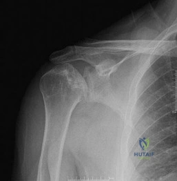
A 45-year-old male falls on an outstretched hand and sustains the injury shown in the radiograph.

During surgical reconstruction of this "terrible triad" of the elbow, the surgeon systematically addresses the structures. Following fixation of the coronoid process, management of the radial head, and repair of the lateral collateral ligament (LCL) complex, the elbow remains unstable in extension. What is the most appropriate next step in management?


Explanation

The standard protocol for treating terrible triad injuries of the elbow (elbow dislocation, radial head fracture, coronoid fracture) involves a sequential approach: 1) Restore the anterior buttress via coronoid fixation or anterior capsular repair; 2) Restore the lateral column via radial head fixation or arthroplasty; 3) Repair the LCL complex to its isometric footprint on the lateral epicondyle. If the elbow remains unstable (subluxates or dislocates in extension) after these three steps are successfully completed, the next appropriate step is to repair the MCL complex. If instability persists even after MCL repair, a hinged external fixator is applied.

Question 22

A 60-year-old osteoporotic female sustains a highly comminuted, intra-articular distal humerus fracture.

You are planning open reduction and internal fixation (ORIF) with dual plating. Based on biomechanical studies comparing parallel versus orthogonal plate configurations for distal humerus fractures, which of the following statements is most accurate?


Explanation

Biomechanical studies have demonstrated that parallel plating constructs for distal humerus fractures are significantly stiffer and provide greater resistance to both axial and torsional loading compared to orthogonal (90-90) plating, especially in the setting of metaphyseal comminution or osteoporotic bone. However, clinical studies have not shown a statistically significant difference in union rates or functional outcomes between the two techniques.

Question 23

A 32-year-old male sustains a closed, oblique fracture of the distal third of the humeral shaft (Holstein-Lewis fracture). Upon examination in the emergency department, he is unable to extend his wrist or fingers, though his triceps function is intact. There is no open wound. What is the most appropriate initial management?


Explanation

A primary radial nerve palsy in the setting of a closed humeral shaft fracture (including Holstein-Lewis distal third oblique fractures) is best managed with a functional brace and observation. More than 70-85% of these primary palsies are neuropraxias and will recover spontaneously. Surgical exploration is indicated for an open fracture, a secondary palsy that occurs after closed reduction, a vascular injury, or if there is no clinical or EMG evidence of nerve recovery by 3 to 4 months.

Question 24

A 28-year-old professional weightlifter presents with a dull, aching pain in his right posterior shoulder and neck. On examination, there is pronounced lateral winging of the scapula, which is exacerbated during resisted external rotation and shoulder abduction. He recently had a minor lymph node biopsy in the posterior triangle of his neck. Injury to which of the following nerves is the most likely cause of his scapular winging?


Explanation

Lateral winging of the scapula is caused by trapezius muscle dysfunction, which is innervated by the spinal accessory nerve (CN XI). This nerve is highly vulnerable to iatrogenic injury during procedures in the posterior triangle of the neck (such as lymph node biopsy). Medial winging of the scapula is caused by serratus anterior dysfunction due to long thoracic nerve injury.

Question 25

A 40-year-old male undergoes a single-incision anterior approach for repair of a distal biceps tendon rupture. Postoperatively, he complains of numbness and tingling along the radial border of his forearm. Which of the following is the most likely etiology of this complication?


Explanation

The most common complication following a single-incision anterior repair of a distal biceps tendon rupture is a neurapraxia of the lateral antebrachial cutaneous nerve (LABCN), which occurs in approximately 10-25% of cases. The LABCN courses superficially in the lateral aspect of the antecubital fossa and is highly susceptible to traction or direct injury from retractors during this approach. Injury to the PIN is less common but is a severe complication associated with poor retractor placement or failure to keep the forearm in supination during the approach.

Question 26

A 68-year-old female sustains a complex proximal humerus fracture.

According to Hertel's radiographic criteria, which combination of findings is the most reliable predictor of subsequent avascular necrosis (AVN) of the humeral head?


Explanation

Hertel identified specific radiographic predictors for ischemia of the humeral head following proximal humerus fractures. The most predictive combination for the development of avascular necrosis includes: 1) an anatomic neck fracture, 2) a short calcar segment (metaphyseal extension < 8 mm attached to the articular segment), and 3) disruption of the medial capsular hinge (> 2 mm displacement of the shaft relative to the head).

Question 27

A 25-year-old mountain biker falls directly onto the point of his shoulder, sustaining an acute high-grade (Type V) acromioclavicular (AC) joint separation.

Surgical reconstruction of the coracoclavicular (CC) ligaments is planned. Biomechanically, which native structure acts as the primary restraint to superior translation of the distal clavicle?


Explanation

The coracoclavicular (CC) ligaments provide primary vertical stability to the AC joint. The conoid ligament is the more medial of the two CC ligaments and is the primary restraint to superior translation of the clavicle. The trapezoid ligament is more lateral and provides the primary restraint against axial compression. The AC capsular ligaments primarily provide anteroposterior (horizontal) stability.

Question 28

A 35-year-old mechanic sustains an Essex-Lopresti injury after falling from a ladder. This injury pattern is characterized by a radial head fracture, disruption of the distal radioulnar joint (DRUJ), and tearing of the interosseous membrane (IOM). If the radial head is completely excised without replacement in this patient, what is the expected biomechanical consequence?


Explanation

An Essex-Lopresti injury involves a longitudinal radioulnar dissociation. The radial head and the interosseous membrane are the primary restraints to proximal migration of the radius. If the radial head is resected in the setting of an IOM tear (Essex-Lopresti lesion), the radius will migrate proximally. This causes positive ulnar variance, leading to severe ulnocarpal impaction, wrist pain, and restricted forearm rotation. Therefore, the radial head must be fixed or replaced with an arthroplasty to restore the longitudinal stability of the forearm.

Question 29

A 45-year-old male sustains a transverse fracture of the olecranon. The surgeon plans to use a tension band wiring technique. To maximize the biomechanical strength of the construct and minimize wire pullout, where should the K-wires be directed and seated?


Explanation

In tension band wiring of olecranon fractures, engaging the K-wires into the anterior ulnar cortex distal to the coronoid process (bicortical fixation) provides significantly greater resistance to wire back-out and loss of fixation compared to placing the wires straight down the intramedullary canal. Intramedullary placement relies solely on friction, which is biomechanically inferior to bicortical purchase.

Question 30

A 22-year-old elite rugby player with recurrent anterior shoulder instability undergoes a Latarjet procedure due to 25% anterior glenoid bone loss. The procedure relies on a "triple blocking" effect. Which of the following provides the most significant contribution to the dynamic stability (the "sling effect") conferred by the Latarjet procedure when the arm is abducted and externally rotated?


Explanation

The Latarjet procedure confers stability through a "triple blocking" mechanism: 1) The osseous block of the coracoid extending the glenoid articular arc; 2) The dynamic "sling effect" of the conjoint tendon (short head of biceps and coracobrachialis) acting on the inferior capsule and lower subscapularis when the arm is in abduction and external rotation; 3) The capsular repair (often using the CA ligament stump). Biomechanical studies show that the dynamic "sling effect" of the conjoint tendon provides 50% to 70% of the stabilizing force.

Question 31

A 50-year-old male is involved in a high-speed motor vehicle accident and sustains a closed fracture of the scapular neck.

According to the criteria described by Goss and Ada, what threshold of medial displacement (medialization) of the glenoid fragment relative to the lateral border of the scapula is widely considered an absolute indication for operative fixation?


Explanation

Most extra-articular scapular neck fractures are managed non-operatively. However, significant displacement alters shoulder biomechanics (rotator cuff tension, impingement). The classic indications for operative management (ORIF) of scapula fractures based on Goss and Ada criteria include: medial/lateral displacement (medialization) greater than 20 mm, angular displacement greater than 40 degrees, or a combination of >10 mm displacement with >40 degrees of angulation. Intra-articular step-off >4-5 mm is also an indication for fixation.

Question 32

A 38-year-old female falls onto an extended arm and presents with severe elbow pain. Radiographs and CT scan reveal a coronal shear fracture of the distal humerus. The fracture fragment includes the capitellum and the lateral half of the trochlea. According to the Bryan and Morrey classification with McKee's modification, what type of fracture is this?


Explanation

In the Bryan and Morrey classification of capitellar fractures: Type I (Hahn-Steinthal) is a large osseous piece of the capitellum. Type II (Kocher-Lorenz) is a sleeve fracture of articular cartilage with minimal subchondral bone. Type III is a comminuted capitellar fracture. McKee modified the classification by adding Type IV, which is a coronal shear fracture that involves the capitellum and extends medially to include a large portion of the trochlea. It is a critical distinction because Type IV fractures require more extensile exposures (often an extended lateral or olecranon osteotomy) for adequate fixation of the trochlear component.

Question 33

A 65-year-old male with chronic kidney disease presents with posterior elbow pain and an inability to actively extend his elbow against gravity following a sudden eccentric load. Radiographs show a small "Fleck sign" avulsed from the proximal ulna. You diagnose a complete triceps tendon rupture and plan for surgical repair. What is the anatomic characteristic of the triceps insertion on the olecranon?


Explanation

The triceps tendon inserts onto the proximal ulna over a broad, dome-shaped footprint. Studies on the anatomic footprint of the triceps reveal it inserts slightly distal to the tip of the olecranon (often 1-2 cm wide). The medial head of the triceps has a deep, fleshy insertion, while the lateral and long heads form a superficial tendinous insertion. Repair techniques aim to restore this broad footprint to maximize biomechanical strength and tendon-to-bone healing.

Question 34

Which of the following variables is considered the STRONGEST independent predictor for the development of nonunion in a midshaft clavicle fracture treated non-operatively?


Explanation

In non-operatively treated midshaft clavicle fractures, fracture shortening (displacement) greater than 2 cm is the strongest predictor of nonunion, with some studies citing a nonunion rate of up to 15-20% in these highly displaced fractures compared to <5% for non-displaced fractures. While comminution, older age, and female gender are also known risk factors, displacement without bone contact (100% displacement / >2 cm shortening) remains the most significant indication to consider operative fixation to prevent nonunion and symptomatic malunion.

Question 35

A 6-year-old boy presents to the emergency department after falling from monkey bars. Radiographs reveal a plastic deformation of the ulnar shaft and an anterior dislocation of the radial head. This corresponds to a Bado Type I Monteggia equivalent lesion.

What is the most appropriate initial management for this injury?


Explanation

A Monteggia fracture-dislocation in a pediatric patient (including equivalent lesions with ulnar plastic deformation) is fundamentally an injury driven by the ulnar deformity. The appropriate initial management is closed reduction to correct the ulnar bowing/angulation. Once the anatomic length and alignment of the ulna are restored, the radial head almost always reduces spontaneously into its anatomic position. Immobilization in a long arm cast (usually in supination for anterior/Type I lesions) is then performed. Open reduction or ulnar osteotomy is reserved for cases where closed reduction fails to restore ulnar alignment or reduce the radial head.

Question 36

A 75-year-old female sustains a displaced 4-part proximal humerus fracture. Given her poor bone quality and fracture complexity, she undergoes a Reverse Total Shoulder Arthroplasty (RTSA). During the procedure, the tuberosities are repaired to the shaft and the prosthesis. Healing of which of the following structures is most strongly associated with improved external rotation and higher overall patient-reported functional outcome scores?


Explanation

In the setting of a Reverse Total Shoulder Arthroplasty (RTSA) performed for a proximal humerus fracture, healing of the greater tuberosity is strongly correlated with significantly improved clinical outcomes, particularly active external rotation, forward elevation, and subjective functional scores. The infraspinatus and teres minor (attached to the greater tuberosity) provide the necessary external rotation capability in a reverse shoulder construct, which lacks an anatomic rotator cuff.

Question 37

A 30-year-old male bodybuilder feels a sudden "pop" and tearing sensation in his anterior chest and axilla while performing a heavy bench press. MRI confirms a complete rupture of the sternal head of the pectoralis major muscle from its humeral insertion. The clavicular head is intact. Anatomically, how does the sternal head of the pectoralis major insert onto the humerus relative to the clavicular head?


Explanation

The pectoralis major tendon undergoes a 90-degree twist before it inserts onto the lateral lip of the bicipital groove of the humerus. The clavicular head fibers run linearly to insert distally and superficially. The lower (sternal/costal) fibers twist behind the upper fibers, resulting in their insertion being deep and proximal. The sternal head is the most commonly ruptured segment during heavy eccentric loading activities like the bench press.

Question 38

An 18-year-old football player presents after a direct blow to the medial clavicle. He is complaining of severe pain, shortness of breath, mild stridor, and difficulty swallowing. Clinical examination and plain radiographs are suggestive of a posterior sternoclavicular (SC) joint dislocation.

What is the most appropriate next step in management?


Explanation

A posterior sternoclavicular joint dislocation is a true orthopedic emergency due to the high risk of compression or injury to posterior mediastinal structures (trachea, esophagus, great vessels), as evidenced by this patient's stridor and dysphagia. The crucial next step is a CT scan of the chest/neck (ideally with IV contrast) to accurately assess the displacement of the medial clavicle and its relationship to the mediastinal anatomy. Closed reduction should be attempted in the operating room (not the ED) with a cardiothoracic surgeon available, given the risk of massive hemorrhage if a great vessel is lacerated upon reduction.

Question 39

A 35-year-old male complains of a painful "clunk" and giving way of his right elbow when he pushes himself out of a chair with his arms. A lateral pivot-shift test of the elbow reproduces his symptoms. This condition is primarily caused by insufficiency of a specific ligamentous structure. What is the normal anatomic origin and insertion of the deficient ligament?


Explanation

The patient is presenting with posterolateral rotatory instability (PLRI) of the elbow. PLRI is caused by insufficiency of the Lateral Ulnar Collateral Ligament (LUCL). The LUCL originates on the lateral epicondyle of the humerus, blends with the fibers of the annular ligament, and inserts onto the supinator crest of the proximal ulna. It acts as the primary restraint to posterolateral rotatory subluxation of the radial head relative to the capitellum.

Question 40

A 42-year-old male is brought to the trauma bay following a motorcycle crash. He is diagnosed with a "floating shoulder," defined as a double disruption of the superior shoulder suspensory complex (SSSC). According to Goss, the SSSC is a bone-and-soft-tissue ring. Which of the following components is NOT considered part of the SSSC?


Explanation

The Superior Shoulder Suspensory Complex (SSSC), as described by Goss, is a continuous structural ring consisting of bone and soft tissue that maintains the relationship between the upper extremity and the axial skeleton. The SSSC ring is composed of the glenoid process, the coracoid process, the coracoclavicular (CC) ligaments, the distal clavicle, the acromioclavicular (AC) joint, and the acromion process. The proximal clavicle is not considered part of this specific suspensory ring (the struts are the middle clavicle and the lateral scapular body). A "floating shoulder" implies disruptions in two places of this complex (e.g., a surgical neck fracture of the glenoid combined with a clavicle fracture or CC ligament tear).

Question 41

A 28-year-old elite volleyball player presents with insidious onset of posterior shoulder pain and weakness. On examination, there is isolated weakness in external rotation with the arm at the side, but abduction is normal. MRI shows a paralabral cyst. Which of the following is true regarding this condition?


Explanation

Isolated weakness of the infraspinatus indicates suprascapular nerve entrapment at the spinoglenoid notch. A paralabral cyst in this location is typically associated with a posterosuperior labral tear (SLAP). Entrapment at the suprascapular notch affects both supraspinatus and infraspinatus. The suprascapular artery does not typically pass through the spinoglenoid notch with the nerve (it usually runs superficial to the spinoglenoid ligament).

Question 42

A 19-year-old rugby player presents to the emergency department after a direct blow to the medial clavicle. He is complaining of chest pain, dyspnea, and dysphagia. Physical examination reveals a palpable defect over the medial clavicle. An AP radiograph and CT scan confirm a posterior sternoclavicular dislocation. What is the most appropriate next step in management?


Explanation

Posterior sternoclavicular dislocations are orthopedic emergencies due to the risk of compression of mediastinal structures (trachea, esophagus, great vessels). Closed reduction should be attempted in the operating room under general anesthesia with cardiothoracic surgery backup in case of great vessel injury during the reduction maneuver.

Question 43

A 45-year-old man falls from a height and sustains a comminuted radial head fracture. During surgery, the radial head is deemed unsalvageable and excised. Postoperatively, the patient develops progressive wrist pain and ulnar-sided prominence.

What is the primary pathomechanical cause of this complication?


Explanation

The clinical presentation is classic for an Essex-Lopresti injury, which consists of a radial head fracture, disruption of the interosseous membrane (IOM), and injury to the distal radioulnar joint (DRUJ). Excision of the radial head in the presence of an IOM injury leads to proximal migration of the radius, causing ulnar impaction syndrome and DRUJ instability. The primary stabilizer against proximal radial migration is the radial head, and the secondary stabilizer is the central band of the IOM.

Question 44

When performing an olecranon osteotomy for the surgical management of an intra-articular distal humerus fracture (AO/OTA type 13C), what is the optimal shape of the osteotomy to maximize stability and surface area for healing?


Explanation

A chevron osteotomy with the apex directed proximally is preferred because it increases the surface area for healing and provides intrinsic rotational stability compared to a transverse osteotomy. It should be directed into the 'bare area' of the greater sigmoid notch, where there is naturally less articular cartilage.

Question 45

A 35-year-old weightlifter undergoes a single-incision anterior approach for a distal biceps tendon rupture repair using suture anchors. Postoperatively, he complains of numbness and tingling over the lateral aspect of his forearm. Which nerve was most likely injured, and what structure does it pierce to become superficial?


Explanation

Numbness over the lateral forearm indicates injury to the lateral antebrachial cutaneous nerve (LABCN), which is the terminal sensory branch of the musculocutaneous nerve. It emerges lateral to the biceps tendon, piercing the deep fascia just proximal to the elbow crease, and is highly susceptible to traction or iatrogenic injury during a single-incision anterior approach to the distal biceps.

Question 46

A 40-year-old woman falls on an outstretched hand and sustains a 'terrible triad' injury of the elbow.

Which of the following describes the most universally accepted sequence of intraoperative repair for this injury?


Explanation

The standard surgical algorithm for a terrible triad injury (elbow dislocation, radial head fracture, coronoid fracture) typically proceeds from deep to superficial: 1. Fixation of the coronoid fracture (or anterior capsule repair). 2. Fixation or replacement of the radial head. 3. Repair of the lateral collateral ligament (LCL) complex to the lateral epicondyle. If the elbow remains unstable after these steps, the medial collateral ligament (MCL) may be repaired or an external fixator applied.

Question 47

What is the strongest predictor of nonunion in nonoperatively managed midshaft clavicle fractures?


Explanation

The lack of cortical contact (i.e., 100% displacement) is widely recognized as the strongest predictive factor for nonunion in diaphyseal clavicle fractures treated conservatively. Other significant risk factors include advanced age, smoking, severe comminution, and shortening > 2 cm.

Question 48

A 22-year-old competitive rugby player undergoes evaluation for recurrent anterior shoulder instability. A 3D CT scan reveals a 20% anterior glenoid bone loss and a Hill-Sachs lesion. According to the 'glenoid track' concept, an 'off-track' Hill-Sachs lesion is defined by which of the following?


Explanation

The glenoid track is defined as 83% of the intact glenoid width minus the anterior glenoid bone loss. An 'off-track' Hill-Sachs lesion occurs when the medial margin of the Hill-Sachs lesion extends further medial than the medial boundary of the glenoid track. This indicates that the lesion will 'engage' the anterior rim of the glenoid during abduction and external rotation, increasing the risk of recurrent dislocation. Such lesions typically require a Latarjet procedure or Remplissage.

Question 49

A 6-year-old boy presents with an elbow injury after falling from monkey bars. Radiographs demonstrate a fracture of the proximal third of the ulna with apex posterior angulation and a posterior dislocation of the radial head. According to the Bado classification, what type of Monteggia lesion is this?


Explanation

The Bado classification of Monteggia fractures depends on the direction of the radial head dislocation. Type I: Anterior dislocation with anterior angulation of the ulnar fracture. Type II: Posterior dislocation with posterior angulation of the ulnar fracture. Type III: Lateral or anterolateral dislocation (most common in children). Type IV: Anterior dislocation with fractures of the radius and ulna at the same level. Therefore, apex posterior angulation with posterior dislocation is Bado Type II.

Question 50

A 28-year-old man sustains a closed, displaced transverse fracture of the middle third of the humeral shaft. On initial evaluation, he is unable to extend his wrist or fingers, and has numbness in the first dorsal web space. What is the most appropriate management regarding the nerve palsy?


Explanation

Primary radial nerve palsy associated with a closed humeral shaft fracture is typically a neurapraxia or axonotmesis. The initial management is conservative (e.g., coaptation splint followed by functional bracing), as >85% will recover spontaneously. Operative exploration is indicated if there is an open fracture, vascular injury requiring repair, or if there is no clinical or EMG evidence of nerve recovery after 3 to 4 months of observation.

Question 51

A 42-year-old woman falls on her outstretched hand and sustains a shear fracture of the distal humerus articular surface. Radiographs reveal a fracture involving the capitellum and the lateral half of the trochlea, with a large piece of subchondral bone attached. According to the Bryan and Morrey classification, what type of fracture is this?


Explanation

The Bryan and Morrey classification describes capitellum fractures. Type I (Hahn-Steinthal): involves a large fragment of osseous capitellum. Type II (Kocher-Lorenz): an articular cartilage fracture with very little subchondral bone attached. Type III (Broberg-Morrey): severely comminuted capitellum fracture. Type IV (added by McKee): involves the capitellum and the lateral half of the trochlea. The presence of the trochlear extension is critical to recognize as it requires more extensive fixation.

Question 52

Which of the following biomechanical changes is most directly responsible for restoring active elevation in a patient with rotator cuff arthropathy undergoing a reverse total shoulder arthroplasty?


Explanation

A reverse total shoulder arthroplasty (rTSA) medializes and distalizes the center of rotation of the glenohumeral joint. Medialization increases the moment arm of the deltoid muscle, and distalization increases the resting tension (and thus the contractile force) of the deltoid. Together, these biomechanical alterations allow the deltoid to effectively substitute for the deficient rotator cuff and initiate arm elevation.

Question 53

A 32-year-old carpenter presents with right shoulder weakness and a dull ache in the shoulder blade. On physical examination, when the patient pushes against a wall with arms extended forward, the medial border of the right scapula becomes prominent. Injury to which nerve is the most likely cause of this finding?


Explanation

Medial winging of the scapula is caused by weakness or paralysis of the serratus anterior muscle, which is innervated by the long thoracic nerve. This is accentuated by asking the patient to push against a wall. In contrast, lateral winging of the scapula is caused by weakness of the trapezius muscle, innervated by the spinal accessory nerve, and is accentuated by resisted abduction.

Question 54

A 25-year-old cyclist falls directly onto his right shoulder. He complains of severe pain at the top of the shoulder. Radiographs show a 200% superior displacement of the distal clavicle relative to the acromion.

Which ligaments are disrupted in this Rockwood Type V injury?


Explanation

In a Rockwood Type V acromioclavicular (AC) joint injury, there is complete disruption of both the AC ligaments and the coracoclavicular (CC) ligaments, along with gross disruption of the deltotrapezial fascia. This extensive soft tissue stripping allows severe superior displacement of the clavicle (100-300% compared to the contralateral side).

Question 55

When performing a shoulder hemiarthroplasty for a severe 4-part proximal humerus fracture, what is the most critical factor for a successful functional outcome?


Explanation

In hemiarthroplasty for proximal humerus fractures, functional outcome is directly related to the anatomic reduction and reliable healing of the greater and lesser tuberosities. Malposition or nonunion of the tuberosities leads to profound weakness, loss of active motion, and poor function. While appropriate version and height are important for tuberosity healing, the actual healing of the tuberosities is the single most critical determinant of clinical success.

Question 56

A 'floating shoulder' typically involves ipsilateral fractures of the clavicular shaft and which other structure?


Explanation

A 'floating shoulder' refers to a double disruption of the superior shoulder suspensory complex (SSSC). The classic description is an ipsilateral fracture of the midshaft clavicle and the scapular neck. This inherently unstable injury can lead to inferior and medial displacement of the glenoid. Operative fixation of at least one of the lesions (most commonly the clavicle) is often indicated to restore the SSSC.

Question 57

A 30-year-old motorcyclist is involved in a high-speed collision and presents with severe swelling of the left shoulder and complete loss of motor and sensory function in the left upper extremity. An AP chest radiograph shows lateral displacement of the left scapula compared to the right, and an intact clavicle. Which associated injury has the highest immediate mortality risk in this condition?


Explanation

The clinical picture and radiograph (lateralization of the scapula with massive swelling) describe a scapulothoracic dissociation. This is a severe, high-energy injury characterized by disruption of the scapulothoracic articulation. It is highly associated with brachial plexus injuries (often complete avulsion) and major vascular injuries (subclavian or axillary artery/vein disruption). The vascular injury is limb- and life-threatening, making it the most critical immediate concern.

Question 58

A 35-year-old man undergoes surgical release for post-traumatic elbow stiffness following a terrible triad injury 1 year ago. To prevent recurrence due to heterotopic ossification (HO), which of the following is the most standard prophylactic regimen?


Explanation

Prophylaxis for heterotopic ossification (HO) around the elbow typically involves either nonsteroidal anti-inflammatory drugs (NSAIDs) such as Indomethacin (e.g., 75 mg sustained release daily or 25 mg TID for 3-6 weeks) or a single fraction of low-dose radiation therapy (700-800 cGy) administered within 24 to 48 hours before or after surgery. Corticosteroids and methotrexate are not standard HO prophylaxis.

Question 59

A 26-year-old baseball pitcher presents with pain deep in his throwing shoulder and a 'dead arm' sensation. MR arthrogram reveals a SLAP lesion.

During diagnostic arthroscopy, the superior labrum and biceps anchor are found to be detached from the superior glenoid. What type of SLAP tear is this according to the Snyder classification?


Explanation

The Snyder classification of SLAP (Superior Labrum Anterior to Posterior) tears: Type I: Fraying and degeneration of the superior labrum with an intact biceps anchor. Type II: Detachment of the superior labrum and biceps anchor from the superior glenoid. Type III: Bucket-handle tear of the superior labrum with an intact biceps anchor. Type IV: Bucket-handle tear of the superior labrum that extends into the biceps tendon. Type II is the most common type requiring surgical repair or biceps tenodesis.

Question 60

A 22-year-old gymnast sustains an anterior sternoclavicular (SC) joint dislocation. She complains of an unsightly bump on her chest but has no respiratory or swallowing difficulties. After a trial of conservative management, she continues to have pain and instability. If surgery is considered, which of the following is the most appropriate procedure?


Explanation

The preferred surgical treatment for symptomatic, chronic anterior sternoclavicular joint instability that has failed conservative management is a ligamentous reconstruction, typically using a soft tissue graft (e.g., semitendinosus or palmaris longus) in a figure-of-eight or similar configuration. Transarticular Kirschner wires are strictly contraindicated in the SC joint due to the high risk of catastrophic migration into the heart or great vessels. Medial clavicle excision without reconstruction can lead to continued instability.

Question 61

Which combination of radiographic findings in a proximal humerus fracture is most highly predictive of humeral head ischemia according to Hertel's criteria?


Explanation

Hertel identified that a calcar segment less than 8 mm, disruption of the medial hinge, and an anatomical neck fracture pattern are the most reliable predictors of humeral head ischemia. Combined, these factors indicate severe disruption of the critical vascular supply via the anterior and posterior humeral circumflex arteries.

Question 62

In reverse total shoulder arthroplasty (rTSA), which of the following glenosphere configurations is most effective at minimizing the risk of inferior scapular notching?


Explanation

Inferior placement and inferior tilt of the glenosphere help lateralize the humerus slightly and clear the inferior scapular neck, significantly reducing the mechanical impingement that causes scapular notching. Superior or medial placement increases impingement risk.

Question 63

A 65-year-old male with an irreparable posterosuperior rotator cuff tear and a positive Hornblower's sign undergoes a latissimus dorsi tendon transfer. During the harvest of the tendon, which neurovascular bundle is at greatest risk and must be carefully protected?


Explanation

The latissimus dorsi is innervated and supplied by the thoracodorsal nerve and artery. This pedicle runs on the deep surface of the muscle and must be carefully identified and protected during tendon harvest and mobilization to ensure the viability and function of the transfer.

Question 64

A 42-year-old female presents with persistent shoulder pain and weakness 4 weeks after undergoing a lymph node biopsy in the posterior cervical triangle. On physical exam, she demonstrates lateral winging of the scapula and an inability to actively abduct the shoulder past 90 degrees. Injury to which of the following nerves is the most likely cause?


Explanation

Lateral scapular winging combined with a history of posterior triangle neck surgery is classic for an iatrogenic spinal accessory nerve injury, leading to trapezius palsy. Medial winging is associated with long thoracic nerve palsy (serratus anterior).

Question 65

A 38-year-old male undergoes surgical repair of a complete distal biceps tendon rupture using a single-incision anterior approach. Postoperatively, he complains of numbness over the radial aspect of his volar forearm. Which structure was most likely injured during the procedure?


Explanation

The lateral antebrachial cutaneous nerve (LABCN) runs closely alongside the cephalic vein and biceps tendon distally. It is the most commonly injured neurologic structure during a single-incision anterior approach for distal biceps repair.

Question 66

A 28-year-old female complains of recurrent clicking and a sense of instability in her elbow when pushing up from a chair. Physical examination reveals apprehension during a pivot-shift test. This condition is primarily due to insufficiency of which of the following structures?


Explanation

Posterolateral rotatory instability (PLRI) of the elbow presents with a positive lateral pivot-shift test and apprehension when extending the elbow with supination and an axial load. It is primarily caused by an incompetent lateral ulnar collateral ligament (LUCL).

Question 67

A 45-year-old male falls from a height and sustains a comminuted radial head fracture, along with significant wrist pain. Radiographs show proximal migration of the radius. If the radial head is resected without replacement in this setting, what is the most likely biomechanical consequence?


Explanation

This presentation describes an Essex-Lopresti injury (radial head fracture, interosseous membrane tear, DRUJ disruption). Resecting the radial head without replacement eliminates the proximal block to migration, resulting in severe proximal radial migration and secondary ulnocarpal impaction.

Question 68

A 78-year-old female with severe rheumatoid arthritis sustains a highly comminuted, osteopenic distal humerus fracture (AO/OTA 13-C3). She is treated with a total elbow arthroplasty (TEA). What is the most critical postoperative restriction she must strictly adhere to?


Explanation

Total elbow arthroplasty (TEA) for distal humerus fractures in the elderly is associated with excellent pain relief but carries strict lifetime lifting restrictions. Patients are generally restricted to a 5 lb repetitive and 10-15 lb singular lifting limit to prevent early aseptic loosening and implant failure.

Question 69

A 30-year-old male sustains a closed, spiral fracture of the distal third of the humeral shaft. Initial examination reveals an intact neurologic profile. Following a closed reduction and application of a coaptation splint, the patient demonstrates an inability to extend his wrist and fingers. What is the most appropriate next step in management?


Explanation

While primary radial nerve palsy in a Holstein-Lewis fracture is typically observed, a secondary radial nerve palsy that develops AFTER a closed reduction attempt indicates possible entrapment of the nerve in the fracture site. Immediate surgical exploration is indicated.

Question 70

A 19-year-old male rugby player presents to the emergency department with severe anterior chest wall pain, dysphagia, and a sensation of choking following a direct blow to the medial clavicle. Suspecting a posterior sternoclavicular dislocation, what is the most appropriate initial imaging modality to confirm the diagnosis and assess associated risks?


Explanation

A posterior sternoclavicular joint dislocation is a true orthopedic emergency due to its proximity to vital mediastinal structures. A CT scan of the chest with IV contrast is the gold standard to assess the dislocation and rule out vascular or tracheal compromise.

Question 71

A 25-year-old male sustains a severe crush injury to the forearm. Several hours later, he develops excruciating pain out of proportion to the injury. Which of the following clinical findings is generally considered the most reliable early indicator of evolving forearm compartment syndrome?


Explanation

Pain with passive stretch of the muscles in the involved compartment (e.g., passive finger extension for the deep volar forearm compartment) is the most reliable and earliest clinical sign of compartment syndrome. Pulselessness and pallor are very late and unreliable signs.

Question 72

According to current literature, which of the following is a widely accepted absolute indication for Open Reduction and Internal Fixation (ORIF) of a scapular fracture?


Explanation

Intra-articular glenoid fractures resulting in glenohumeral instability or those involving greater than 25% of the articular surface with displacement are absolute indications for ORIF. Most body and neck fractures are managed non-operatively unless highly displaced or angulated.

Question 73

A 22-year-old male collegiate wrestler presents with recurrent anterior shoulder instability. Advanced imaging reveals an anterior glenoid bone defect measuring 28% of the glenoid width, along with an engaging Hill-Sachs lesion. Which of the following surgical procedures is most appropriate?


Explanation

For patients with critical anterior glenoid bone loss (>20-25%), a soft tissue repair alone (Bankart) has unacceptably high failure rates. A bony augmentation procedure, such as the Latarjet (coracoid transfer), is required to restore stability via both the bone block and the sling effect of the conjoint tendon.

Question 74

Following an unsalvageable radial head fracture, a metallic radial head arthroplasty is planned. To prevent overstuffing the radiocapitellar joint, the proximal articular surface of the radial head implant should ideally be placed at which anatomic landmark relative to the proximal radioulnar joint (PRUJ)?


Explanation

To restore proper elbow kinematics and avoid overstuffing, the proximal rim of the radial head implant should be placed flush with, or no more than 1-2 mm proximal to, the lateral edge of the lesser sigmoid notch of the ulna.

Question 75

A 35-year-old cyclist sustains a midshaft clavicle fracture. Which of the following radiographic parameters is most strongly associated with an increased risk of symptomatic nonunion if treated nonoperatively?


Explanation

Significant fracture shortening (greater than 2 cm) and comminution are the strongest predictive risk factors for nonunion and poor functional outcomes in midshaft clavicle fractures managed conservatively.

Question 76

A 45-year-old male presents with a closed, isolated scapula fracture following a high-energy fall. Which of the following radiographic parameters is a widely accepted indication for operative fixation?


Explanation

A glenopolar angle (GPA) of less than 22 degrees is a recognized indication for open reduction and internal fixation of scapular neck fractures. Medialization greater than 20 mm, angulation greater than 45 degrees, and intra-articular step-off greater than 4 mm are also typical operative indications.

Question 77

A 6-year-old boy presents with an anterior dislocation of the radial head and a fracture of the ulnar diaphysis. What is the most commonly injured nerve associated with this specific injury pattern?


Explanation

This patient has a Bado Type I Monteggia fracture-dislocation. The posterior interosseous nerve (PIN) is the most commonly injured nerve in this fracture pattern due to traction or direct trauma from the anteriorly displaced radial head.

Question 78

Recent quantitative perfusion studies have redefined the vascularity of the proximal humerus. According to these studies, which artery provides the predominant blood supply to the humeral head?


Explanation

Historically, the anterior humeral circumflex artery (via the arcuate artery) was thought to provide the main blood supply to the humeral head. However, recent studies (e.g., Hettrich et al.) demonstrate that the posterior humeral circumflex artery supplies approximately 64% of the humeral head.

Question 79

A 32-year-old female sustains a coronal shear fracture of the distal humerus that involves the capitellum and the lateral half of the trochlea. Which classification accurately describes this fracture pattern?


Explanation

The McKee modification of the Bryan-Morrey classification describes a Type IV fracture as a coronal shear fracture that involves the capitellum and extends medially to include the lateral portion of the trochlea. This creates a pathognomonic 'double-arc' sign on a lateral radiograph.

Question 80

A 50-year-old female with poorly controlled type 2 diabetes presents with progressive, painful restriction of active and passive shoulder motion. What is the predominant histological finding in the joint capsule of a patient with this condition?


Explanation

Adhesive capsulitis is characterized primarily by fibroplasia rather than acute inflammation. The predominant histological finding is a dense proliferation of fibroblasts and myofibroblasts within the joint capsule, leading to contracture.

Question 81

A 28-year-old cyclist sustains a closed midshaft clavicle fracture. If managed nonoperatively, which of the following fracture characteristics is most strongly associated with an increased risk of nonunion?


Explanation

Fracture shortening greater than 2 cm is a well-established risk factor for nonunion and symptomatic malunion in completely displaced midshaft clavicle fractures. Other risk factors include advancing age, comminution, and female sex.

Question 82

A 40-year-old male undergoes radial head arthroplasty for a severely comminuted radial head fracture. Overstuffing the radiocapitellar joint during this procedure will most likely lead to which of the following complications?


Explanation

Overstuffing the radiocapitellar joint with an oversized radial head implant increases radiocapitellar contact pressures. This leads to capitellar erosion, stiffness, early osteoarthritis, and gapping of the medial elbow compartment.

Question 83

During arthroscopic evaluation of a 22-year-old baseball pitcher, a superior labral anterior-posterior (SLAP) tear is identified with detachment of the biceps anchor. Which of the following physical examination findings was most likely positive preoperatively?


Explanation

The O'Brien (active compression) test is commonly used to evaluate for SLAP tears. Pain deep in the shoulder with internal rotation that is relieved by external rotation is considered a positive result for labral pathology.

Question 84

When utilizing an olecranon osteotomy for open reduction and internal fixation of a multi-fragmentary distal humerus fracture (AO/OTA 13C3), which osteotomy type is biomechanically superior for minimizing nonunion?


Explanation

A distally pointing chevron osteotomy created at the bare area of the sigmoid notch provides maximum articular surface contact area and superior rotational stability compared to a transverse osteotomy, thereby minimizing the risk of nonunion.

Question 85

A 30-year-old competitive weightlifter ruptures his pectoralis major tendon during a bench press. When performing an anatomic primary repair, where should the sternal head insert on the humerus relative to the clavicular head?


Explanation

The pectoralis major tendon undergoes a 180-degree twist before inserting onto the humerus. This causes the sternal head to insert deep (posterior) and proximal to the clavicular head.

Question 86

Which tendon is most commonly and primarily involved in the underlying pathoanatomy of lateral epicondylitis (tennis elbow)?


Explanation

Lateral epicondylitis is characterized by angiofibroblastic hyperplasia primarily occurring at the origin of the extensor carpi radialis brevis (ECRB) tendon.

Question 87

A 19-year-old male presents to the trauma bay with a posterior sternoclavicular joint dislocation following a rugby tackle. He has mild dysphagia but stable vitals. What is the most appropriate initial management?


Explanation

Posterior sternoclavicular dislocations can compress critical mediastinal structures. Closed reduction should be attempted but must be performed in the operating room under general anesthesia with a cardiothoracic surgeon available due to the risk of catastrophic retrosternal vascular injury during reduction.

Question 88

A 26-year-old female presents with lateral scapular winging and an inability to actively abduct her arm past 90 degrees. She recently underwent a lymph node biopsy in the posterior cervical triangle. Which nerve was most likely injured?


Explanation

Injury to the spinal accessory nerve (CN XI) paralyzes the trapezius muscle, leading to a drooping shoulder, lateral winging of the scapula, and weakness in shoulder abduction. It is a known complication of surgical procedures in the posterior cervical triangle.

Question 89

A 42-year-old male sustains an Essex-Lopresti injury characterized by a comminuted radial head fracture, rupture of the interosseous membrane, and disruption of the distal radioulnar joint (DRUJ). In the acute setting, what is the most appropriate management of the radial head?


Explanation

In an Essex-Lopresti injury, longitudinal forearm stability is lost due to interosseous membrane rupture. The radial head must be preserved or replaced (radial head arthroplasty) to prevent proximal migration of the radius and chronic wrist pain. Radial head excision is absolutely contraindicated.

None

Dr. Mohammed Hutaif
Medically Verified Content by
Prof. Dr. Mohammed Hutaif
Consultant Orthopedic & Spine Surgeon
Chapter Index