Open Fractures: Why Continuous Pressure Monitoring is Crucial

Key Takeaway
This topic focuses on Open Fractures: Why Continuous Pressure Monitoring is Crucial, The biggest concern following high-energy tibial fractures is compartment syndrome. This critical condition is monitored through careful assessment of clinical signs and symptoms, but definitive diagnosis often involves continuous pressure monitoring within the muscle compartments. Early detection and intervention are crucial to prevent permanent damage and optimize patient outcomes.
Comprehensive Introduction and Patho-Epidemiology
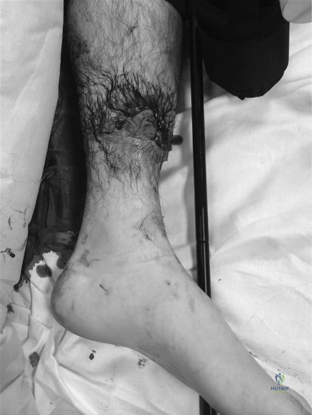
Open fractures of the long bones, particularly the tibia, represent a profound challenge in orthopedic trauma surgery, characterized by the simultaneous disruption of the osseous architecture and the surrounding soft tissue envelope. The bimodal epidemiological distribution of these injuries typically involves high-energy mechanisms—such as motor vehicle collisions and industrial accidents—in young adult males, and low-energy osteoporotic falls in the elderly population. The hallmark of a severe open fracture is the extensive zone of injury, which extends far beyond the visible wound margins. This zone is characterized by profound microvascular thrombosis, periosteal stripping, and soft tissue contusion, creating an ischemic environment highly susceptible to bacterial colonization and subsequent deep infection. The initial clinical presentation often features a large transverse or stellate wound over the subcutaneous border of the tibia, with exposed, devitalized bone protruding through the defect, accompanied by profound local tissue destruction.
A critical and historically misunderstood aspect of open fracture patho-epidemiology is the relationship between fascial disruption and the development of acute compartment syndrome (ACS). A dangerous, yet pervasive, myth in traumatology is that the traumatic rent in the fascial envelope created by an open fracture confers a prophylactic auto-decompression, thereby eliminating the risk of compartment syndrome. Contemporary trauma registries and prospective studies have unequivocally debunked this fallacy. The fascial laceration is rarely sufficient to accommodate the massive volumetric expansion of the compartment driven by reactive edema, hematoma formation, and post-ischemic reperfusion injury. Consequently, open fractures, particularly high-energy Gustilo-Anderson Type II and III injuries, carry a substantial risk of ACS, with incidence rates reported between 3% and 9% in open tibial shaft fractures.
The insidious nature of compartment syndrome in the setting of open fractures necessitates a paradigm shift in postoperative monitoring, particularly for patients who are unable to provide reliable clinical feedback. In the polytraumatized patient—often obtunded, intubated, or suffering from a concomitant traumatic brain injury—the classic clinical hallmarks of ACS (pain out of proportion, pain with passive stretch, paresthesia) are completely masked. In these highly vulnerable cohorts, reliance on clinical examination is not merely insufficient; it is an abrogation of surgical responsibility. Continuous intracompartmental pressure monitoring emerges not as an adjunct, but as a mandatory, limb-saving diagnostic modality. Utilizing slit catheters or solid-state transducer systems allows for the real-time calculation of the delta pressure (diastolic blood pressure minus intracompartmental pressure), providing an objective physiological parameter to guide emergent fasciotomy before irreversible myonecrosis occurs.
The overarching philosophy of managing these catastrophic injuries has evolved from arbitrary temporal mandates to a more nuanced, physiologically driven approach. The integration of Advanced Trauma Life Support (ATLS) principles ensures that life-threatening hemorrhage and systemic instability are addressed prior to limb-specific interventions. However, once the patient is stabilized, the orthopedic surgeon must meticulously balance the competing demands of radical tissue debridement, skeletal stabilization, and vigilant monitoring for ischemic complications. The subsequent sections of this chapter will exhaustively detail the anatomical, surgical, and physiological principles required to master the management of open fractures, with a specific emphasis on the indispensable role of continuous pressure monitoring.
Detailed Surgical Anatomy and Biomechanics
The osteology and regional anatomy of the lower extremity predispose the tibia to severe open fractures and subsequent compartmental catastrophes. The tibia is a long, triangular bone with a distinctly subcutaneous anteromedial face that lacks any muscular coverage from the tibial tubercle down to the medial malleolus. This anatomical vulnerability means that even moderate-energy direct impacts can easily breach the thin cutaneous envelope, resulting in an open fracture. Furthermore, the diaphyseal blood supply is heavily reliant on the nutrient artery, a branch of the posterior tibial artery, which enters the posterolateral cortex at the proximal third of the bone. In high-energy trauma, the nutrient artery is frequently sheared, rendering the diaphyseal cortex highly dependent on the periosteal microcirculation. When severe periosteal stripping occurs—as is typical in Gustilo-Anderson Type IIIB and IIIC injuries—the underlying cortical bone becomes completely avascular, functioning essentially as a necrotic sequestrum that serves as a nidus for biofilm-forming pathogens.
The lower leg is partitioned into four distinct, unyielding osseofascial compartments: the anterior, lateral, superficial posterior, and deep posterior compartments. The anterior compartment, bounded by the tibia, fibula, interosseous membrane, and the rigid anterior crural fascia, is the most frequently implicated in acute compartment syndrome. It contains the tibialis anterior, extensor hallucis longus, extensor digitorum longus, and peroneus tertius muscles, along with the deep peroneal nerve and anterior tibial artery. The deep posterior compartment, often considered the most difficult to clinically assess and surgically release, contains the tibialis posterior, flexor hallucis longus, and flexor digitorum longus, safeguarding the critical posterior tibial artery and tibial nerve. The inelasticity of these fascial boundaries means that any increase in intracompartmental volume—whether from hemorrhage from fractured bone ends or interstitial edema from soft tissue trauma—results in an exponential rise in intracompartmental pressure.

From a biomechanical and microvascular perspective, the pathophysiology of compartment syndrome is governed by the critical closing pressure of the capillary beds. Normal tissue perfusion requires a hydrostatic pressure gradient between the arterial supply and the venous drainage. As intracompartmental pressure rises, it first exceeds venous pressure, leading to venous collapse and subsequent venous hypertension. This exacerbates capillary hydrostatic pressure, driving more fluid into the interstitial space and creating a vicious, self-propagating cycle of edema and pressure elevation. Once the interstitial pressure surpasses the local capillary perfusion pressure (typically when the intracompartmental pressure comes within 30 mmHg of the diastolic blood pressure), the microcirculation completely collapses. Cellular hypoxia ensues, leading to the failure of the ATP-dependent sodium-potassium pumps, intracellular swelling, and ultimately, irreversible myonecrosis and nerve infarction.
Understanding this biomechanical threshold underscores the absolute necessity of the delta pressure concept (ΔP = Diastolic BP - Compartment Pressure) in continuous pressure monitoring. Absolute intracompartmental pressure values (e.g., a rigid threshold of 30 mmHg) are notoriously unreliable, as they fail to account for the patient's systemic perfusion pressure. A hypotensive polytrauma patient may experience catastrophic muscle ischemia at an absolute compartment pressure of 25 mmHg, whereas a hypertensive patient may easily tolerate a pressure of 40 mmHg. By employing continuous slit-catheter monitoring, the orthopedic surgeon obtains a dynamic, real-time assessment of the delta pressure. A sustained delta pressure of less than 30 mmHg for more than two hours is universally recognized as an absolute indication for emergent four-compartment fasciotomy, superseding any ambiguous clinical findings.
Exhaustive Indications and Contraindications
The decision-making matrix for the management of high-energy open fractures, particularly regarding the timing of surgery, the method of stabilization, and the deployment of continuous pressure monitoring, requires a sophisticated understanding of precise indications and contraindications. The initial management in the emergency department is universally standardized: immediate administration of broad-spectrum intravenous antibiotics (e.g., Cefuroxime 1.5 g IV, supplemented with an aminoglycoside for severe contamination), tetanus prophylaxis, gross decontamination, application of a sterile saline-soaked dressing, and provisional splinting. However, the subsequent surgical pathway diverges based on the physiological status of the patient and the local characteristics of the wound.
The indications for immediate, emergent surgical exploration (within 6 hours of injury) are strictly defined. According to the British Orthopaedic Association Standards for Trauma (BOAST) guidelines, immediate surgery is mandated only if there is an associated vascular injury resulting in limb ischemia, or if the wound is heavily contaminated with marine, agricultural, or sewage matter. In the absence of these critical factors, the historical "6-hour rule" has been largely abandoned in favor of prudent, early surgery within 24 hours. This paradigm shift, heavily supported by the Lower Extremity Assessment Project (LEAP) study, dictates that surgery should be performed during normal daylight working hours by a dedicated ortho-plastic multidisciplinary team. Proceeding to the operating theater in the middle of the night with an exhausted, non-specialized team for a non-ischemic open fracture is now considered a relative contraindication, as it significantly increases the risk of inadequate debridement and suboptimal soft tissue handling.
Regarding continuous intracompartmental pressure monitoring, the indications are heavily weighted toward patients who lack the cognitive capacity to participate in a reliable clinical examination. The absolute indications include intubated and ventilated patients, those with severe traumatic brain injuries (GCS < 8), patients under the influence of profound systemic analgesia or illicit narcotics, and polytraumatized patients with distracting injuries. In these cohorts, an invasive monitoring device such as a slit catheter or a solid-state transducer must be inserted into the anterior and deep posterior compartments. Conversely, contraindications for continuous monitoring are few but clinically relevant. They include patients who are fully alert, cooperative, and capable of articulating the progression of their symptoms, as well as limbs with established, delayed-presentation compartment syndrome (e.g., >24 hours of ischemia) where monitoring is moot and immediate fasciotomy or amputation is the only recourse.
| Parameter | Indications | Contraindications |
|---|---|---|
| Immediate Surgery (< 6 hours) | Vascular compromise (ischemic limb); Gross agricultural/marine/sewage contamination; Compartment syndrome. | Hemodynamically unstable polytrauma (requires damage control); Nighttime presentation without specialist ortho-plastic team (if limb is well-perfused). |
| Continuous Pressure Monitoring | Obtunded/intubated patients; Traumatic brain injury; Distracting injuries; Equivocal clinical exam in high-energy trauma. | Fully alert, cooperative patient with reliable serial exams; Missed/late compartment syndrome (>24h) where muscle is already dead. |
| Definitive IM Nailing (Acute) | Gustilo I, II, or IIIA with adequate soft tissue coverage; Hemodynamically stable patient; Complete, aggressive debridement achieved. | Gustilo IIIB/IIIC requiring complex free flaps; Hemodynamic instability (Damage Control Orthopedics); Inadequate initial debridement. |
| Provisional External Fixation | Damage Control Orthopedics in polytrauma; Severe soft tissue loss requiring serial debridements; Gross contamination. | Simple, low-energy open fractures with pristine soft tissue; Situations where immediate definitive internal fixation is definitively safe. |
Pre-Operative Planning, Templating, and Patient Positioning
Thorough pre-operative planning is the cornerstone of successful outcomes in the management of complex open fractures. Following the initial ATLS resuscitation and primary survey, the secondary survey must include a meticulous neurovascular examination of the affected extremity. The presence of palpable pulses does not preclude a significant arterial intimal tear; therefore, any asymmetry in pulses or an Ankle-Brachial Index (ABI) of less than 0.9 mandates an immediate CT angiography. Digital photography of the wound in the emergency department is a critical step; it allows the surgical team to assess the injury morphology without subjecting the patient to repeated, painful dressing takedowns that increase the risk of nosocomial bacterial inoculation.
Radiographic templating requires high-quality orthogonal views of the entire injured long bone, explicitly encompassing the joints above and below the fracture to rule out contiguous injuries or intra-articular extensions. In the context of a tibial shaft fracture, the surgeon must measure the isthmic diameter, assess the canal morphology, and determine the optimal entry point (suprapatellar versus infrapatellar) if intramedullary nailing is planned. For complex, comminuted fractures, templating for a spanning external fixator is equally critical. The surgeon must plan the trajectory of the Schanz pins to ensure they remain outside the zone of injury, avoid planned future surgical incisions (such as fasciotomy lines or local rotational flap donor sites), and provide adequate multi-planar stability to protect the soft tissues during transport and nursing care.
Patient positioning in the operating theater must facilitate both the orthopedic skeletal stabilization and the plastic surgical soft tissue reconstruction. For tibial fractures, the patient is typically positioned supine on a radiolucent Jackson or OSI table. A gel bump is placed under the ipsilateral hip to correct the natural external rotation of the lower extremity, ensuring the patella faces directly anteriorly. This neutral alignment is paramount for assessing rotational reduction during intramedullary nailing. The entire limb is prepped and draped freely from the toes to the proximal thigh, allowing for unrestricted manipulation, visual assessment of mechanical axis alignment, and access to potential skin graft donor sites. A sterile tourniquet may be applied to the proximal thigh but should remain uninflated during the initial debridement to allow for accurate assessment of tissue perfusion and bleeding.

If the patient meets the criteria for continuous compartment pressure monitoring, the setup and calibration of the apparatus should occur prior to the commencement of the formal surgical debridement, or immediately post-operatively if the risk of ACS remains high. The slit catheter system must be flushed with sterile saline to eliminate air bubbles and zeroed at the level of the compartment being measured. Insertion is typically performed under sterile conditions, utilizing a trocar to introduce the catheter into the muscle belly of the anterior compartment (approached laterally to the tibial crest) and the deep posterior compartment (approached medially, posterior to the medial tibial border). The catheter is then secured to the skin, and the transducer is connected to the anesthesia monitor, providing a continuous, real-time readout of the intracompartmental pressure, which is constantly cross-referenced against the patient's arterial line diastolic pressure.
Step-by-Step Surgical Approach and Fixation Technique
The surgical management of an open fracture is fundamentally an exercise in radical, systematic debridement, often referred to as a "cancer operation for trauma." The procedure begins with the extension of the traumatic wound along extensile surgical planes. Transverse traumatic lacerations must be extended longitudinally to provide adequate exposure without creating ischemic skin flaps. The debridement proceeds systematically from superficial to deep: outside-to-in. The skin edges are sharply excised until healthy, bleeding dermal margins are encountered. Subcutaneous fat, which is highly susceptible to necrosis and has poor resistance to infection, must be aggressively resected if it appears contused, avulsed, or impregnated with foreign debris.
Muscle viability is the most critical determinant of postoperative infection and is assessed using the classic "4 Cs": Color, Consistency, Contractility, and Capacity to bleed. Healthy muscle is dark red (Color), firm and resilient to the touch (Consistency), twitches briskly when stimulated with electrocautery or forceps (Contractility), and bleeds freely from its cut surface (Capacity to bleed). Any muscle that is pale, dusky, mushy, non-contractile, or avascular must be ruthlessly excised. Retained necrotic muscle serves as an ideal culture medium for clostridial and other virulent pathogens, leading to catastrophic gas gangrene or deep-seated osteomyelitis. Unlike skin, which may sometimes be left for a "second look" if its viability is borderline, necrotic muscle must be removed entirely during the index procedure.
Following soft tissue debridement, attention is turned to the skeletal elements. The bone ends must be delivered into the wound to allow for circumferential inspection and curettage of the medullary canal, which is often packed with contaminated marrow and debris. The "tug test" is employed to assess the viability of cortical fragments; any cortical bone that is completely devoid of soft tissue attachments and can be easily pulled away with forceps is devitalized and must be discarded, regardless of its size or structural importance. Once the debridement is complete, the wound and fracture site are subjected to copious, low-pressure irrigation using a minimum of 6 liters of warmed normal saline. The use of high-pressure pulsatile lavage is generally discouraged, as biomechanical studies have demonstrated that it can drive particulate matter and bacteria deeper into the cancellous bone architecture.
The final phase of the index procedure involves skeletal stabilization. If the patient is physiologically stable, the soft tissue envelope is adequately debrided, and definitive coverage can be achieved (either primarily or via a planned flap within 72 hours), definitive internal fixation with a reamed intramedullary nail is the gold standard for diaphyseal tibial fractures. However, in the setting of hemodynamic instability (Damage Control Orthopedics), massive contamination, or extensive bone loss requiring later reconstruction, a spanning external fixator is applied. If continuous pressure monitoring indicates a delta pressure of less than 30 mmHg, or if the clinical index of suspicion is overwhelmingly high, a prophylactic or therapeutic four-compartment fasciotomy is performed using a standard dual-incision technique (anterolateral and posteromedial incisions), ensuring the complete release of the fascial envelopes from the knee to the ankle.
Complications, Incidence Rates, and Salvage Management
Despite meticulous adherence to surgical principles, high-energy open fractures are fraught with severe, limb-threatening complications. The most devastating acute complication is a missed compartment syndrome. When intracompartmental pressures remain elevated above the capillary perfusion threshold for more than 6 to 8 hours, irreversible myonecrosis and nerve infarction occur. This leads to Volkmann's ischemic contracture, a catastrophic condition characterized by rigid, fibrotic, non-functional muscle tissue and profound neuropathic pain. Systemically, the breakdown of massive amounts of muscle tissue releases myoglobin into the circulation, precipitating rhabdomyolysis and subsequent acute kidney injury. In severe cases of missed ACS, the only viable salvage option is a major limb amputation to prevent fatal septicemia and renal failure.
Deep infection and chronic osteomyelitis represent the most challenging subacute and chronic complications. The incidence of deep infection in Gustilo Type III open tibial fractures can range from 10% to 25%, heavily dependent on the adequacy of the initial debridement and the timing of soft tissue coverage. Pathogens, particularly Staphylococcus aureus and Pseudomonas aeruginosa, adhere to devitalized bone and orthopedic implants, secreting an exopolysaccharide glycocalyx to form a highly resistant biofilm. Once a biofilm is established, systemic antibiotics are rendered largely ineffective. Management requires a return to the operating room for radical excision of all infected bone and soft tissue, removal of retained hardware, and the placement of local antibiotic-impregnated polymethylmethacrylate (PMMA) spacers to manage the resultant dead space.
Non-union and mal-union are frequent sequelae of the profound biological and mechanical disruption inherent in open fractures. The destruction of the periosteal blood supply, combined with the structural void left by the debridement of devitalized cortical fragments, creates an environment highly hostile to osteogenesis. Hypertrophic non-unions, characterized by abundant but unbridged callus, indicate adequate biology but insufficient mechanical stability, typically requiring hardware revision or dynamization. Atrophic non-unions, conversely, represent a failure of biology. Salvage management for atrophic non-unions or massive segmental bone defects often involves the Masquelet technique (a two-stage procedure utilizing an induced biomembrane and massive autologous bone grafting) or distraction osteogenesis using an Ilizarov circular frame for bone transport.

When complex reconstruction fails, or when the initial injury is so devastating that reconstruction would result in a painful, functionless limb, amputation remains a critical salvage strategy. The Lower Extremity Assessment Project (LEAP) study provided landmark data indicating that, at two years post-injury, there is no significant difference in functional outcomes or quality of life between patients who underwent successful complex limb salvage and those who underwent early below-knee amputation. This data is crucial when counseling patients with severe Gustilo Type IIIC injuries, where multiple operations, years of rehabilitation, and chronic pain must be weighed against the definitive, albeit ablative, solution of amputation and early prosthetic fitting.
| Complication | Estimated Incidence (Type III) | Pathophysiology & Risk Factors | Salvage Management Strategy |
|---|---|---|---|
| Missed Compartment Syndrome | 3% - 9% | Failure to monitor obtunded patients; Microvascular collapse; Myonecrosis. | Late fasciotomy (controversial due to infection risk); Excision of necrotic muscle; Amputation. |
| Deep Infection / Osteomyelitis | 10% - 25% | Retained necrotic tissue; Biofilm formation on implants; Delayed soft tissue coverage. | Radical debridement; Hardware removal; Antibiotic PMMA spacers; Long-term IV antibiotics. |
| Atrophic Non-Union | 15% - 30% | Severe periosteal stripping; Segmental bone loss; Inadequate biological environment. | Masquelet technique (induced membrane); Autologous iliac crest bone grafting; Ilizarov bone transport. |
| Amputation (Early or Late) | 5% - 15% | Irreparable vascular injury; Overwhelming sepsis; Intractable chronic pain; Failed reconstruction. | Trans-tibial or Trans-femoral amputation; Targeted muscle reinnervation (TMR); Prosthetic fitting. |
Phased Post-Operative Rehabilitation Protocols
The rehabilitation of a patient following a severe open fracture is a protracted, multi-disciplinary endeavor that extends far beyond the acute surgical phase. The process is broadly divided into distinct phases, each tailored to the biological progression of tissue healing and the mechanical stability of the fixation construct. Phase I (0-2 weeks post-injury) represents the acute post-operative period. The primary goals during this phase are the protection of the surgical site, the management of profound edema, and the prevention of deep vein thrombosis. Strict limb elevation is mandatory. If continuous pressure monitoring was utilized, the catheters are typically removed once the delta pressure has remained safely above 30 mmHg for 24-48 hours and the clinical trajectory is improving. Wound care is paramount; Negative Pressure Wound Therapy (NPWT) is frequently employed over closed incisions or open wounds awaiting delayed primary closure or flap coverage, as it promotes angiogenesis, reduces interstitial edema, and provides a closed, sterile environment. Early passive and active-assisted range of motion (ROM) of the adjacent joints (knee and ankle) is initiated immediately to prevent arthrofibrosis, provided it does not compromise the soft tissue reconstruction.
Phase II (2-6 weeks post-injury) focuses on the maturation of the soft tissue envelope and the early phases of endochondral ossification. For patients who have undergone complex plastic surgical reconstruction, such as free tissue transfer, flap monitoring is critical, and dependent positioning of the limb is introduced gradually to condition the microvasculature of the flap. Weight-bearing status during this phase is highly variable and dictated entirely by the orthopedic surgeon's assessment of the fracture pattern and the rigidity of the fixation. In cases of length-stable fractures treated with a robust intramedullary nail, partial weight-bearing may be permitted. However, in cases of severe comminution, bone loss, or reliance on external fixation, the patient remains strictly non-weight-bearing to prevent hardware failure or loss of reduction. Physical therapy intensifies, focusing on isometric muscle strengthening and progressive ROM.
Phase III (6-12 weeks post-injury) is characterized by the radiographic appearance of bridging callus and the transition toward functional loading. Clinical assessment of fracture stability is performed in conjunction with orthogonal radiographs. If an intramedullary nail was utilized and there is evidence of delayed union (lack of progressive callus formation), the surgeon may elect to dynamize the nail by removing the static interlocking screws, thereby allowing axial micromotion to stimulate osteogenesis according to Wolf’s Law. Weight-bearing is progressively advanced from partial to full, utilizing assistive devices such as crutches or a walker. Proprioceptive training and closed-kinetic-chain exercises are integrated into the physical therapy regimen to restore neuromuscular control and balance, which are invariably compromised following prolonged immobilization.
Phase IV (3-6 months and beyond) represents the functional restoration and return-to-activity phase. The bone is typically clinically and radiographically united, allowing for the discontinuation of external supports. Rehabilitation focuses on advanced strengthening, cardiovascular conditioning, and work-hardening or sports-specific drills. It is crucial to acknowledge that the rehabilitation of a severe open fracture is not solely physical. The psychological trauma associated with a mangled extremity, prolonged hospitalization, and the loss of independence frequently manifests as depression, anxiety, or Post-Traumatic Stress Disorder (PTSD). Comprehensive rehabilitation must therefore include access to psychological support services, pain management specialists, and occupational therapists to ensure a holistic recovery and successful reintegration into society.
Summary of Landmark Literature and Clinical Guidelines
The modern management of open fractures is heavily dictated by a synthesis of landmark clinical trials and consensus guidelines that have fundamentally reshaped orthopedic dogma. Foremost among these is the Lower Extremity Assessment Project (LEAP) study, a multicenter, prospective, observational study that evaluated the outcomes of severe lower extremity trauma. The LEAP study was instrumental in dispelling the rigid "6-hour rule" for surgical debridement. By analyzing hundreds of open fractures, the investigators demonstrated that there was no statistically significant difference in infection rates whether the initial debridement was performed within 6 hours or up to 24 hours post-injury, provided that early, appropriate intravenous antibiotics were administered in the emergency department. This finding revolutionized trauma systems, allowing complex cases to be safely deferred to daytime operating lists where specialized ortho-plastic teams and optimal resources are available.
The British Orthopaedic Association Standards for Trauma (BOAST) guidelines for the management of open fractures serve as the definitive framework for clinical practice in many healthcare systems. These guidelines codify the necessity of a multidisciplinary approach, mandating that severe open fractures (Gustilo IIIB and IIIC) be managed jointly by consultant orthopedic and plastic surgeons. The BOAST guidelines also standardize the antibiotic prophylaxis protocols, recommending the immediate administration of Cefuroxime 1.5g IV (or a suitable alternative based on local microbiology) and emphasizing that antibiotics should not be continued beyond 72 hours or definitive wound closure, whichever occurs first, to mitigate the risk of antimicrobial resistance and Clostridium difficile infection. Furthermore, the guidelines explicitly outline the indications for urgent surgery (vascular compromise or severe marine/
