Fracture Healing Understanding: How Bones Repair & What Treatments Help
Key Takeaway
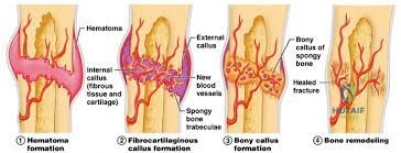
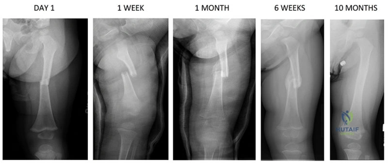
In this comprehensive guide, we discuss everything you need to know about Fracture Healing Understanding: How Bones Repair & What Treatments Help. Fracture healing understanding involves a complex, sequential process restoring injured bone. It progresses through stages: inflammation (hematoma formation, cytokine release from immune cells like macrophages, neutrophils, and platelets), followed by reparative phases. Healing type (primary via Haversian remodeling or secondary via endochondral ossification) is dictated by mechanical stability and strain at the fracture site.
We are discussing the biological environment of bone healing. Could you explain the concept of Perren's Strain Theory and how it influences your choice of fixation construct for a diaphyseal fracture?
Candidate: Perren's strain theory states that the strain at the fracture site determines what tissue forms. High strain requires soft callus, while low strain allows for primary healing. For a diaphyseal fracture, I would use an intramedullary nail to provide relative stability, which is appropriate for secondary healing.
Candidates often define strain vaguely without mentioning the quantitative thresholds (2% and 10%). They frequently fail to link the "working length" of the implant to the strain environment, and may confuse primary healing with "better" healing, ignoring that primary healing is actually slower and more biologically demanding.
The candidate defines strain as the relative change in gap length/original length. 1. Thresholds: <2% strain allows primary/direct bone healing; 2-10% allows secondary healing (callus); >10% leads to nonunion. 2. Application: In diaphyseal fractures, we aim for relative stability (2-10%) via bridge plating or intramedullary nailing. By increasing the "working length" of the implant, we decrease strain, protecting the healing tissue while allowing the micromotion required for robust endochondral ossification.
You are treating a patient with a femoral shaft fracture. You opt for a locked intramedullary nail. The patient returns at 4 months with minimal radiographic callus and a persistent fracture line. How do you classify this, and what is your management strategy?
Candidate: This is a delayed union or nonunion. I would check if it is hypertrophic or atrophic. If hypertrophic, I would dynamize the nail. If atrophic, I would bone graft it.
Failing to distinguish between Hypertrophic (Biological capacity present, mechanical instability) vs Atrophic (Biological deficit). Examiners want to hear the "Diamond Concept" mentioned here, as it provides the framework for why the bone failed to heal.
First, I differentiate the nonunion type. Hypertrophic (Elephant foot): Indicates good biology but poor stability. Management is increasing stability (e.g., exchange nailing to a larger diameter nail or compression plating). Atrophic: Indicates failed biology. Management requires addressing the "Diamond Concept": providing osteogenic cells, osteoconductive scaffold, and osteoinductive growth factors (e.g., BMPs/Bone graft), combined with stable fixation. I would also investigate systemic factors like smoking or DM.
