Nonunion Fractures: Orthopedic Epidemiology, Biomechanics, & Classification
Key Takeaway
Nonunion fractures are failures of bone healing, persisting beyond expected time, affecting 5-10% of fractures. They are classified into hypertrophic (biologically active with mechanical instability, often abundant callus) and atrophic (biologically deficient with minimal callus, compromised vascularity). Understanding their distinct pathophysiology is crucial for effective orthopedic management and restoring function.
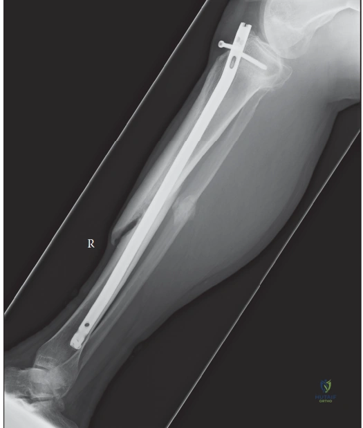
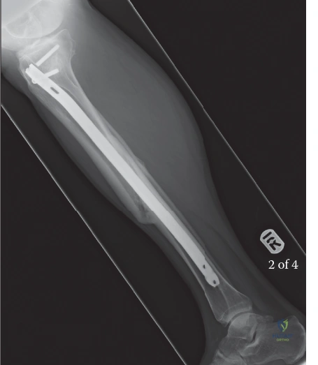
A 45-year-old smoker presents with persistent pain and an inability to bear weight on his left leg 9 months following a closed tibial shaft fracture treated with an intramedullary nail. Clinical examination reveals local tenderness and palpable motion at the fracture site. Radiographs are provided below.
How do you classify this nonunion, and what are the underlying biomechanical implications?
Candidate: This is a hypertrophic nonunion. It's caused by excessive motion at the site, which prevents bridging. The radiographs show plenty of callus, which is the "elephant's foot" appearance. I would stabilize it better to stop the motion.
The candidate fails to use formal classification terminology or discuss the biological versus mechanical nature of the nonunion. Simply saying "stabilize it better" is too vague; examiners look for specific surgical strategies like compression or reamed exchange nailing.
This is a hypertrophic nonunion, characterized by abundant callus formation (often referred to as 'elephant's foot' or 'horse hoof'). Biomechanically, this indicates the bone has high biological potential but suffers from mechanical instability. The interfragmentary strain is too high, preventing endochondral ossification. The management priority is to increase mechanical stability and compression—often achieved via exchange nailing (which also provides biological stimulation via reaming) or compression plating—rather than focusing on biological augmentation.
You have decided to proceed with surgery. During your debridement of a complex, recalcitrant tibial nonunion, you encounter the appearance shown below. What are the essential next steps in your surgical management to ensure a successful outcome?
Candidate: I would clean out all that fibrous tissue until I see bleeding bone. Then I'd probably put in some bone graft to help it heal and fix it more securely, maybe with a new plate or a larger nail.
Candidates often forget the "Infection Workup" aspect of a revision case. If the nonunion looks like this, it could be infected; placing hardware back into an infected site without proper tissue sampling is a major error.
First, I must rule out infection. I would take multiple deep tissue biopsies for aerobic, anaerobic, and fungal cultures. Secondly, I need a radical debridement of all non-viable, sclerotic, and fibrous tissue until 'punctate bleeding' (the Papineau technique principle) is achieved. Once the bed is 'biologically clean,' I would provide structural stability—ideally via a reamed intramedullary nail for load-sharing—and augment the gap with autologous bone graft (iliac crest) to provide osteogenic and osteoinductive properties.
The patient asks about the role of "bone stimulation" devices like LIPUS (Low-Intensity Pulsed Ultrasound) to avoid a second surgery. How would you counsel this patient in the context of an established atrophic nonunion?
Candidate: I'd tell them it might work, but surgery is usually better. It’s an option if they really don't want an operation.
Lack of evidence-based reasoning. The candidate fails to distinguish between the clinical utility of physical adjuncts in delayed vs. atrophic nonunions, and fails to mention the high success rate of surgery compared to these devices.
I would explain that while LIPUS is an FDA-approved adjunct for nonunions, it is most effective in delayed unions or stable, hypertrophic cases. For an established atrophic nonunion, the biological deficit and mechanical instability typically require surgical intervention (debridement and grafting) to achieve union. I would advise that relying on non-invasive stimulation alone in this scenario carries a low success rate, and surgical correction remains the standard of care for restoring both function and structural integrity.
