Proximal Femoral Varus Osteotomy with 90-Degree Blade Plate: An Intraoperative Masterclass
Key Takeaway
Join us in the operating theater for a masterclass on proximal femoral varus osteotomy using a 90-degree blade plate. We'll meticulously cover patient selection, comprehensive preoperative planning, intricate surgical anatomy, and a granular, real-time breakdown of every intraoperative step. Learn essential pearls, avoid common pitfalls, and understand critical postoperative management for optimal patient outcomes. This guide is designed for surgical fellows seeking to master this complex procedure.
Introduction and Epidemiology
Proximal femoral varus osteotomy (PFVO) remains a cornerstone in the surgical management of a wide spectrum of pediatric and adult hip pathologies. Executed frequently with a 90-degree blade plate, this procedure is highly versatile and is indicated for the correction of coxa valga deformity, hip subluxation across nearly all etiologies, and as a primary containment strategy for Legg-Calvé-Perthes disease. Furthermore, it serves a critical role in the management of early degenerative arthrosis secondary to structural dysplasia.

The pathogenesis of proximal femoral deformities is intimately linked to the development of normal femoral anatomy. The resolution of fetal bone alignment requires the attainment of gross motor activities at a typical age and is heavily dependent on normal musculoskeletal forces. In neuromuscular conditions such as cerebral palsy or myelomeningocele, muscle imbalance, spasticity, and delayed or absent ambulation disrupt these forces. This disruption prevents the normal physiological decrease in the neck-shaft angle and femoral anteversion, culminating in persistent coxa valga and excessive anteversion.
The natural history of these deformities, particularly in neuromuscular populations, is progressive. If femoral head uncoverage exceeds 50% based on the Reimers migration index, further subluxation and eventual dislocation are highly probable. In the context of Legg-Calvé-Perthes disease, femoral head uncoverage during the critical resorption and reossification stages places the hip at severe risk for permanent structural deformity, hinge abduction, and subsequent early-onset osteoarthritis. A poor radiographic outcome inevitably predisposes the joint to premature degeneration, necessitating complex reconstructive or arthroplasty procedures later in life.
Proximal femoral varus osteotomy can be accomplished at any age, provided the appropriate preoperative planning is executed, as satisfactory implants are available for all bone sizes. In complex situations, particularly neuromuscular disease, it is often necessary to address the etiology of the proximal femoral deformity and the hip disease simultaneously. This may involve concomitant soft tissue releases or pelvic osteotomies to achieve concentric, stable reduction.
Surgical Anatomy and Biomechanics
A profound understanding of the osseous and muscular anatomy of the proximal femur is requisite for the safe and effective execution of a varus-producing osteotomy.
Osseous Anatomy and Angular Parameters
The normal femoral neck-shaft angle (caput-collum-diaphyseal or CCD angle) in a mature adult is approximately 135 degrees, with a physiological range of 120 to 150 degrees. At birth, the neck-shaft angle is typically 150 degrees, gradually decreasing to the adult average by skeletal maturity in response to normal weight-bearing and abductor muscle forces.

Similarly, femoral anteversion undergoes a physiological reduction. Normal anteversion at birth is approximately 45 degrees. Through normal growth and motor development, this decreases to roughly 10 degrees in boys and 15 degrees in girls by 8 years of age. In the structurally normal hip, the tip of the greater trochanter lies precisely at the level of the center of the femoral head. This relationship is critical; significant alteration of this relationship during osteotomy can profoundly impact abductor mechanics.
Radiographic Anatomy
The true neck-shaft angle cannot be directly assessed from a standard anteroposterior (AP) pelvis radiograph unless femoral anteversion is explicitly compensated for. This is achieved by internally rotating the femur during image acquisition to eliminate the anteversion profile. Failure to account for anteversion results in a projected neck-shaft angle that appears falsely valgus.
Biomechanical Principles of Varus Osteotomy
The primary biomechanical objective of a proximal femoral varus osteotomy is to improve the containment of the femoral head within the acetabulum, thereby increasing the contact area and decreasing peak focal articular cartilage contact stresses.

By varusizing the proximal femur, the surgeon effectively rotates the femoral head medially into the depth of the acetabulum. In Legg-Calvé-Perthes disease, patients may present with a subluxated or uncovered femoral head despite proximal femoral anatomy that is otherwise normal (excluding the avascular segment). With intact neuromuscular function, varusizing the femur is well tolerated and significantly improves the containment of the diseased epiphysis, promoting spherical healing.
Furthermore, varus osteotomy alters the abductor lever arm. While extreme varus can elevate the greater trochanter relative to the center of rotation—potentially weakening the abductors and causing a Trendelenburg gait—a meticulously planned osteotomy optimizes the abductor moment arm and normalizes joint reaction forces. Correction in other planes can be accomplished simultaneously; derotation corrects excessive anteversion, while extension or flexion osteotomies address sagittal plane deformities.
Indications and Contraindications
Patient selection for proximal femoral varus osteotomy requires careful consideration of the underlying pathology, the patient's physiological age, and the presence of concomitant deformities.
Primary Indications
The procedure is primarily indicated for hip subluxation or dislocation secondary to neuromuscular disorders (e.g., cerebral palsy, spina bifida), where spasticity and lack of weight-bearing have led to persistent coxa valga and excessive anteversion. It is also a primary intervention for containment in severe Legg-Calvé-Perthes disease, particularly in children over the age of 6 to 8 years presenting with lateral pillar B or B/C involvement. Additional indications include developmental dysplasia of the hip (DDH) with residual subluxation, and early degenerative arthrosis where joint preservation is prioritized over arthroplasty.

Contributing factors to hip joint pathology often include musculotendinous contractures, ligamentous laxity, and coexistent acetabular dysplasia. These factors frequently require direct, simultaneous treatment. Adductor lengthening, psoas lengthening, open reduction of the hip with capsulorrhaphy, and acetabuloplasty (e.g., Dega, Pemberton, or San Diego osteotomies) must be considered as part of a comprehensive surgical strategy.
Contraindications
Absolute contraindications include active joint infection, severe and irreversible degenerative joint disease (Kellgren-Lawrence grade 4) where arthroplasty is more appropriate, and medical comorbidities precluding major orthopedic surgery. Relative contraindications include fixed pelvic obliquity driven by severe scoliosis (which should be addressed prior to or concurrent with hip reconstruction) and severe osteopenia that may compromise hardware fixation.
| Indication Category | Operative Management (PFVO) | Non-Operative Management |
|---|---|---|
| Neuromuscular Hip Dysplasia | Reimers Migration Percentage >40-50%, progressive subluxation, pain, difficulty with perineal care. | Reimers Migration Percentage <30%, stable serial radiographs, optimized seating/positioning. |
| Legg-Calvé-Perthes Disease | Age >8 years, lateral pillar B or B/C, loss of containment, hinge abduction amenable to correction. | Age <6 years, lateral pillar A, maintenance of spherical congruency, full range of motion. |
| Developmental Dysplasia of the Hip | Residual coxa valga/anteversion causing subluxation, failed closed reduction. | Concentric reduction maintained in Pavlik harness or spica cast, normalizing acetabular index. |
| Degenerative Arthrosis | Early-stage disease, localized joint space narrowing, congruent joint with functional ROM. | End-stage osteoarthritis, inflammatory arthropathy, severe restriction of motion. |
Pre Operative Planning and Patient Positioning
Thorough clinical evaluation and meticulous radiographic templating are paramount to achieving the desired angular correction and avoiding intraoperative complications.
Clinical Evaluation
There are no isolated physical findings that are exclusively diagnostic for coxa valga. The typical history for neuromuscular conditions, DDH, or Perthes disease will dictate the index of suspicion. In these cases, associated musculotendinous or joint contractures are frequently present on physical examination. These may include hip flexion contractures (assessed via the Thomas test), hip adduction contractures, or altered transverse plane rotation.
In Perthes disease, restricted internal hip rotation and abduction are classic findings, often secondary to muscle spasm, synovitis, or mechanical impingement from an extruded anterolateral femoral head.
Femoral anteversion is evaluated clinically by palpation of the greater trochanter with the patient in the prone position (Craig's test or trochanteric prominence test). The examiner rotates the hip internally and externally until the trochanter is most prominent laterally; at this point, the femoral neck is parallel to the floor. In the absence of tibial deformity, the angular difference between the tibial shaft and a vertical line indicates the degree of femoral anteversion. In an otherwise normal hip, clinical anteversion is typically about 20 degrees less than the maximum internal rotation range of motion.
Radiographic Templating
Standard preoperative imaging includes a standing (or simulated weight-bearing) AP pelvis, a frog-leg lateral of the hips, and a true lateral of the affected femur.

Templating determines the exact size of the wedge to be resected (if any), the angle of the seating chisel, and the appropriate blade length. When utilizing a 90-degree blade plate, the entry angle of the blade into the greater trochanter dictates the final neck-shaft angle. For example, if a final neck-shaft angle of 110 degrees is desired, and a 90-degree plate is used, the blade must be inserted at an angle of 20 degrees relative to the femoral shaft axis.
Patient Positioning
The patient is typically positioned supine on a completely radiolucent operating table to facilitate unimpeded fluoroscopic imaging in both the AP and lateral planes.

A bump is placed under the ipsilateral hemipelvis to internally rotate the operative limb, aiding in the assessment of true anteversion. The entire operative limb is prepped and draped free to allow for dynamic assessment of hip range of motion and intraoperative manipulation during the reduction of the osteotomy. Intraoperative fluoroscopy must be positioned to allow swift transitions between AP and cross-table lateral views without compromising the sterile field.
Detailed Surgical Approach and Technique
The execution of a proximal femoral varus osteotomy using a 90-degree blade plate requires precise geometric calculation and rigid internal fixation.
Surgical Approach
A direct lateral approach to the proximal femur is standard. The incision is centered over the greater trochanter and extended distally along the femoral shaft. The fascia lata is incised longitudinally. The vastus lateralis is elevated off the intermuscular septum and retracted anteriorly, or a vastus-splitting approach is utilized, exposing the proximal femoral shaft and the base of the greater trochanter.

Guidewire Placement and Chisel Seating
The crucial first step in osseous preparation is the accurate placement of the seating guidewire. This wire determines the trajectory of the blade plate. The wire is advanced into the femoral neck and head under strict fluoroscopic guidance. It must be perfectly centered in the lateral plane to avoid anterior or posterior cortical violation. In the AP plane, the angle of the guidewire relative to the femoral shaft must equal the desired degree of varus correction minus the angle of the plate (for a 90-degree plate, the math is simplified as discussed in preoperative planning).

Once the guidewire is confirmed to be in the optimal position, the seating chisel is driven into the proximal femur over the wire. The rotation of the chisel must be carefully controlled, as the orientation of the chisel handle will dictate the sagittal alignment (flexion/extension) of the proximal fragment.
The Osteotomy Cut
The level of the osteotomy is typically intertrochanteric, located just proximal to the lesser trochanter. This maximizes the cancellous bone surface area for rapid healing and provides adequate bone stock for plate fixation.

Prior to completing the osteotomy, longitudinal alignment marks are made on the anterior femur using an osteotome or electrocautery. These marks are essential for judging the exact degree of derotation applied later in the procedure. The bone is then transected using an oscillating saw. If shortening is required (often necessary in severe neuromuscular dislocations to reduce tension on the reconstructed joint), a measured cylindrical or wedge segment of bone is resected.
Implantation and Reduction
The seating chisel is extracted, and the 90-degree blade plate is carefully inserted into the prepared tract. The blade must be seated fully so that the shoulder of the plate rests flush against the lateral cortex of the proximal fragment.

The distal femoral fragment is then manipulated to align with the side plate. The pre-marked alignment lines are used to dial in the planned degree of external rotation of the distal fragment (which effectively internally rotates the proximal fragment, correcting the anteversion). The distal fragment is translated medially to restore the mechanical axis and reduce the bending moment on the implant.
Fixation and Compression
Once alignment, rotation, and translation are optimized, the plate is clamped to the distal femoral shaft. Fixation is achieved using cortical screws.

An articulated tension device or dynamic compression principles are utilized to apply axial compression across the osteotomy site. This compression is critical for primary bone healing and for increasing the construct's rigidity. Final fluoroscopic images are obtained in both AP and lateral planes to confirm hardware placement, osteotomy reduction, and hip concentricity.
Complications and Management
While proximal femoral varus osteotomy is generally highly successful, the procedure involves significant alteration of proximal femoral geometry and carries inherent risks. Meticulous surgical technique mitigates, but does not eliminate, these complications.

Hardware failure, including blade cutout or plate fracture, is a recognized complication, particularly in patients with severe osteopenia or those who are non-compliant with postoperative weight-bearing restrictions. Over-varusization (creating a neck-shaft angle <100 degrees) can lead to a severe abductor lurch, functionally shortening the limb and creating impingement of the greater trochanter against the ilium. Conversely, under-correction fails to address the primary pathology, leading to recurrent subluxation.
| Complication | Estimated Incidence | Salvage Strategy |
|---|---|---|
| Hardware Failure / Cutout | 2% - 5% | Immediate revision internal fixation; may require conversion to a locking plate construct or valgus-producing osteotomy if cutout damages superior neck. |
| Nonunion / Delayed Union | 1% - 3% | Optimization of metabolic parameters; revision fixation with bone grafting; exchange to a more stable construct if instability is the primary driver. |
| Over-correction (Severe Varus) | 3% - 6% | Valgus-producing osteotomy or distal and lateral transfer of the greater trochanter to restore abductor tension and clear impingement. |
| Avascular Necrosis (AVN) | < 1% | Observation for partial head involvement; core decompression for early symptomatic lesions; eventual arthroplasty for end-stage collapse. |
| Deep Surgical Site Infection | 1% - 4% | Aggressive surgical debridement, implant retention if stable, targeted intravenous antibiotic therapy based on intraoperative cultures. |
Post Operative Rehabilitation Protocols
Postoperative management is highly individualized, depending heavily on the patient's age, bone quality, cognitive status, and the security of the internal fixation achieved intraoperatively.

In the pediatric neuromuscular population, where compliance with weight-bearing restrictions is often impossible, a one-and-a-half spica cast may be utilized for 4 to 6 weeks to protect the osteotomy and any concomitant soft tissue or acetabular procedures.
In older, neurotypical patients where rigid compression plating was achieved, early mobilization is encouraged. Patients are typically restricted to toe-touch or partial weight-bearing (approximately 20% of body weight) on the operative limb for the first 6 weeks. Physical therapy focuses on early passive and active-assisted range of motion to prevent capsular adhesions and preserve articular cartilage health.
Radiographic evaluation is performed at 2, 6, and 12 weeks postoperatively. Once bridging callus is visualized across the osteotomy site (typically around 6 to 8 weeks), weight-bearing is progressively advanced to full. Abductor strengthening is a critical component of the late rehabilitation phase, as the altered geometry of the proximal femur requires neuromuscular re-education to eliminate Trendelenburg gait.
Summary of Key Literature and Guidelines
The efficacy of proximal femoral varus osteotomy is well-supported in orthopedic literature, particularly regarding its role in altering the natural history of dysplastic and neuromuscular hip conditions.

Reimers' foundational work on the migration percentage remains the gold standard for monitoring neuromuscular hips, establishing the threshold of >40-50% uncoverage as an absolute indication for varus derotation osteotomy to prevent the disastrous sequelae of chronic dislocation.
In the realm of Legg-Calvé-Perthes disease, multicenter prospective cohort studies have demonstrated that varus osteotomy yields superior radiographic outcomes compared to non-operative management in children over the age of 8 with lateral pillar B or B/C involvement. The procedure successfully contains the extruded epiphysis, allowing the femoral head to remodel concentrically within the acetabular mold during the reossification phase.
Recent biomechanical studies comparing the traditional 90-degree blade plate to modern pediatric locking proximal femoral plates show comparable union rates. However, the blade plate provides a distinct advantage in its ability to generate dynamic compression across the osteotomy site, a feature that remains highly valued among experienced pediatric orthopedic surgeons. Ultimately, the choice of implant and the precise execution of the osteotomy must be tailored to the individual patient's pathoanatomy to ensure durable hip stability and function.
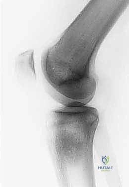
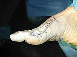
Clinical & Radiographic Imaging
You Might Also Like