Full Question & Answer Text (for Search Engines)
Question 1:
The incidence of compartment syndrome following calcaneus fracture is:
Options:
Correct Answer: 10%
Explanation:
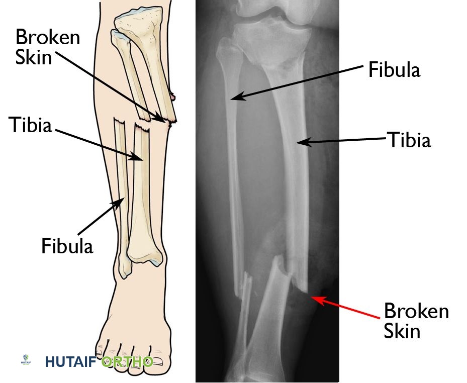
In a review article by Myerson, compartment syndrome was described to occur in 10% of calcaneal fractures. Of these, half will develop clawing, stiffness, or neurologic dysfunction. Diagnosis is confirmed by multistick invasive catheterization, especially the calcaneal compartment.
Question 2:
Posterior antiglide plating of AO type B lateral malleolar fractures may be associated with:
Options:
- Early loss of fixation
- Greater wound healing complications
- Syndesmotic irritation
- Peroneal tendonitis or peroneal tendon lesions
- Greater risk for nonunion
Correct Answer: Peroneal tendonitis or peroneal tendon lesions
Explanation:
Posterior antiglide plating is associated with an increased need for hardware removal (43%) and an increased incidence of peroneal tendon lesions. The highest risk for peroneal tendon lesions was with distal placement of the plate and a protruding screw head in the most distal hole.
Question 3:
Displaced talar neck fractures should be treated:
Options:
- Emergently within 6 hours to minimize the risk of avascular necrosis
- Urgently within 1 day to minimize the risk of avascular necrosis
- There is no correlation between emergent or urgent fixation of talar neck fractures and risk of talar avascular necrosis.
- Emergently within 1 hour of injury
- Emergently within 3 hours of injury
Correct Answer: There is no correlation between emergent or urgent fixation of talar neck fractures and risk of talar avascular necrosis.
Explanation:
A retrospective review of 102 talar neck fractures that underwent open reduction internal fixation showed no decrease in the development of osteonecrosis in fractures that were treated earlier. The mean time to fixation was 3.4 days for patients who had development of osteonecrosis, compared with 5 days for patients who did not have development of osteonecrosis.
Question 4:
How many weeks following open reduction and internal fixation of a right ankle fracture can patients resume driving with normal braking times:
Options:
- 6 weeks
- 9 weeks
- 12 weeks
- 16 weeks
- 18 weeks
Correct Answer: 9 weeks
Explanation:
Total braking time following open reduction and internal fixation of right ankle fractures was tested at 6, 9, and 12 weeks postoperatively. These patients were managed with a functional brace, non-weight bearing, and early range of motion in the postoperative period. Braking time was significantly slower than normal at 6 weeks, but had returned to near normal by 9 weeks postoperatively.
Question 5:
Time to radiographic fusion following arthroscopic ankle arthrodesis is:
Options:
- Longer than following an open technique arthrodesis
- Shorter than following an open technique arthrodesis
- The same as open technique
- Is affected by whether external bone stimulation is utilized
- Is affected by whether two-screw or three-screw fixation is utilized
Correct Answer: Shorter than following an open technique arthrodesis
Explanation:
Time to radiographic fusion following arthroscopic ankle arthrodesis is shorter than following open ankle arthrodesis. Theoretically, the decreased dissection and soft-tissue stripping contributes to greater vascular inflow to heal the fusion site.
Question 6:
Superficial peroneal nerve injury following ankle fracture:
Options:
- Does not occur with nonoperative treatment
- Can best be avoided during open reduction internal fixation with a posterolateral approach to the fibula
- Did not ultimately affect the final AOFAS ankle-hindfoot score
- Occurs in fewer than 5% of operatively fixed fibula fractures
- Can best be avoided during open reduction internal fixation with an anterolateral approach to the fibula
Correct Answer: Can best be avoided during open reduction internal fixation with a posterolateral approach to the fibula
Explanation:
One hundred twenty patients with ankle fractures were evaluated. Symptomatic superficial peroneal nerve injury was identified in 21% of patients who underwent open reduction internal fixation and 9% of nonoperatively treated patients. AOFAS scores were decreased in patients with symptomatic superficial peroneal nerve injury. No injuries to the superficial peroneal nerve occurred in patients who underwent surgery involving a posterolateral approach to the fibula.
Question 7:
Which of the following is the most reliable way to determine that a deltoid ligament injury is associated with a Weber B level lateral malleolus fracture:
Options:
- The presence of medial tenderness on clinical examination
- The presence of medial ecchymosis on clinical examination
- The presence of significant medial swelling on clinical examination
- Evidence of medial clear space widening on stress radiographs
- The presence of lateral malleolus tenderness
Correct Answer: Evidence of medial clear space widening on stress radiographs
Explanation:
Weber B supination, external rotation ankle fractures were evaluated to determine the reliability of medial tenderness, ecchymosis, and swelling in predicting deltoid incompetence. These clinical signs were poorly predictive, and stress radiographs were recommended for an accurate diagnosis of instability.
Question 8:
The optimal position for ankle arthrodesis is:
Options:
- 5° plantarflexion, 5° valgus, 5° external rotation
- Neutral flexion, 5° valgus, 5° external rotation
- Neutral flexion, 0° varus/valgus, 5° external rotation
- Neutral flexion, 5° valgus, 5° internal rotation
- 5° dorsiflexion, 5° valgus, 5° external rotation
Correct Answer: Neutral flexion, 5° valgus, 5° external rotation
Explanation:
The optimal position for ankle arthrodesis is neutral flexion, 5° valgus, and 5° external rotation. Historically, surgeons thought that women should be fused in some amount of equinus to better allow them to wear heeled shoes. However, this can increase the development of neighboring joint arthritis and also create a knee recurvatum deformity when ambulating barefoot. Currently it is recommended that all patients are fused in neutral dorsi- /plantarflexion.
Question 9:
Varus malunion following talar neck fracture is best corrected by:
Options:
- Subtalar arthrodesis
- Rotational calcaneal osteotomy with a bone block
- Deltoid ligament release and lateral ligament reconstruction
- Talar neck osteotomy with lengthening or by triple arthrodesis
- Lateral column lengthening
Correct Answer: Talar neck osteotomy with lengthening or by triple arthrodesis
Explanation:
The best way to address varus malunion in talar neck fractures and maintain motion is by talar neck osteotomy. However, there is a further possible risk of talar avascular necrosis with this procedure. The other acceptable treatment is a triple arthrodesis, although this eliminates all hindfoot motion.
Question 10:
Neighboring joint arthritis following ankle arthrodesis has not been found in the:
Options:
- Knee joint
- Naviculocuneiform joint
- First metatarsophalangeal joint
- Subtalar joint
- Hindfoot joint
Correct Answer: Knee joint
Explanation:
Long-term follow-up of ankle fusions show that nearly all patients develop arthritis in the hindfoot, midfoot, and 1st metatarsophalangeal joint. There is no evidence to show that the hip or knee is at greater risk for developing arthritis following ankle fusion.
Question 11:
Following calcaneus fracture, risk factors for later need for subtalar arthrodesis due to painful posttraumatic arthritis include all of the following except:
Options:
- Bohlers angle <0°
- Sanders type IV fractures
- Workers compensation
- Initial nonoperative care
- Female gender
Correct Answer: Female gender
Explanation:
Buckley conducted a series of large prospective studies following calcaneus fracture outcomes in Canada. All of the above factors were associated with the need for later subtalar fusion except female gender. In his other studies, it was demonstrated that male gender was a risk factor for not having a significantly better clinical outcome with surgery versus nonsurgical treatment.
Question 12:
Range of motion following total ankle replacement is closely correlated with:
Options:
- Amount of osteophytes resected during surgery
- Meticulous ligament balancing
- Level of tibial and talar saw cuts
- Preoperative range of motion
- Size of implant
Correct Answer: Preoperative range of motion
Explanation:
A radiographic study comparing preoperative to postoperative tibio-talar range of motion as measured by radiographs showed that the amount of motion that patients had following ankle replacement was most dependent upon the motion they had before surgery.
Question 13:
Patients sustaining a crushing injury to the foot with midfoot tenderness but without any radiographic signs of fracture or dislocation:
Options:
- Should be managed with a postoperative shoe and early physical therapy until the tenderness resolves
- Should be splinted and kept non-weight bearing until nontender
- Should be protected in a cast boot with early weight bearing to tolerance
- Requires open reduction internal fixation to prevent long-term arthritis
- C an be discharged with no further follow-up
Correct Answer: Should be splinted and kept non-weight bearing until nontender
Explanation:
Patients who sustain a foot injury and have clinical midfoot tenderness should be assumed to have a serious midfoot sprain until proven otherwise. These patients should be protected non-weight bearing until the tenderness is gone before weight-bearing and physical therapy begins.
Question 14:
The distinguishing factor in a Hawkins type 4 talar neck fracture is:
Options:
- The presence of an incongruent ankle joint
- Incongruity of the ankle and/or subtalar joint with the presence of a talonavicular dislocation.
- The presence of an incongruent subtalar joint
- The presence of an associated talar body fracture
- The presence of a posterior process of the talus fracture
Correct Answer: The presence of an incongruent ankle joint
Explanation:
Hawkins type 1 fractures are nondisplaced. Hawkins type 2 fractures have an incongruent subtalar joint. Hawkins type 3 fractures have an incongruent ankle and subtalar joint. Hawkins type 4 fractures have the above injuries and incongruent talo-navicular joint.
Question 15:
The calcaneal compartment of the foot contains all of the following structures except:
Options:
- Quadratus plantae muscle
- Posterior tibial nerve, artery, and vein
- Lateral plantar nerve, artery, and vein
- Interossei muscles
- 1st dorsal metatarsal artery
Correct Answer: Interossei muscles
Explanation:
The four interossei muscles are contained in their respective interosseous compartments. The calcaneal compartment may also variably contain the medial plantar nerve. The remaining compartments of the foot are the adductor, medial, lateral, and superficial.
Question 16:
Gustilo-Anderson type I and type IIA open calcaneal fractures with a medial wound can be treated:
Options:
- With initial washout and subsequent open reduction internal fixation with a lateral plate once the soft tissues and swelling have stabilized
- With initial washout and external fixation only due to the risk of osteomyelitis
- With initial washout and late reconstruction once the soft tissue has healed to address the malunion
- Washout is unnecessary for type I and IIA open calcaneal fractures
- With immediate fasciotomy
Correct Answer: With initial washout and subsequent open reduction internal fixation with a lateral plate once the soft tissues and swelling have stabilized
Explanation:
Forty-three open calcaneal fractures were studied, showing that open reduction internal fixation with plate and screws of type I and type IIA fractures with medial wounds had outcomes similar to closed injuries. Type IIIB open calcaneal fractures should undergo early flap coverage. Early internal fixation should be avoided in these injuries due to the high rates of osteomyelitis and amputation.
Question 17:
Take-down of ankle arthrodesis and conversion to total ankle replacement:
Options:
- Is impossible if the fibula has been resected
- Is a dependable procedure with a rate of complications similar to primary ankle replacement
- Has a poor clinical success rate if there is no clear underlying cause of pain from the ankle fusion
- Results in minimal gains in ankle range of motion
- Requires custom made prosthetic implants
Correct Answer: Has a poor clinical success rate if there is no clear underlying cause of pain from the ankle fusion
Explanation:
This article studied the success rates of revising previous ankle fusions to ankle replacement. The authors found that if the etiology of a patientâ s pain was unclear, the patients did poorly. Patients with prior fibula resection could still be revised to ankle replacement with allograft bone to support the lateral side of the implant. Range of motion following revision to arthroplasty was comparable to primary replacement.
Question 18:
The distinction between a Lauge-Hansen supination-external rotation III injury and a Lauge-Hansen supination-external rotation IV injury is:
Options:
- A spiral oblique fracture of the lateral malleolus
- Anteroinferior tibiofibular ligament (AITFL) disruption
- Posteroinferior tibiofibular ligament (PITFL) disruption or posterior malleolus fracture
- Deltoid ligament disruption or medial malleolus fracture
- Anterior talo-fibular ligament disruption
Correct Answer: A spiral oblique fracture of the lateral malleolus
Explanation:
The sequence of injury according to the Lauge-Hansen classification system in supination-external rotation injuries is AITFL disruption, spiral oblique fracture of the lateral malleolus, PITFL disruption or posterior malleolus fracture, and finally stage IV, which is a deltoid ligament disruption or medial malleolus fracture.C orrect Answer: Deltoid ligament disruption or medial malleolus fracture
Question 19:
Development of hindfoot arthritis following total ankle replacement is seen in:
Options:
- 0% of patients
- <25% of patients
- 50% of patients
- 75% of patients
- >75% of patients
Correct Answer: <25% of patients
Explanation:
Although it is felt that the retention of some degree of ankle motion with ankle replacement can help prevent the development of hindfoot arthritis, in a 9-year follow-up study nearly 25% of patients still had radiographic signs of arthritis.
Question 20:
When using external fixation in the treatment of tibial pilon fractures, distal transfixation wires:
Options:
- Should always traverse the distal tibia-fibula joint to get optimal fixation
- Should remain >12.2 mm above the subchondral plate of the distal tibia
- Are not at risk for causing joint infection
- Are least at risk for penetrating the joint capsule over the anterolateral aspect of the ankle
- C annot be olive wires because of a higher risk for pin-tract infection
Correct Answer: Should remain >12.2 mm above the subchondral plate of the distal tibia
Explanation:
In cadaver specimens, the anterolateral capsular reflection of the ankle joint extended proximally the highest with an average of 9.3 mm and a maximum of 12.2 mm. There was a 100% communication between the distal tibia- fibula joint and the ankle joint.
Question 21:
C linical improvement following ankle distraction arthroplasty:
Options:
- Typically reaches its maximal improvement by the end of 1 year
- Is accompanied by major gains in ankle range of motion
- C an take up to 5 years to reach maximal improvement
- Is not accompanied by improvement in radiographic joint space
- Is usually realized within the first month following removal of the frame
Correct Answer: C an take up to 5 years to reach maximal improvement
Explanation:
Distraction arthroplasty with an Ilizarov external fixator is usually associated with half of the clinical improvement occurring within the first year, and the other half happening over the next 5 years.
Question 22:
Failure following supramalleolar osteotomy for ankle arthritis is associated with:
Options:
- Inadequate correction and poor cartilage on initial arthroscopy
- Opening wedge supramalleolar osteotomy with bone graft
- Early weight bearing postoperatively
- Addition of a fibular osteotomy to the procedure
- Use of internal fixation
Correct Answer: Inadequate correction and poor cartilage on initial arthroscopy
Explanation:
In their clinical series, Takakura and colleagues showed that inadequate correction and initial chondromalacia were predictors of poor outcome following supramalleolar osteotomy.
Question 23:
Isolated talonavicular fusion:
Options:
- Decreases subtalar motion by 25%
- Decreases subtalar motion by 50%
- Locks subtalar motion
- Has no effect on subtalar motion
- Decreases subtalar motion by 10%
Correct Answer: Locks subtalar motion
Explanation:
This cadaver study examined the motion that remained in the hindfoot joints following sequential immobilization of the talonavicular, subtalar, and calcaneo-cuboid joints. Fixing the talo-navicular joint virtually locked all subtalar motion.
Question 24:
Following triple arthrodesis, ankle range of motion is:
Options:
- Unaffected
- Increased
- Decreased
- Improves over time
- Increases initially, but then returns to preoperative levels
Correct Answer: Decreased
Explanation:
This clinical study following triple arthrodesis patients for 10 years showed a 27% loss of ankle plantarflexion but no loss of dorsiflexion.
Question 25:
Triple arthrodesis is associated with:
Options:
- Long-term clinical stability with respect to pain relief
- High rates of nonunion
- Worse patient satisfaction when ankle arthritis is present
- Development of ankle arthritis over time
- No increased risk for ankle arthritis
Correct Answer: Development of ankle arthritis over time
Explanation:
Saltzman and colleagues followed 67 patients who underwent triple arthrodesis at 44-year follow-up. Nearly all patients had ankle arthritis at final follow-up. C linical relief of pain deteriorated over time between intermediate 25-year follow-up and 44-year follow-up in the same group of patients.
Question 26:
Isolated subtalar arthrodesis:
Options:
- Increases transverse tarsal joint over time
- Decreases talonavicular motion less than calcaneocuboid motion
- Decreased talonavicular joint motion but increases calcaneocuboid joint motion
- Decreases talonavicular motion more than calcaneocuboid motion
- Increases subtalar motion
Correct Answer: Decreases talonavicular motion less than calcaneocuboid motion
Explanation:
Subtalar fusion decreased talonavicular motion more so than calcaneocuboid motion in this cadaver study. Isolated talonavicular fusion is the most influential of the hindfoot joints, locking hindfoot motion.
Question 27:
Isolated subtalar fusion:
Options:
- Is not associated with development of ankle or transverse tarsal joint arthritis
- Is associated only with development of transverse joint arthritis, but the ankle joint is spared
- Is associated only with development of ankle arthritis, but the transverse tarsal joints are spared
- Is associated with the development of both ankle and transverse tarsal joint arthritis
- Is associated with knee joint degenerative arthritis
Correct Answer: Is associated only with development of transverse joint arthritis, but the ankle joint is spared
Explanation:
In 48 subtalar fusions followed for 5 years, 36% of patients developed ankle arthritis and 41% of patients developed transverse tarsal joint arthritis.
Question 28:
Following anatomic open reduction and internal fixation of a Lisfranc fracture-dislocation:
Options:
- Development of tarsometatarsal arthritis will not occur.
- Tarsometatarsal arthritis may still arise in approximately 25% of patients.
- If tarsometatarsal arthritis develops, then subsequent arthrodesis is required.
- The screws should be routinely removed at 12 weeks.
- The screws should be routinely removed at 6 weeks.
Correct Answer: Tarsometatarsal arthritis may still arise in approximately 25% of patients.
Explanation:
In a series of patients who underwent open reduction internal fixation of Lisfranc fracture dislocations, 25% of patients developed midfoot arthritis at final follow-up, but only half of these patients required eventual midfoot arthrodesis.
Question 29:
Which injury is likely to have a worse clinical outcome:
Options:
- A purely ligamentous Lisfranc injury
- A Lisfranc fracture-dislocation Purely ligamentous Lisfranc injuries have a worse clinical outcome than injuries associated with bony fractures.C orrect Answer: A purely ligamentous Lisfranc injury
Correct Answer: A purely ligamentous Lisfranc injury
Question 30:
Hallux rigidus is associated with:
Options:
- Metatarsus primus elevatus
- First ray hypermobility
- Long first metatarsal
- Flat- or chevron-shaped metatarsal head
- Bipartate sesamoid
Correct Answer: Metatarsus primus elevatus
Explanation:
In a large series of patients with hallux rigidus, risk factors were evaluated. The only factor that had a positive correlation with having hallux rigidus was the radiographic shape of the 1st metatarsal head. Metatarsus primus elevatus, first ray hypermobility, or long first metatarsal head were not significantly associated with hallux rigidus.C orrect Answer: Flat- or chevron-shaped metatarsal head
Question 31:
C urrently recommended indications for surgical management of hallux rigidus with an arthrodesis include:
Options:
- Positive axial grind test on preoperative clinical examination
- >50% of the cartilage on the metatarsal head remaining
- Osteophytes over the dorsolateral head of the first metatarsal
- Osteophytes over the dorsal aspect of the proximal phalanx
- Normal first metatarsophalangeal joint motion
Correct Answer: Positive axial grind test on preoperative clinical examination
Explanation:
Coughlin and colleagues recommend that when pain with axial grind testing of the metatarsophalangeal joint is present or >50% loss of articular cartilage occurs intraoperatively, then first metatarsophalangeal arthrodesis should be performed.
Question 32:
A Moberg procedure for hallux rigidus is:
Options:
- An oblique first metatarsal shortening osteotomy
- An ostectomy of the medial eminence of the metatarsal
- A medial closing wedge osteotomy of the proximal phalanx
- A dorsal closing wedge osteotomy of the proximal phalanx
- A lateral closing wedge osteotomy of the proximal phalanx
Correct Answer: A dorsal closing wedge osteotomy of the proximal phalanx
Explanation:
The Moberg procedure involves a dorsal closing wedge osteotomy of the proximal phalanx. This sets the hallux higher off the floor, allowing for easier toe-off with less dorsal impingement during gait.
Question 33:
The optimal position for hallux interphalangeal joint arthrodesis is:
Options:
- 5° to 10° of plantarflexion
- 5° to 10° of dorsiflexion
- Neutral flexion
- Slight supination of the toe
- 10° of valgus
Correct Answer: 5° to 10° of plantarflexion
Explanation:
The optimal position for hallux interphalangeal joint arthrodesis is 5° to 10° of plantarflexion, neutral varus-valgus, and neutral rotation. The plantarflexion helps the toe pad to contact the ground during gait.
Question 34:
First metatarsophalangeal prosthetic joint replacements:
Options:
- Significantly increase joint range of motion
- Have less complications than first metatarsophalangeal arthrodesis
- Provide less pain relief than first metatarsophalangeal arthrodesis
- Have not been found to undergo osteolysis or loosening
- Provide greater pain relief than first metatarsophalangeal arthrodesis
Correct Answer: Provide less pain relief than first metatarsophalangeal arthrodesis
Explanation:
First metatarsophalangeal joint replacement in this prospective comparative study performed poorly compared to arthrodesis. Patients with arthroplasties had greater pain and little improvement in range of motion.
Question 35:
Deep infection following open reduction internal fixation (ORIF) for tibial pilon fractures is most commonly associated with:
Options:
- Open fractures
- Postoperative wound dehiscence
- Anterior incision
- Medial and lateral plating
- Low energy injury
Correct Answer: Postoperative wound dehiscence
Explanation:
Deep infection following ORIF of pilon fractures is correlated with postoperative wound dehiscence or skin slough but not with the presence of an open fracture in a series of 60 pilon fractures treated by ORIF.
Question 36:
Talar body fractures are best classified by a fracture line:
Options:
- That extends superiorly into the trochlea
- That extends anywhere posterior to the talar neck
- That extends inferiorly, posterior to the lateral process
- That extends inferiorly, anterior to the lateral process
- That extends into the talar head
Correct Answer: That extends superiorly into the trochlea
Explanation:
Talar neck and body fractures can be difficult to distinguish, especially when they extend superiorly into the anteromedial aspect of the trochlea. These two fractures have a different prognosis. The authors recommend classification of these fractures based on the inferior fracture line; if anterior to lateral process of the talus, then it is a neck fracture; if posterior to lateral process of the talus, then it is a body fracture.
Question 37:
The most effective fixation technique that will ensure adequate visualization (imaging) of avascular necrosis changes following talar neck fracture is:
Options:
- Fixation with 0.062-inch K-wires
- C losed reduction
- Fixation with stainless steel mini-fragment screws
- Fixation with titanium screws
- Fixation with a stainless steel locking plate
Correct Answer: Fixation with 0.062-inch K-wires
Explanation:
High-quality magnetic resonance images of the talus can consistently be obtained in the presence of titanium screws in contrast to images obtained with stainless steel implants. Magnetic resonance imaging is better than plain radiographs at assessing the volume of talar avascular necrosis.
Question 38:
The plantar ecchymosis sign is:
Options:
- An indication of possible compartment syndrome
- Related to a specific bacterial infection
- An indication of possible Lisfranc fracture or sprain
- Described as a sign of plantar fascia rupture
- Requires immediate fasciotomy
Correct Answer: An indication of possible compartment syndrome
Explanation:
The plantar ecchymosis sign is described as an ecchymotic area on the plantar midfoot that is indicative of possible injury to the plantar tarsometatarsal ligaments.
Question 39:
The joint contact area of the second tarsometatarsal joint after Lisfranc dislocation diminishes the greatest with:
Options:
- Dorsolateral subluxation
- Dorsal subluxation
- Lateral subluxation
- Medial subluxation
- Plantar subluxation
Correct Answer: Dorsolateral subluxation
Explanation:
Minor degrees of displacement not apparent on plain radiographs lead to significant decrease in the contact area of the second tarsometatarsal joint. Dorsolateral subluxation of the second tarsometatarsal joint suffers a loss of contact area more severely than pure dorsal or lateral subluxation. Just 3 mm of dorsolateral subluxation causes a 38% loss of contact area.
Question 40:
The â fleck signâ in midfoot injuries is a result of avulsion of the:
Options:
- Lisfranc ligament that extends from the first metatarsal base to the second metatarsal base
- Lisfranc ligament that extends from the middle cuneiform to the first metatarsal base
- Lisfranc ligament that extends from the medial cuneiform to the first metatarsal base
- Lisfranc ligament that extends from the medial cuneiform to the second metatarsal base
- Lisfranc ligament that extends from the lateral cuneiform to the third metatarsal base
Correct Answer: Lisfranc ligament that extends from the first metatarsal base to the second metatarsal base
Explanation:
The fleck sign was described as an avulsion of the ligament that runs from the medial cuneiform to the base of the second metatarsal, the so-called Lisfranc ligament. It is considered pathognomonic for a tarsometatarsal injury.
Question 41:
Delayed unions and nonunions of base of fifth metatarsal fractures have been demonstrated to heal by:
Options:
- Prolonged cast immobilization and non-weight bearing
- Pulsed electromagnetic fields
- C ontinued use of a fracture boot with protected weight-bearing
- Injection of demineralized bone matrix
- Rigid carbon fiber shoe inserts
Correct Answer: Pulsed electromagnetic fields
Explanation:
Nine delayed unions and nonunions of the proximal fifth metatarsal were treated with pulsed electromagnetic fields. All fractures healed in a mean of 4 months (follow-up 39 months, no refractures).
Question 42:
The strongest hardware configuration for fixation of talar neck fractures is:
Options:
- Two crossed screws from distal to proximal
- Two parallel screws inserted from distal to proximal
- One large screw from posterior to anterior
- Two parallel screws from posterior to anterior
- One oblique screw from distal to proximal
Correct Answer: Two parallel screws inserted from distal to proximal
Explanation:
Biomechanical cadaveric testing of several screw configurations showed two parallel screws from proximal to distal as the strongest fixation. The screws can be inserted either open or percutaneously. All screw configurations were stronger than K-wire configurations.
Question 43:
According to Sandersâ computed tomography (C T) classification for calcaneus fractures, a Sanders III fracture has:
Options:
- One fracture line in the posterior facet
- Two fracture lines in the posterior facet
- Three fracture lines in the posterior facet
- Three fracture lines in the posterior facet
- Five fracture lines in the posterior facet
Correct Answer: Two fracture lines in the posterior facet
Explanation:
The Sanders C T classification is determined on coronal C T scans of the calcaneus at the level where the posterior facet is widest. A Sanders I is a nondisplaced fracture; Sanders II consists of a single fracture line splitting the posterior facet into two main fragments; Sanders III has two fracture lines with three main posterior facet fragments; and a Sanders IV has four or more articular fragments present.
Question 44:
The incidence of compartment syndrome following calcaneus fracture is:
Options:
Correct Answer: 10%
Explanation:
In a review article by Myerson, compartment syndrome was described to occur in 10% of calcaneal fractures. Of these, half will develop clawing, stiffness, or neurologic dysfunction. Diagnosis is confirmed by multistick invasive catheterization, especially the calcaneal compartment.
Question 45:
Posterior antiglide plating of AO type B lateral malleolar fractures may be associated with:
Options:
- Early loss of fixation
- Greater wound healing complications
- Syndesmotic irritation
- Peroneal tendonitis or peroneal tendon lesions
- Greater risk for nonunion
Correct Answer: Peroneal tendonitis or peroneal tendon lesions
Explanation:
Posterior antiglide plating is associated with an increased need for hardware removal (43%) and an increased incidence of peroneal tendon lesions. The highest risk for peroneal tendon lesions was with distal placement of the plate and a protruding screw head in the most distal hole.
Question 46:
Displaced talar neck fractures should be treated:
Options:
- Emergently within 6 hours to minimize the risk of avascular necrosis
- Urgently within 1 day to minimize the risk of avascular necrosis
- There is no correlation between emergent or urgent fixation of talar neck fractures and risk of talar avascular necrosis.
- Emergently within 1 hour of injury
- Emergently within 3 hours of injury
Correct Answer: There is no correlation between emergent or urgent fixation of talar neck fractures and risk of talar avascular necrosis.
Explanation:
A retrospective review of 102 talar neck fractures that underwent open reduction internal fixation showed no decrease in the development of osteonecrosis in fractures that were treated earlier. The mean time to fixation was 3.4 days for patients who had development of osteonecrosis, compared with 5 days for patients who did not have development of osteonecrosis.
Question 47:
Superficial peroneal nerve injury following ankle fracture:
Options:
- Does not occur with nonoperative treatment
- Can best be avoided during open reduction internal fixation with a posterolateral approach to the fibula
- Did not ultimately affect the final AOFAS ankle-hindfoot score
- Occurs in fewer than 5% of operatively fixed fibula fractures
- Can best be avoided during open reduction internal fixation with an anterolateral approach to the fibula
Correct Answer: Can best be avoided during open reduction internal fixation with a posterolateral approach to the fibula
Explanation:
One hundred twenty patients with ankle fractures were evaluated. Symptomatic superficial peroneal nerve injury was identified in 21% of patients who underwent open reduction internal fixation and 9% of nonoperatively treated patients. AOFAS scores were decreased in patients with symptomatic superficial peroneal nerve injury. No injuries to the superficial peroneal nerve occurred in patients who underwent surgery involving a posterolateral approach to the fibula.
Question 48:
Which of the following is the most reliable way to determine that a deltoid ligament injury is associated with a Weber B level lateral malleolus fracture:
Options:
- The presence of medial tenderness on clinical examination
- The presence of medial ecchymosis on clinical examination
- The presence of significant medial swelling on clinical examination
- Evidence of medial clear space widening on stress radiographs
- The presence of lateral malleolus tenderness
Correct Answer: Evidence of medial clear space widening on stress radiographs
Explanation:
Weber B supination, external rotation ankle fractures were evaluated to determine the reliability of medial tenderness, ecchymosis, and swelling in predicting deltoid incompetence. These clinical signs were poorly predictive, and stress radiographs were recommended for an accurate diagnosis of instability.
Question 49:
Varus malunion following talar neck fracture is best corrected by:
Options:
- Subtalar arthrodesis
- Rotational calcaneal osteotomy with a bone block
- Deltoid ligament release and lateral ligament reconstruction
- Talar neck osteotomy with lengthening or by triple arthrodesis
- Lateral column lengthening
Correct Answer: Talar neck osteotomy with lengthening or by triple arthrodesis
Explanation:
The best way to address varus malunion in talar neck fractures and maintain motion is by talar neck osteotomy. However, there is a further possible risk of talar avascular necrosis with this procedure. The other acceptable treatment is a triple arthrodesis, although this eliminates all hindfoot motion.
Question 50:
Following calcaneus fracture, risk factors for later need for subtalar arthrodesis due to painful posttraumatic arthritis include all of the following except:
Options:
- Bohlerâ s angle <0°
- Sanders type IV fractures
- Workersâ compensation
- Initial nonoperative care
- Female gender
Correct Answer: Female gender
Explanation:
Buckley conducted a series of large prospective studies following calcaneus fracture outcomes in C anada. All of the above factors were associated with the need for later subtalar fusion except female gender. In his other studies, it was demonstrated that male gender was a risk factor for not having a significantly better clinical outcome with surgery versus nonsurgical treatment.