Full Question & Answer Text (for Search Engines)
Question 1:
Which of the following problems is most common in achondroplasia:
Options:
- Atlantoaxial instability
- Basilar invagination
- Symptomatic kyphosis
- Thoracolumbar stenosis
- Spondylolisthesis
Correct Answer: Thoracolumbar stenosis
Explanation:
Symptomatic stenosis of the thoracic and lumbar spine is seen in almost half of all achondroplastic patients, although not all patients require surgery. Atlantoaxial instability is rare in achondroplasia, although it is not uncommon in other dysplasias. Basilar invagination is not present in achondroplasia. Kyphosis is often transient in achondroplasia and rarely persists beyond the second year. Kyphosis is rarely symptomatic. Spondylolisthesis is rare in achondroplasia.
Question 2:
A 2-year-old girl with diastrophic dysplasia is brought into the office for an overall examination. A lateral radiograph of the spine shows a kyphosis of 35° from C 3 to C 6. A neurologic exam is normal, although she does have stiff joints. The patient is not yet walking. For management of this kyphosis, recommended treatment includes:
Options:
- Observation
- C ervicothoracic orthosis
- Halo traction
- Posterior fusion
- Anterior and posterior fusion
Correct Answer: Observation
Explanation:
Many of these kyphoses will correct spontaneously if the curve does not exceed 50°. In this patient, the inability to walk is most likely due to other skeletal factors. There is no evidence that orthosis will change the natural history of the disorder. There is no need for traction given the high chance of spontaneous resolution and the dangers of traction. Posterior fusion is only indicated if the kyphosis is continually progressive, or if neurologic signs or symptoms develop. Anterior and posterior surgery is only indicated in cases with severe pre- existing neurologic deficit.
Question 3:
A baby born with diastrophic dysplasia today may eventually require all of the following orthopedic procedures during childhood or adulthood except:
Options:
- C orrection of equinus or varus feet
- Arthrodesis from the occiput to the atlas or axis
- Posterior spinal fusion for scoliosis
- Arthroplasty of the hips
- Arthroplasty of the knees
Correct Answer: Arthrodesis from the occiput to the atlas or axis
Explanation:
Patients with diastrophic dysplasia rarely have instability of the upper cervical spine. Babies with diastrophic dysplasia often have rigid equinovarus feet that require surgery to become plantigrade and wear normal shoes. A number of patients with diastrophic dysplasia develop progressive scoliosis that requires surgical treatment. Degenerative disease of the hips is common and often requires arthroplasty in early adulthood. Degenerative disease of the knees is common and often requires arthroplasty in early adulthood.
Question 4:
A patient with spondyloepiphyseal dysplasia congenita reaches the age of 5 without being able to walk with a walker. She has five beats of clonus in both ankles. Her reflexes are brisk and her toes are upgoing. The most likely problem that accounts for these conditions is:
Options:
- Severe scoliosis of the thoracic spine
- Foramen magnum stenosis
- Lumbar stenosis
- Thoracolumbar kyphosis
- Atlantoaxial instability
Correct Answer: Atlantoaxial instability
Explanation:
Atlantoaxial instability, sometimes combined with stenosis of the atlas, is a frequent cause of myelopathy in spondyloepiphyseal dysplasia congenita. Scoliosis does not account for developmental delay or myelopathy. Foramen magnum stenosis is rare in spondyloepiphyseal dysplasia congenita. Lumbar stenosis is rare with spondyloepiphyseal dysplasia congenita and would not account for myelopathy. Thoracolumbar kyphosis severe enough to cause myelopathy is rare in spondyloepiphyseal dysplasia congenita condition.
Question 5:
Scoliosis in cleidocranial dysplasia is frequently associated with which of the following conditions:
Options:
- Syringomyelia
- Atlantoaxial instability
- Spondylolisthesis
- Spinal stenosis
- Spinal decompensation
Correct Answer: Syringomyelia
Explanation:
Scoliosis with cleidocranial dysplasia (C C D) is frequently associated with syringomyelia. Cervical instability is rare in C C D. Spondylolisthesis is rare in C C D. Spinal stenosis is rare in C C D. Spinal decompensation is rare in C C D.
Question 6:
A 3-year-old girl with Larsen syndrome is brought into the office for examination. A spinal radiograph demonstrates a 50° kyphosis of the cervical spine. Her neurologic examination is normal. Recommended treatment includes:
Options:
- Observation
- Halo traction
- C ervical-thoracic orthosis
- Posterior cervical fusion
- Anterior and posterior fusion
Correct Answer: Posterior cervical fusion
Explanation:
Posterior cervical fusion has been proven effective in curves of 60°or less, in preventing progression, and allowing correction with anterior growth. This curve is much more likely to worsen than to spontaneously improve, so preventive surgery is indicated. There is no role for halo traction in this situation. Bracing has not been proven effective in helping patients with Larsen syndrome. Addition of anterior fusion is not needed for this degree of curve in a neurologically normal child.
Question 7:
A 10-year-old patient with Hurler syndrome has undergone a bone marrow transplant and is currently medically stable. He has developed a painful thoracolumbar kyphosis that measures 50° with 25% subluxation T12 on L1. Recommended treatment includes which of the following:
Options:
- Exercise program for the trunk extensor muscles
- Thoracolumbar orthosis
- Halo traction followed by orthosis
- Posterior spinal fusion
- Anterior and posterior spinal fusion
Correct Answer: Posterior spinal fusion
Explanation:
Anterior and posterior fusion will correct the translation, instability, and ensure a solid fusion. Exercises will not correct the subluxation, which is the cause of the pain. A thoracolumbar orthosis is not corrective or well tolerated. There is no need for halo traction. Posterior fusion alone is not enough to control this focal instability if the patient is well enough to tolerate a more involved procedure.
Question 8:
Aneurysmal bone cyst of the spine is most common in which of the following regions:
Options:
- C ervical
- Upper thoracic
- Lower thoracic
- Lumbar
- Sacral
Correct Answer: Lumbar
Explanation:
Aneurysmal bone cyst of the spine is most common in the lumbar spine, followed by the cervical spine. Aneurysmal bone cyst of the spine is treated with embolization and/or surgical resection and reconstruction.
Question 9:
Aneurysmal bone cyst of the spine is most likely in this age group:
Options:
- First decade
- Second decade
- Third decade
- Fourth decade
- Fifth decade
Correct Answer: Second decade
Explanation:
The most common age is the second decade; the mean age is 13 years old.
Question 10:
A 14-year-old girl is examined because of a pain in her left flank. The radiographs of the lumbar spine show loss of the pedicle with expansion of the lateral wall of the third lumbar vertebral body. Magnetic resonance imaging shows multiple fluid levels in the vertebral body with no additional areas of involvement. She is neurologically normal. The least invasive, effective treatment is which?
Options:
- Observation
- Radiation therapy
- Selective arterial embolization
- Radical en bloc resection
- C urettage plus radiation therapy
Correct Answer: Selective arterial embolization
Explanation:
This patient has an aneurysmal bone cyst of the vertebra. Selective arterial embolization is a minimally invasive treatment that often succeeds in arresting the lesions. Many times it is the only treatment needed. Selective arterial embolization can also be used as part of a strategy to be followed by curettage and reconstruction to decrease operative bleeding. This lesion will continue to expand and might cause neurologic compromise or mechanical instability. Radiation therapy poses risks of later malignant degeneration. There are other ways of treating this lesion. Radical en bloc resection may unnecessarily injure neurologic structures. While curettage is often necessary, there is no reason to introduce the risk of radiation therapy.
Question 11:
A 15-year-old girl has severe hip pain 3 years after a slipped capital femoral epiphysis that was complicated by avascular necrosis. Recommended treatment is a hip arthrodesis. In response to questions about late effects, after surgery the patient should be told that she is most likely to experience:
Options:
- Low back pain
- Marked limitation of activity
- Pain in the contralateral hip
- C ontinued severe pain in the ipsilateral hip
- Significant continued limp
Correct Answer: Low back pain
Explanation:
Low back pain, followed closely by ipsilateral knee pain, is the most common late effect of hip arthrodesis in young patients. The tolerable pain usually occurs much later but may be treated by conversion to arthroplasty, if needed. Ipsilateral hip pain should be minimal or absent if the fusion is successful. Activity following arthrodesis is not significantly limited. Sports and heavy physical activities are feasible. The limp is usually minimal because the loss of hip motion is masked by lumbar motion. Pain in the contralateral hip is rare and is often minimal after hip arthrodesis.
Question 12:
Which of the following is an appropriate position for arthrodesis of the hip in a young person:
Options:
- Flexion of 45°
- Abduction of 15° if there is shortening
- Adduction of 0°
- External rotation of 25°
- Shortening of at least 3 cm
Correct Answer: Adduction of 0°
Explanation:
Neutral abduction is important in preventing back pain. The flexion should be between 25° and 35°. Any abduction beyond neutral poses increased risk of back pain. External rotation beyond approximately 5° is not needed. Arthrodesis often produces some shortening; therefore, intentional shortening is not needed.
Question 13:
A 12-year-old girl is brought into the office for an examination because of hip pain. She is able to bear weight on the involved limb while using crutches for stability. Radiographs reveal a grade III slip of the capital femoral epiphysis. Recommended treatment for this patient is:
Options:
- Skeletal femoral traction in order to improve the position
- Manipulate the hip under anesthesia in order to improve the position of the head
- Osteotomy of the femoral neck to improve the alignment
- Application of a hip spica
- In situ fixation
Correct Answer: In situ fixation
Explanation:
In situ fixation provides the best results no matter what the grade of slip.
Question 14:
A 9-year-old boy with cerebral palsy has trouble sitting. His mother states that whenever his diapers are changed or his hips are moved, he begins to cry. Radiographs demonstrate high dislocations of both femoral heads. The femoral heads have an ovoid shape and superolateral flattening. Recommended treatment is:
Options:
- Botulinum toxin injected into the adductors
- Bilateral open adductor tenotomy
- Bilateral femoral osteotomies with acetabuloplasty
- Bilateral proximal femoral resection
- Bilateral C olonna arthroplasty
Correct Answer: Bilateral proximal femoral resection
Explanation:
Bilateral proximal femoral resection is the recommended treatment. Femoral head dislocations may become painful in cerebral palsy at a much earlier age than in nonspastic individuals. Botulinum toxin or adductor tenotomy will not solve the problem. Replacing the deformed femoral heads into the acetabulum will not achieve the long-term goal of good hip range of motion.
Question 15:
A 9-year-old boy is examined due to a closed distal forearm fracture. The radius and ulna are both fractured and translated 100%. After manipulation twice with sedation, the translation cannot be reduced. There is 10-mm shortening of the radius and 5- mm shortening of the ulna. The distal radial angulation on the anteroposterior view is 5° less than normal. The least invasive treatment which would produce acceptable results is:
Options:
- Closed reduction in the operating room under general anesthesia
- Open reduction and cast application
- Open reduction and percutaneous pin fixation
- Open reduction and plate fixation
- Acceptance of the reduction and maintenance with a cast
Correct Answer: Acceptance of the reduction and maintenance with a cast
Explanation:
The translation and shortening are not problems and the amount of angulation will easily remodel with this fracture. There is nothing to be gained from operative reduction.
Question 16:
When applying a halo for postoperative immobilization in a skeletally mature teenager, which of the following is the proper torque for the pins:
Options:
- 2 inch-pounds
- 4 inch-pounds
- 8 inch-pounds
- 10 inch-pounds
- 12 inch-pounds
Correct Answer: 8 inch-pounds
Explanation:
This patient should be treated like an adult. Eight inch-pounds is the currently recommended torque to provide optimal biomechanical fixation while minimizing pin penetration.
Question 17:
A posterior spine fusion with segmental hook fixation from T4-L4 is performed for idiopathic scoliosis in a 15-year-old girl. Somatosensory evoked potential monitoring is normal throughout the procedure. The patient awakens and is unable to move either lower extremity, but she does have some sensation in the lower extremities. Recommended treatment includes:
Options:
- Removal of instrumentation
- Myelogram
- Laminectomy above the conus medullaris
- Administration of corticosteroids and observation for 6 hours
- Full heparinization of the patient
Correct Answer: Removal of instrumentation
Explanation:
Spinal cord injury occurs in approximately 1% of patients operated upon for idiopathic scoliosis. In some cases, sensory spinal cord monitoring may be unchanged, especially if the injury preserves the dorsal columns. The instrumentation should be removed as soon as possible in case spinal traction or derotation or implant protrusion is producing effects on the cord or its blood supply. Corticosteroids should be administered at spinal cord injury doses, but this should not be the only measure. Obtaining a myelogram may delay the removal of instrumentation and should not be the first step. Heparinization has no proven effect.
Question 18:
A 12-year-old boy with achondroplasia has a gradual 40° thoracolumbar kyphosis. He is unable to walk more than two blocks. Magnetic resonance imaging reveals spinal stenosis, and the patient is scheduled to undergo posterior decompression from T12- S1. In addition to this procedure, you recommend:
Options:
- Observation with serial radiographs every 4 months
- Postoperative brace for 6 months
- In situ fusion with bone graft
- Posterior fusion across the kyphosis with instrumentation
- Anterior corpectomy and fusion of T12
Correct Answer: Posterior fusion across the kyphosis with instrumentation
Explanation:
Extensive posterior decompression poses a high risk of postoperative increase in kyphosis because of both the patientâ s age and pre-existing kyphosis. Observation would not be a good idea because the risk is already known to be high. Neither a brace nor an uninstrumented fusion would prevent the deformity from developing. Corpectomy is not indicated because the kyphosis is not focal. Posterior instrumented fusion at the time of decompression is indicated.
Question 19:
Which of the following is true regarding brace treatment for Scheuermann kyphosis:
Options:
- The Milwaukee brace is not indicated.
- Permanent improvement is usually obtainable if compliant.
- Bracing is effective in curves over 75°.
- Bracing is ineffective in curves having an apex at or above T8.
- The brace should be worn for 1 year after starting brace treatment.
Correct Answer: Permanent improvement is usually obtainable if compliant.
Explanation:
Brace treatment is effective for Scheuermann kyphosis. Unlike idiopathic scoliosis, permanent improvement of the deformity is the goal. The Milwaukee brace is often indicated. Brace treatment is ineffective for curves over 74°. The brace should be worn until skeletal maturity.
Question 20:
Which of the following statements is true about bone marrow transplantation in mucopolysaccharidoses:
Options:
- Bone marrow transplantation is contraindicated.
- Bone marrow transplantation does not affect the orthopedic problems.
- Bone marrow transplantation reverses the orthopedic manifestations.
- Graft-versus-host disease is rare.
- Bone marrow transplantation should be deferred until skeletal maturity.
Correct Answer: Bone marrow transplantation is contraindicated.
Explanation:
Bone marrow transplantation is effective in minimizing the deposition of mucopolysaccharides in solid organs. Transplantation should be done early to prevent organ damage. Because the lysosomal enzyme does not cross the cell membrane of osteocartilaginous cells, it does not affect the orthopedic aspects. The risk of graft-versus-host disease is high but may be treated in most cases. Survival rate is 61% at 2 years for Hurler disease, which is otherwise fatal before maturity.
Question 21:
A 7-year-old boy is brought in for an examination due to back pain. He has limited forward bending. Neurologic examination is normal. Radiographs reveal a uniform flattening of the third lumbar vertebra to 10% of its normal height. His temperature is 37.1° C and his white blood count is 11,000. The erythrocyte sedimentation rate is 18. The most likely diagnosis is:
Options:
- Osteogenesis imperfecta
- Eosinophilic granuloma
- Tuberculosis
- Bacterial infection
- C ompression fracture
Correct Answer: Eosinophilic granuloma
Explanation:
Eosinophilic granuloma often produces complete flattening of a single vertebral body in the absence of trauma or neurologic deficit. Osteogenesis imperfecta produces a more uniform flattening of the vertebrae but not by this degree. Tuberculosis rarely produces this much flattening of a single vertebra. The erythrocyte sedimentation rate is elevated in this condition. Bacterial infection rarely produces this much flattening of a single vertebral body without associated disk changes. The lab studies should suggest an inflammatory process. C ompression fracture produces less complete flattening of the vertebral bodies.
Question 22:
A 12-year-old patient with osteogenic sarcoma metastatic to the spine is noted to have new onset of weakness of both lower extremities. Magnetic resonance imaging shows a mass expanding posteriorly and encroaching on the spinal cord. The recommended initial step is:
Options:
- Radiation therapy and steroids
- Increasing the dose of chemotherapy
- Surgical resection
- Steroids and observation alone
- Observation only
Correct Answer: Radiation therapy and steroids
Explanation:
Radiation therapy combined with steroids should be tried first to try to halt progression of the tumor. Unfortunately, the prognosis for this child is extremely poor. Increasing the dose of chemotherapy is not likely to work because the metastasis has already progressed despite initial treatment. Surgical resection must be tried if radiation does not produce improvement. Steroids are an adjunct to treatment but not sufficient alone. The patient is likely to have progressive paraparesis and loss of bowel function. In order to improve the quality of life remaining, surgical resection should be offered to the patient.
Question 23:
The primary purpose of osteotomy in the closure of classic exstrophy of the bladder is to:
Options:
- Decrease the tension on the closure of the abdominal wall and bladder
- Decrease the strain on the sacroiliac joints
- Prevent degenerative disease of the hip
- Normalize the gait
- Allow reconstruction of a normal symphysis pubis
Correct Answer: Decrease the tension on the closure of the abdominal wall and bladder
Explanation:
The primary purpose of osteotomy is to improve the chance of a successful urologic reconstruction. This is achieved by decreasing the tension on the closure of the abdominal wall and bladder. The strain on the sacroiliac joints has not been measured with or without closure; this is not a primary purpose of the osteotomy. There is no conclusive evidence that the hips are at increased risk of degenerative disease in patients with exstrophy, or that osteotomy will alter the condition. In patients with exstrophy, the gait progressively normalizes over time. It is not possible to reconstruct a normal symphysis pubis in exstrophy.
Question 24:
Which of the following is not a common finding in cloacal exstrophy:
Options:
- Omphalocele
- Spinal dysrhaphism
- Hydrocephalus
- Dysplasia of the sacroiliac joints
- Dislocation of the hip(s)
Correct Answer: Hydrocephalus
Explanation:
Hydrocephalus is rare because most patients have lipomeningocele, not myelomeningocele. Omphalocele is common in cloacal exstrophy. Most patients with cloacal exstrophy have a lipomeningocele that is a form of spinal dysrhaphism. Many patients have malformations of the sacroiliac joints. Approximately 25% of patients have dislocations of at least one hip.
Question 25:
The thickness of a flexible intramedullary nail used in pediatric femur fractures should be which of the following percentages of the diameter of the femoral isthmus:
Options:
Correct Answer: 40%
Explanation:
It is recommended that the intramedullary nail be 40% of the diameter of the femoral isthmus.
Question 26:
An infant is born with fibular hemimelia and has 20% shortening of the involved below-knee segment. Four rays are present on the foot, and the ankle is in slight valgus. Limb lengthening is likely to be superior to Syme disarticulation in which of the following parameters:
Options:
- Pain
- Function
- Psychological acceptance
- Number of procedures
- Prosthetic costs
Correct Answer: Prosthetic costs
Explanation:
The cost of prosthetics is greater in the disarticulation group. Pain is greater in the lengthened group. Function is equal to or better in the group who had disarticulation. Psychological acceptance is greater in the disarticulation group. The lengthened group requires more than twice the number of procedures.
Question 27:
Which of the following is the best discriminator for risk of nonaccidental (child abuse) injury in young children with femoral shaft fractures:
Options:
- Pattern of the fracture
- Level of the fracture on the femur
- Socioeconomic class
- Ability to walk
- C oexisting disability in the child
Correct Answer: Ability to walk
Explanation:
Nonaccidental injury was a factor in 29% of patients who were unable to walk vs 3% for patients who were able to walk. The fracture pattern and level of fracture do not help determine nonaccidental injury. Socioeconomic class is not the best discriminator and, generally, should not be factored into the decision process. Coexisting disability is not a significant discriminator.
Question 28:
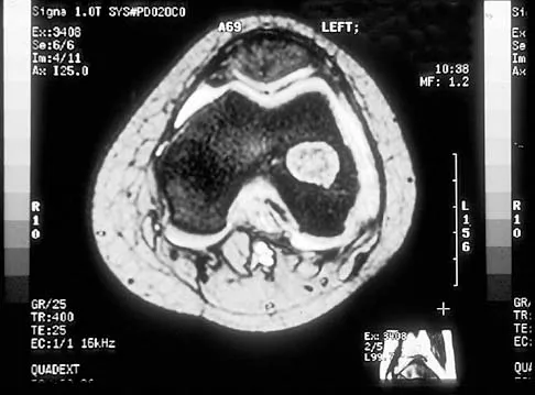
A 12-year-old boy with hemophilia A has a painless mass in his thigh. The femur is eroded anterolaterally and there is a large overlying soft tissue mass. Magnetic resonance imaging shows a 5 cm x 7 cm mass arising from the bone. The most likely diagnosis is:
Options:
- Telangiectatic osteosarcoma
- Aneurysmal bone cyst
- Infection
- Pseudotumor
- Lymphangioma
Correct Answer: Pseudotumor
Explanation:
A pseudotumor is a hemophilic subperiosteal hematoma. The pseudotumor expands by repeated bleeds and increasing osmotic pressure. There was no periosteal reaction or intralesional calcification. The bone wall itself is not expanded as in aneurysmal bone cyst. There is nothing in the physical examination or patient history to point to infection.
Question 29:
In classic hemophilia, a natural factor-VIII level of less than what percentage will lead to severe bleeding and complications:
Options:
Correct Answer: 50%
Explanation:
A surprisingly small amount of circulating factor-VIII (approximately 5%) is necessary to protect a patient from severe bleeding complications.
Question 30:
A 1-week-old female infant with arthrogryposis multiplex congenital has hips that are stiff in flexion and abduction, and her knees have a range of flexion from 20° to 40°. In addition, her right thigh has become swollen and tender. The most likely cause of this latter problem is:
Options:
- Osteomyelitis of the femur
- Septic arthritis of the hip
- Dislocation of the hip
- Deep vein thrombosis
- Fracture of the femur
Correct Answer: Fracture of the femur
Explanation:
Fracture is common in this condition because of osteopenia and the stress concentration due to joint stiffness. Osteomyelitis is uncommon in the diaphysis and much less common in this scenario than fracture. Septic arthritis of the hip is uncommon in this disease. Dislocation of the hip would not cause pain and swelling in this setting. Deep vein thrombosis is uncommon at this age.
Question 31:
Which of the following is a true statement regarding the results of surgery for a contracted joint in arthrogryposis:
Options:
- The joint range of motion can easily be doubled.
- The joint cannot be changed.
- The beginning and end of the range may change, but the total amount of motion remains about the same.
- The joint usually becomes stiffer.
- There is not an indication for such surgery.
Correct Answer: The beginning and end of the range may change, but the total amount of motion remains about the same.
Explanation:
The beginning and end of the range may change, but the total amount of motion remains about the same. The amount of the range cannot be significantly increased. The endpoint can change, but not the amount of the range. The joint does not usually become stiffer. There may be an indication for surgery to put the joints in a functional position.
Question 32:
Which of the following statements is true regarding scoliosis in cerebral palsy (C P):
Options:
- Scoliosis is most common in hemiplegic C P because of muscle imbalance.
- A thoracolumbosacral orthosis is usually successful in halting curve progression.
- Scoliotic curves over 50° are likely to worsen even if the children are mature.
- Surgery for scoliosis will prolong life expectancy.
- The surgical complication rate is lower in C P than idiopathic scoliosis.
Correct Answer: Scoliotic curves over 50° are likely to worsen even if the children are mature.
Explanation:
Curves greater than 50° usually progress. Scoliosis is most common in totally involved C P patients. Scoliosis is rare in patients with hemiplegia. Braces rarely halt curves in CP. Surgery has no proven effect on prolonging life expectancy. The complication rate is higher in CP.
Question 33:
A 12-year-old girl is referred because of a positive school scoliosis screen. She has a curve of 16° from T5 to T12, convex to the right. She incidentally also complains of mild back pain over the region of the curve several times per month. Neurologic examination is normal. Recommended treatment includes:
Options:
- Magnetic resonance imaging
- Technetium bone scintigraphy with SPEC T
- Treatment with a thoracolumbosacral orthosis
- Computed tomography of the thoracic spine
- Home exercises and re-examination in follow-up
Correct Answer: Home exercises and re-examination in follow-up
Explanation:
Home exercises and re-examination in follow-up is the most appropriate treatment in view of lack of any worrisome features. If this child had severe pain or significant night pain, then further imaging studies would be warranted. The magnetic resonance imaging is not indicated in this situation. The bone scan has a low likelihood of being positive. Bracing is not indicated for the curve or the pain. Computer tomography is unlikely to demonstrate any pathology.
Question 34:
A 10-year-old boy undergoes biopsy of a spinal cord tumor through a laminectomy of C7-T2. The most likely complication of this procedure is:
Options:
- Progressive cervicothoracic kyphosis
- Progressive cervicothoracic lordosis
- Progressive scoliosis
- Degenerative disk disease
- Progressive C 7 radiculopathy
Correct Answer: Progressive cervicothoracic kyphosis
Explanation:
The removal of posterior restraints in the young and growing flexible spine usually leads to cervicothoracic kyphosis.
Question 35:
A patient with neurofibromatosis and a 55° scoliosis may be treated with a posterior fusion and instrumentation alone in which of the following situations:
Options:
- He has a kyphosis of 75°.
- He is also undergoing multilevel laminectomy for tumor.
- He has a prior pseudarthrosis.
- He has a kyphosis of 35°.
- He has a bone age of 9.
Correct Answer: He has a kyphosis of 75°.
Explanation:
He has a kyphosis of 35°. This degree of kyphosis increases the risk of pseudarthrosis with posterior fusion alone. The laminectomy increases the risk of pseudarthrosis. Anterior fusion should be added when there is a history of pseudarthrosis. A 9-year-old boy has a high risk of crankshift phenomenon with posterior fusion alone.
Question 36:
Which of the following statements is true regarding school screening for scoliosis:
Options:
- The American Academy of Orthopaedic Surgeons (AAOS) no longer recommends it.
- The AAOS recommends screening each year.
- The AAOS recommends screening boys and girls at age 9.
- The AAOS recommends screening boys and girls at age 11.
- The AAOS recommends screening only boys at age 16.
Correct Answer: The AAOS recommends screening each year.
Explanation:
All children should be screened at age 11. The AAOS still recommends school screening for scoliosis. The AAOS recognizes that yearly screening is counterproductive. Screening at age 9 is too early.. Screening at age 16 is too late.
Question 37:
Treatment of a patient with lumbar level myelomeningocele who has a vertical talus should consist of:
Options:
- Observation only
- Talectomy
- Achilles tenotomy
- Open reduction of the vertical talus
- Triple arthrodesis in a reduced position
Correct Answer: Open reduction of the vertical talus
Explanation:
Open reduction of the vertical talus will most likely prevent problems. With observation only, the patient is likely to stand or walk and develop pressure problems. Talectomy will not produce the most usable foot. Achilles tenotomy will not produce significant correction by itself. Triple arthrodesis will concentrate stress and lead to ulcers.
Question 38:
What percentage of the human genome represents the actual genes:
Options:
Correct Answer: 50%
Explanation:
The percentage of the genome that represents the sequence of our genes is approximately 5%. The rest of the genome codes are for initiator and termination sequences, maintenance functions, and unknown functions.
Question 39:
In studying a newly recognized disorder using a large population of affected individuals, geneticists discover that although the disorder often affects siblings, it is rarely found in any of their ancestors. This disorder most closely follows which pattern of inheritance:
Options:
- Autosomal dominant
- Autosomal recessive
- Sex-linked
- Multifactorial
- Anticipation
Correct Answer: Autosomal recessive
Explanation:
Autosomal recessive conditions classically show â horizontalâ inheritance. Ancestors do not display the gene because they would likely have only one copy of the mutant allele. Only when two carriers reproduce is the phenotype manifest in approximately onefourth of their offspring. Autosomal dominant inheritance is characterized by vertical transmission. Many generations manifest the trait because it takes only a single copy of a mutant allele to display the phenotype. Sex-linked conditions are often traced back in a family. Normally the males are affected and the females are carriers. Multifactorial conditions are thought to result from the combination of different genes. Although the risk of recurrence in kindred is somewhat greater than the population as a whole, it is still quite low (only a few percent). It is rare for siblings to be affected. Anticipation refers to the phenomenon in which successive generations are likely to display more severe forms of a given disorder. Myotonic dystrophy is a classic example of this phenomenon.
Question 40:
Diseases caused by enzyme deficiency are commonly inherited by which of the following patterns:
Options:
- Autosomal dominant
- Autosomal recessive
- X-linked dominant
- Multifactorial
- Non-mendelian
Correct Answer: Autosomal recessive
Explanation:
Two copies of a mutant allele are required to reduce enzyme function to levels that cause clinical impairment. Enzyme defects are rarely inherited by an autosomal dominant pattern because even half of the normal activity of most enzymes is adequate to maintain normal function. Enzyme defects are rarely inherited in an X-linked dominant pattern because one copy of a mutant allele is usually sufficient. Multifactorial inheritance refers to the interaction of multiple, or different genes, to produce a disorder. Enzyme deficiencies are typically the result of a defect in a single gene. Because enzymes are typically coded by a single gene, they follow mendelian patterns.
Question 41:
Morquio syndrome is caused by a deficiency in:
Options:
- Alpha-L-iduronidase
- Galactose-6-sulfatase
- Beta-glucuronidase
- Fibroblast growth factor receptor protein
- Sulfate transport protein
Correct Answer: Galactose-6-sulfatase
Explanation:
Morquio syndrome is a member of the family of mucopolysaccharidoses. Morquio syndrome is a deficiency in the enzyme galactose-6-sulfatase. A deficiency in galactose-6-sulfatase results in increased urinary excretion of keratosulfate. Alpha-L-iduronidase is deficient in Hurler syndrome. Beta-glucuronidase is deficient in some rare mucopolysaccharidoses. Fibroblast growth factor receptor protein is deficient in achondroplasia. Sulfate transport protein is deficient in diastrophic dysplasia.
Question 42:
Polymerase chain reaction (PC R) is best characterized by which of the following descriptions:
Options:
- Use of enzymes to link chains of deoxyribonucleic acid (DNA) together
- Use of viral vectors to insert new DNA into a cell
- Denaturing and reannealing DNA multiple times with known primers
- Use of high temperatures to create ultra-high molecular weight polyethylene
- The process by which a cell-surface receptor turns on the transcription process
Correct Answer: Denaturing and reannealing DNA multiple times with known primers
Explanation:
Polymerase chain reaction refers to denaturing DNA, isolating a segment of interest with known primers, and reannealing the strands multiple times to produce exponential copies of a segment.
Question 43:
Pleiotropy is demonstrated by which of the following examples:
Options:
- Patients with osteogenesis imperfecta differ in the number of fractures they have received.
- Patients with hemophilia A have different target joints.
- Hurler syndrome is usually not present in prior generations of an affected patient.
- Some patients with Marfan syndrome have scoliosis or pectus carinatum, while other patients with Marfan syndrome do not.
- Patients with Ollier disease often have more involvement on one side of the body.
Correct Answer: Patients with osteogenesis imperfecta differ in the number of fractures they have received.
Explanation:
The term pleiotropy refers to a disease taking different shapes in various patients. Variation in the severity of a given problem is better termed "variable expressivity." Target joints are not genetically determined. Hurler syndrome usually not being present in prior generations of an affected patient is an example of autosomal recessive inheritance. The term pleiotropy refers to a disease taking different shapes in different subjects, whereas the cause of patients with Ollier disease having more involvement on one side of the body is unknown.
Question 44:
Which of the following is the most common concern regarding anesthesia for a patient with juvenile rheumatoid arthritis:
Options:
- Basilar invagination
- Rotatory subluxation of C1-C2
- Subaxial subluxation
- Small, stiff jaw
- C ervical stenosis
Correct Answer: Small, stiff jaw
Explanation:
Stiffness and mandibular hypoplasia are fairly common in juvenile rheumatoid arthritis (JRA) due to inflammation of the temporomandibular joint that affects the growth plates of the mandibles. Basilar invagination is rare in JRA. Rotatory subluxation of C 1-C 2 is rare in JRA. Subaxial subluxation is rare in JRA. Cervical stenosis is not a clinical problem in JRA.
Question 45:
A 4-year-old boy is brought to a clinic because he has been fussy, febrile, and unable to bend over for the past 4 days. In the office, his temperature is 38.2° C and his neurologic examination is normal. His lumbar lordosis is flattened and he resists flexion or extension. He has normal range of hip motion. Plain films of the lumbar spine are normal. The next imaging study should be:
Options:
- Magnetic resonance imaging of the spine
- Hip arthrogram
- Spinal ultrasound
- C omputed tomograms of the lumbar spine
- Indium labeled white blood cell scan
Correct Answer: Magnetic resonance imaging of the spine
Explanation:
Magnetic resonance imaging should be the next step to rule out pyogenic spondylitis. Ultrasound has not been proven effective in evaluation of anterior spinal pathology. Computed tomograms do not have a greater sensitivity than plain films in early diagnosis of infection. An indium labeled scan may yield diagnostic information but would not be the preferred test because of the time needed and inability to provide other diagnostic information. Hip arthrogram would not be the next step because the hip range of motion is normal. Even if hip pathology were suspected, the next step would be a plain film and an ultrasound.
Question 46:
A 6-year-old child suffers a displaced fracture of the distal humerus in the supracondylar region. The surgeon decides to reduce and pin the fracture. Which of the following risks increases if the procedure is delayed more than 8 hours?
Options:
- Brachial artery damage
- Median nerve palsy
- Radial nerve palsy
- Need for an open reduction
- No risks increase
Correct Answer: No risks increase
Explanation:
A retrospective comparison study has shown no increase of risks in delayed treatment of supracondylar fractures as long as the neurovascular examination is within normal limits.
Question 47:
Which of the following statements is true regarding the growth plates around the ankle:
Options:
- The distal fibula grows more than the distal tibia.
- The distal tibia grows more than the distal fibula.
- The anterolateral portion of the tibial physis ceases growing first.
- The two physes should be at an even level.
- The two growth plates are part of a common physis.
Correct Answer: The distal fibula grows more than the distal tibia.
Explanation:
The distal tibia grows more than the distal fibula. The anterolateral portion of the tibial physis ceases growing last, thus explaining the phenomenon of the Tillaux fracture. The physis of the distal fibula is always located more distally than the distal tibia. The two physes are not conjoined.
Question 48:
Which of the following is the most common final attribution of back pain in children and adolescents after all appropriate diagnostic studies are performed:
Options:
- Spondylolysis
- Osteoid osteoma
- Infection
- Herniated nucleus pulposus
- No identifiable cause
Correct Answer: No identifiable cause
Explanation:
The majority of children and adolescents do not have an identifiable cause of back pain after all appropriate tests are performed. Many times the diagnosis is "musculo-ligamentous strain." the most common identified cause is spondylolysis.
Question 49:
Which of the following is the most definitive means of making a diagnosis of active skeletal tuberculosis:
Options:
- Positive tuberculin tine test
- Negative tuberculin tine test
- Positive culture and histological exam
- Magnetic resonance imaging
- Enzyme linked immunosorbent assay (ELISA) test
Correct Answer: Positive culture and histological exam
Explanation:
An early histology confirmed later by a culture is considered the definitive means of diagnosis for active skeletal tuberculsosis. The tuberculin tine tests do not indicate active disease, only exposure. Magnetic resonance imaging is not specific for a particular infectious organism. The enzyme linked immunosorbent assay (ELISA) test is used to diagnose Lyme disease.
Question 50:
The most common region of the spine affected by tuberculosis is:
Options:
- Cervical
- Upper thoracic
- Lower thoracic-upper lumbar
- Lower lumbar
- Cacral
Correct Answer: Lower thoracic-upper lumbar
Explanation:
The lower thoracic-upper lumbar spine is most commonly affected by tuberculosis. Multiple vertebrae are often involved in tuberculosis spondylitis.