Non-Ossifying Fibroma vs. Metastasis: A Diagnostic Dilemma in Oncology Patients
Key Takeaway
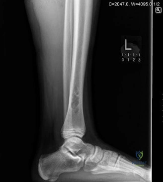
Differentiating Non-Ossifying Fibroma (NOF) from metastatic lesions in oncology patients is crucial. NOF typically presents with a well-defined, eccentric lytic lesion, often multiloculated with a sclerotic rim on radiographs and CT. MRI shows peripheral enhancement and less surrounding marrow edema, aiding distinction from aggressive metastasis.
You are reviewing this 65-year-old patient who presents with vague distal thigh pain. A lateral radiograph of the distal femur is shown. Given the patient’s history of malignancy, what is your systematic approach to assessing this lesion, and how would you utilize the Mirels' criteria in your clinical decision-making?
Candidate: I would start by assessing the patient's general oncological status and stability. I would use Mirels' scoring to evaluate the risk of fracture, looking at the site, lesion type, size, and pain levels. If the score is 8 or above, I would consider prophylactic fixation. For the lesion itself, I need a biopsy to rule out metastasis, as a lytic lesion in this age group is suspicious regardless of the radiographic appearance.
Candidates often fail to mention the need for systemic staging (CT CAP, PET/Bone scan) or immediately jump to recommending surgery without mentioning the necessity of histological confirmation. They may also list Mirels' criteria variables inaccurately or fail to mention that a biopsy tract must be planned to allow for future definitive excision.
A high-scoring candidate will categorize the response: 1. Systemic Workup: Full history, physical exam, CT Chest/Abdomen/Pelvis, and isotope bone scan to check for disseminated disease. 2. Radiographic Analysis: Evaluate the lesion using the Lodwick classification; note the cortical involvement and the presence of a pathological fracture. 3. Mirels' Criteria Application: Systematically calculate the score (Site, Nature, Size, Pain). A score ≥ 8 is a standard indication for prophylactic fixation, but clinical judgment (e.g., patient prognosis) is paramount. 4. Oncological Principles: Emphasize that biopsy is mandatory, specifically the biopsy tract planning (biopsy in the line of the future surgical incision) to avoid contaminating healthy tissue planes.
The biopsy returns a diagnosis of metastatic adenocarcinoma. You decide to proceed with intralesional curettage and stabilization. How do you decide between a retrograde intramedullary nail and a lateral locked plating system for this patient?
Candidate: The choice depends on the extent of the disease. A retrograde nail is good because it addresses the whole femur, but it violates the knee. A plate is safer if I want to avoid knee issues, but it might not provide as much rotational stability as a nail.
Ignoring the potential for "medullary seeding" of tumor cells with an IM nail or failing to mention the biomechanical advantage of PMMA cement as part of the construct in either scenario.
The candidate should contrast the two: Retrograde Nail: Offers load-sharing stability for the entire diaphysis, which is advantageous if there is multi-focal disease. However, it risks articular seeding and requires an intra-articular entry point. Lateral Locked Plate: Avoids the knee joint and allows for direct visualization of the curettage and cementation. It is superior for distal metaphyseal lesions where distal screw purchase is critical. Crucial add-on: Both should be combined with intralesional PMMA cementation, which acts as a load-transfer mechanism and provides local oncological control via thermal necrosis.
