Solving Oncology Cases: Metastatic Bone Diagnosis
Key Takeaway
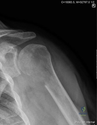
This article provides essential research regarding Solving Oncology Cases: Metastatic Bone Diagnosis. For a 66-year-old female with shoulder pain, weight loss, and destructive imaging features, an impending fracture through a metastatic lesion in the proximal humerus is the most likely diagnosis. In **oncology cases metastatic bone** disease is a common reason for destructive bone lesions in adults. Primary cancer sites frequently metastasizing to bone include breast, lung, kidney, prostate, and thyroid.
A 68-year-old male presents with acute thigh pain and an inability to bear weight after a minor fall. He has a known history of prostate cancer. Physical exam reveals a shortened, externally rotated leg with gross instability in the mid-thigh. Radiographs are presented below. What is your immediate assessment and initial management plan?
Candidate: This looks like a pathological subtrochanteric fracture secondary to metastatic prostate cancer. I would secure the limb in a traction splint, manage pain, obtain bloods including PSA and calcium levels, and order a CT scan of the chest, abdomen, and pelvis to look for the primary. Once stabilized, I would plan for surgical fixation with an intramedullary nail.
Candidates often jump straight to surgical technique without addressing systemic workup (e.g., ruling out myeloma or hypercalcemia) or the necessity of a formal oncology MDT. Failure to mention Mirels' criteria for the contralateral side or the need for a biopsy prior to definitive fixation is a significant omission.
A structured response is essential: 1) Clinical suspicion: Pathological fracture secondary to metastatic disease; prioritize ABCs and pain management. 2) Systemic Workup: Full bloods (Ca, PSA, SPEP to rule out myeloma), and staging (CT TAP). 3) Mechanical Assessment: Calculate Mirels' score to assess for prophylactic needs in the contralateral femur. 4) Pre-operative Planning: Tissue diagnosis (biopsy) is mandatory prior to surgery. 5) Surgical Strategy: Long cephalomedullary nail to bypass the lesion + adjuvant PMMA cement, highlighting the importance of the oncology MDT for post-operative radiation and systemic therapy.
You have decided to proceed with surgery. Given the extensive lytic destruction in the subtrochanteric region, what technical considerations will you incorporate into your surgical approach to ensure durability and local tumor control?
Candidate: I would use a long cephalomedullary nail to ensure I can bridge the entire tumor and the fracture. I would also perform a limited open reduction to ensure anatomic alignment. I plan to use PMMA cement to augment the construct and potentially help with local tumor control through thermal necrosis.
Forgetting to specify the extent of the nail (it must extend at least two cortical diameters beyond the distal margin of the tumor). Candidates also often neglect to mention the risk of iatrogenic fracture during reaming in compromised bone or the risk of tumor seeding if reaming is done too aggressively.
The perfect answer emphasizes: 1. Construct Length: The nail must span the entire lesion, extending at least two cortical diameters beyond the distal extent of the tumor. 2. Mechanical Augmentation: Use of PMMA cement to fill the lytic defect and the fracture gap, providing structural load-sharing. 3. Technical Safety: Careful, low-pressure reaming to avoid further fracture propagation and preventing tumor cell dissemination. 4. Post-operative Adjuvant therapy: Emphasizing that surgery is only half the battle; timely referral for radiotherapy is critical for preventing local recurrence.
