
العربية
Comprehensive Master Guide · Medically Reviewed

Unraveling Metastatic Bone Disease: Key Orthopedic Case Insights

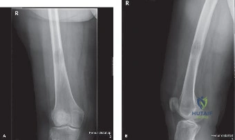
A 60-year-old female with a history of breast cancer treated 5 years ago with mastectomy, chemotherapy, and radiation is seeing you for knee pain. X-rays of th…

15 Detailed Chapters
28 min read
Updated: Jun 2026
Dr. Mohammed Hutaif Clinic
Medically Reviewed by
Prof. Dr. Mohammed Hutaif Clinic
Verified Content Expert Reviewed

Quick Medical Answer

In this comprehensive guide, we discuss everything you need to know about Unraveling Metastatic Bone Disease: Key Orthopedic Case Insights. Metastatic bone disease is when cancer cells spread from their primary site to the bone. Initial workup for a suspicious bone lesion, especially with a cancer history, includes a bone scan and CT chest/abdomen/pelvis. Biopsy is crucial to confirm metastatic bone disease, identify the primary source, and determine receptor expression, which guides specific, targeted oncologic treatments.

Illustration of metastatic bone disease - Dr. Mohammed Hutaif
🎓

FRCS Masterclass: Clinical Viva

Interactive Examiner Scenario • Test your knowledge before revealing the answers.

👨‍⚕️ Examiner Scenario

You are in the acute trauma intake. A 62-year-old female is brought in with severe left thigh pain after a simple trip. She has a known history of Stage IV breast cancer. Radiographs show a displaced subtrochanteric fracture with an underlying lytic lesion involving over 50% of the cortex. How do you approach this case?

Clinical Image
Figure 1: Plain radiograph of the left femur

Candidate: I would stabilize the patient's airway, breathing, and circulation, ensuring adequate analgesia. Given the clinical and radiological findings, I suspect a pathological fracture. I would order an urgent CT of the femur for surgical planning, bloods including a bone profile and calcium, and a systemic staging workup if not already done. I would discuss the case with the Orthopedic Oncology team for definitive fixation.

❌ Common Pitfall (Poor Answer)

Candidates often jump immediately to "I will perform an IM nail." They fail to demonstrate the "Oncological" mindset—missing the mandatory screening for hypercalcemia, failing to mention pre-operative systemic staging, or forgetting to mention that the biopsy must be performed in a way that doesn't compromise future limb salvage.

⭐ The Gold Standard (Perfect Answer)

A high-scoring answer follows a structured approach: 1) **Stabilization & Analgesia** (including hypercalcemia management); 2) **Diagnosis** (Confirming it's pathological vs. incidental trauma via CT and labs); 3) **Oncological Staging** (PET-CT/Bone scan); 4) **Decision Making** (Utilizing the Mirels' score to confirm risk and Tokuhashi/prognostic scores to determine if we are performing palliative fixation vs. reconstruction); 5) **Surgical Planning** (Long cephalomedullary nail, cement augmentation, and intraoperative biopsy).

👨‍⚕️ Examiner Scenario

You have decided to proceed with surgery. Why is a long cephalomedullary nail (CMN) preferred over a plate for this specific subtrochanteric pathological fracture, and what is the role of PMMA cement in this procedure?

Candidate: A long cephalomedullary nail is preferred because it is a load-sharing device. In metastatic disease, bone healing is unreliable, so a device that spans the entire femoral canal and allows for immediate weight-bearing is crucial. PMMA cement is used to fill the lytic void, increasing mechanical stability and providing a local anti-tumor effect through the exothermic reaction.

❌ Common Pitfall (Poor Answer)

Failing to emphasize that the nail must *bypass* the entire lesion and extend into healthy distal bone. Candidates often forget the "prophylactic" nature of the nail—treating the entire femur because the whole bone is a "field" for potential metastatic progression.

⭐ The Gold Standard (Perfect Answer)

Focus on the "Load-Sharing" principle. Explain that the nail functions as an internal splint. PMMA cement is essential for "void filling" in large lytic lesions where internal fixation would otherwise have poor purchase. Mention that the exothermic reaction aids local control, and clarify that the nail must span the entire length to protect against future "skip" lesions in the ipsilateral femur.

Detailed Chapters & Topics

Dive deeper into specialized chapters regarding orthopedic-oncology-cases-metastatic-bone-disease1

15 Chapters
01
Chapter 1 49 min

Orthopedic Board Review: Musculoskeletal Tumors, Ankylosing Spondylitis, Synovial Chondromatosis | Part 15

Prepare for your orthopedic board exam with Part 15 of our review series. Master musculoskeletal tumors, atypical lipom…

02
Chapter 2 49 min

ABOS Orthopedic Board Review: Bone Neoplasms, Chondromas, & Sarcoma Metastasis | Part 9

Ace your ABOS Orthopedic Board Review with Part 9! Explore bone neoplasms, chondromas, and sarcoma metastasis. Test you…

03
Chapter 3 48 min

Orthopedic Oncology Board Review: Soft Tissue Sarcomas, Chondroblastoma & Fibromatosis | Part 17

Ace your Orthopedic Oncology Board Review with Part 17! Test your knowledge on soft tissue sarcomas, chondroblastoma, a…

04
Chapter 4 102 min

ABOS Orthopaedic Oncology Review: Lipomas, Osteosarcomas, HME, Atypical Lipomas | Part 14

Ace your ABOS exam with our Orthopaedic Oncology Review! Part 14 covers lipomas, osteosarcomas, HME, and atypical lipom…

05
Chapter 5 21 min

Orthopedic Board Review: Bone Tumors & Osteochondromas

Test your knowledge on bone tumors, focusing on osteochondromas, multiple hereditary exostoses, malignant transformatio…

06
Chapter 6 45 min

Orthopaedic Board Review: Synovial Chondromatosis, Charcot Joint, Fibrous Dysplasia, Bone Metastases | Part 21

Master Orthopaedic Board Review Part 21! Practice high-yield MCQs on synovial chondromatosis, Charcot joint, fibrous dy…

07
Chapter 7 16 min

Humeral Metastasis Management: An Intraoperative Masterclass in Resection & Reconstruction

Master the surgical management of humeral metastases. Explore expert techniques for bone resection, functional reconstr…

08
Chapter 8 22 min

Optimizing the Management of Difficult Metastatic Lesions

A 47 year old female with metastatic thyroid cancer has progressive left hip pain. X-rays are shown in Figure 8–34A – D…

09
Chapter 9 21 min

Mastering Renal Cell Carcinoma Skeletal Metastasis Cases

A 71-year-old male with back pain has been referred to your office. His primary care physician ordered x-rays of the th…

10
Chapter 10 27 min

PVNS Diagnosis: Clinical and Radiographic Clues You Can't Miss

A 30-year-old female presents to your office with a chief complaint of episodic right knee pain, swelling, and stiffnes…

11
Chapter 11 22 min

Solving Oncology Cases: Metastatic Bone Diagnosis

A left-hand-dominant, 66-year-old female is sent to your office for shoulder pain, possibly a rotator cuff tear. She ha…

12
Chapter 12 13 min

Non-Ossifying Fibroma vs. Metastasis: A Diagnostic Dilemma in Oncology Patients

Learn how to differentiate a benign Non-Ossifying Fibroma from a solitary lytic bone metastasis in older oncology patie…

13
Chapter 13 26 min

Medical Myeloma Treatment: Uncover Key Prognosis & Survival Factors

A 61-year-old female with progressive functional right hip pain is brought to your office by her daughter. She has rece…

14
Chapter 14 15 min

Masterclass: Extra-Articular Resection and Reconstruction of Supra-acetabular Metastatic Lesions

Master extra-articular resection of supra-acetabular metastatic lesions. Learn how to preserve the native hip joint and…

15
Chapter 15 11 min

Operative Management of Metastatic Carcinoma in Orthopaedics

Master the operative management of metastatic bone disease. Discover why aggressive orthopaedic care and proper staging…

Dr. Mohammed Hutaif Clinic
Medically Verified Content by
Prof. Dr. Mohammed Hutaif Clinic
Consultant Orthopedic & Spine Surgeon
Guide Overview