High-Energy Diaphyseal Tibia Fractures: Epidemiology, Anatomy & Biomechanics Guide
Key Takeaway
High-energy diaphyseal tibia fractures result from severe trauma, often presenting with comminution, soft tissue damage, and high complication risks like compartment syndrome and non-union. Their complexity stems from the tibia's precarious subcutaneous location, vital neurovascular structures, and the need for meticulous management integrating anatomy, biomechanics, and soft tissue considerations to optimize outcomes.
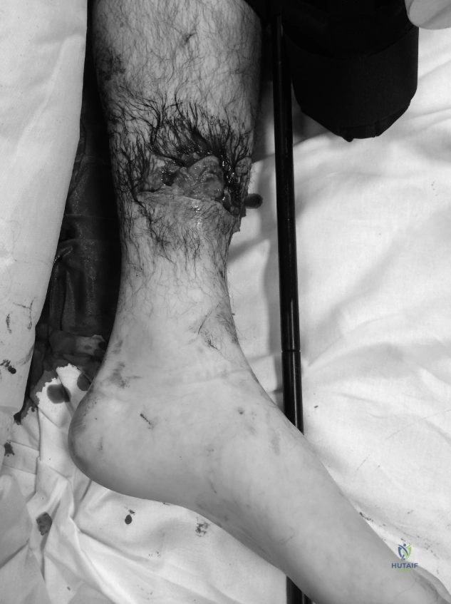
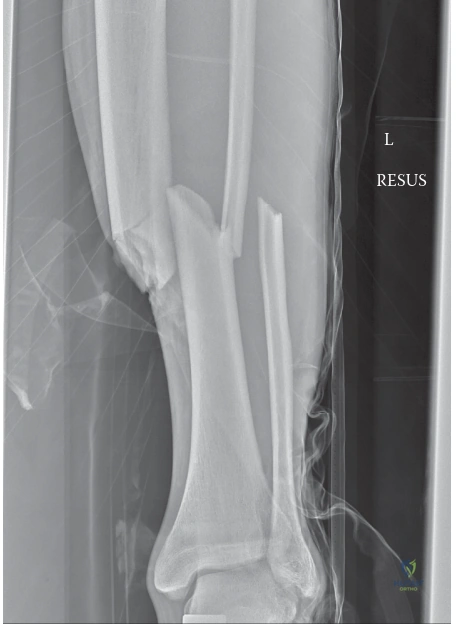
A 28-year-old male presents to the Emergency Department following a high-energy motorcycle collision. He has an obvious closed, angulated deformity of the right midshaft tibia. He complains of severe, disproportionate pain in the calf. How do you approach the initial assessment and stabilization of this limb?
Candidate: I would follow ATLS protocols to ensure the patient is stable. I would perform a neurovascular exam, check for open wounds, and keep a high index of suspicion for compartment syndrome. I would request AP and lateral radiographs of the tibia, including the knee and ankle joints, and place the limb in a splint.
Failing to explicitly mention the "four-compartment" examination of the leg or missing the need for urgent fasciotomy if the clinical diagnosis is suspected. Candidates often forget to mention imaging of the joints above and below, which is vital for detecting floating knee injuries.
I would adhere to ATLS, prioritizing systemic stability. My orthopaedic assessment includes: 1) Neurovascular status: documenting pulses, capillary refill, and nerve function. 2) Soft tissue assessment: excluding open fractures (Gustilo-Anderson) and signs of compartment syndrome (pain out of proportion, pain on passive stretch, tenseness). 3) Imaging: Radiographs of the entire tibia, including knee and ankle joints to rule out occult fractures. 4) Provisional stabilization: Splinting and, if signs of compartment syndrome are present, emergent four-compartment fasciotomy, not just waiting for pressure monitoring.
The patient has been stabilized and is deemed a candidate for definitive fixation. You are planning an intramedullary nail. Discuss the technical considerations for the entry point and the role of reaming in this specific injury pattern.
Candidate: I would choose a medial paratendinous or splitting approach to the patellar ligament for the entry point. I would ream the canal to allow for a larger nail, which improves stability. Reaming also creates bone graft material.
Neglecting to mention the "Suprapatellar" (transpatellar) entry technique, which is now state-of-the-art for better alignment control, or failing to acknowledge the risk of reaming in contaminated open fractures.
Entry point is crucial: the medial paratendinous or split-tendon approach is standard, but the suprapatellar approach offers improved sagittal plane control and is excellent for obese patients or those with high-riding patellae. Regarding reaming: I prefer reamed nails for closed fractures as they maximize contact area, allow for larger, stiffer nails, and provide autologous bone graft (osteoconductive/inductive). However, in severely contaminated open fractures, I would consider an unreamed nail to minimize thermal necrosis and endosteal blood supply disruption, thereby reducing infection risk.
Intraoperatively, you achieve reduction, but there is significant comminution in the midshaft. Your fluoroscopy shows a tendency for the nail to translate into the wide medullary canal, causing a malalignment. How do you resolve this?
Candidate: I would try to adjust the reduction using clamps or reposition the leg. If it's still unstable, I might consider using a larger nail or a plate instead.
Abandoning the nail for a plate in a midshaft fracture is often unnecessary and carries higher risk. The candidate fails to demonstrate knowledge of "Poller screws" (blocking screws).
I would maintain the IM nail as the gold standard for midshaft fixation. To control the deformity, I would utilize blocking screws (Poller screws). By placing these screws adjacent to the nail in the metadiaphyseal region, I can effectively "block" the nail from migrating into the widened canal, thereby correcting the alignment. This is a powerful, minimal-access technique that avoids the need for open plating.
