Complex Pilon Fracture (AO/OTA C3): Detailed Case Study & Diagnostic Approach
Key Takeaway
Diagnosing a complex AO/OTA C3 pilon fracture requires clinical assessment for soft tissue and neurovascular status. Crucially, CT scans with 3D reconstructions precisely map articular fragmentation and metaphyseal defects. This detailed imaging ensures accurate classification and informs comprehensive pre-operative surgical planning.
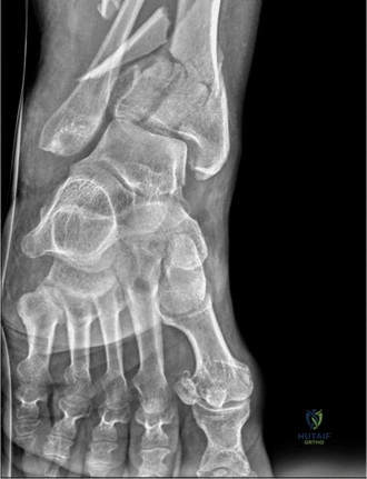
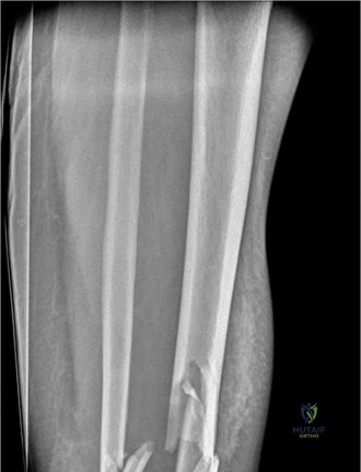
You are on-call in the Major Trauma Centre. A 45-year-old construction worker presents following a high-energy MVC. He has a deformed, swollen, and tense right ankle. Radiographs confirm a closed, multi-fragmentary intra-articular fracture of the distal tibia (AO/OTA 43-C3) with associated fibular comminution. The skin is tense, shiny, and shows early hemorrhagic fracture blisters.
How do you approach the immediate management of this patient in the Emergency Department?
Candidate: I would immediately assess his neurovascular status and ensure the limb is splinted. I'd perform a primary survey according to ATLS protocols. Given the high-energy nature and soft tissue compromise (Tscherne Grade III), I would avoid open reduction at this stage. I would arrange for a CT scan to map the fracture, place him in a temporary spanning external fixator, and elevate the limb to optimize the soft tissues before planning definitive surgery.
Candidates often fail to explicitly mention neurovascular monitoring, gloss over the severity of the soft tissue injury, or fail to articulate the "Span, Scan, and Plan" protocol. Mentioning "plaster casting" for a tense, swelling limb is a major safety error that reflects a misunderstanding of compartment syndrome risk.
I would approach this by: 1. Assessment: Confirm neurovascular integrity and monitor compartment pressures, noting the Tscherne III status. 2. Stabilization: Apply a temporary spanning external fixator (knee-to-foot) to restore length and reduce articular tension. 3. Imaging: Utilize high-resolution CT with 2D/3D reconstructions to guide articular mapping. 4. Soft Tissue Optimization: Admit, elevate, strictly control diabetes/smoking risk factors, and monitor for compartment syndrome. Definitive surgery will be delayed until the 'wrinkle sign' returns, typically 10–21 days post-injury.
The patient has now reached the 14-day mark. The swelling has subsided, and he has a positive "wrinkle sign." You are planning for definitive internal fixation. Based on the imaging provided, how do you approach the surgical reconstruction, specifically regarding incision placement?
Candidate: I would use a dual-approach technique—an anterolateral and a posteromedial approach—to address the articular fragments. I would ensure at least 7 cm of skin bridge between these incisions to prevent necrosis. I would address the fibula first to restore length, then use a femoral distractor for articular visualization.
Failing to emphasize the mandatory skin bridge (which is the most common cause of wound breakdown) or suggesting a single approach for a comminuted 43-C3 fracture. Ignoring the role of the fibula as a lateral buttress is also a critical oversight.
My strategy is: 1. Approach: Dual approaches (Anterolateral and Posteromedial) with a minimum 7cm bridge. 2. Sequence: (a) Open reduction of the fibula to restore the lateral column/length. (b) Use a femoral distractor to gain joint visualization. (c) Reduce articular fragments 'posterior to anterior' (Volkmann to Chaput). 3. Fixation: Secure the articular block to the metaphysis/diaphysis using locking plates. 4. Bone Defect: Use bone graft/substitutes to fill the metaphyseal void to prevent secondary subsidence of the articular reconstruction.
