Mastering the Chiari Medial Displacement Osteotomy: An Intraoperative Masterclass for Acetabular Dysplasia
Key Takeaway
This intraoperative masterclass details the Chiari medial displacement osteotomy for painful, unstable hip dysplasia. We cover comprehensive anatomy, meticulous preoperative planning, and granular, real-time surgical execution. Learn precise osteotomy techniques, neurovascular protection, and crucial pearls for optimal femoral head coverage and stability. Understand postoperative rehabilitation and complication management to achieve stable, pain-free hip function.
Introduction and Epidemiology
The Chiari medial displacement osteotomy is a foundational salvage procedure in the surgical armamentarium for complex hip dysplasia. Originally described by Karl Chiari in 1955, this single pericapsular osteotomy of the ilium is designed to address painful, unstable hips where concentric, congruous reduction of the femoral head into the true acetabulum is impossible. Unlike redirectional osteotomies (such as the Bernese periacetabular osteotomy, Salter innominate osteotomy, or triple pelvic osteotomy), which reorient the native hyaline cartilage to cover the femoral head, the Chiari osteotomy relies on the metaplastic transformation of the interposed hip capsule into fibrocartilage beneath a newly created bony iliac shelf.

The primary goal of the procedure is to provide immediate structural stability by medializing the hip joint center, thereby improving lateral and posterior coverage of the subluxated femoral head. This medialization fundamentally alters the biomechanical environment of the hip, reducing joint reactive forces and providing a stable fulcrum for the abductor musculature.
Epidemiologically, the demand for salvage osteotomies arises from late-diagnosed or refractory developmental dysplasia of the hip (DDH), severe Legg-Calvé-Perthes disease resulting in coxa magna and asphericity, and neuromuscular hip dysplasia, particularly in patients with spastic cerebral palsy. In the spastic hip, the posterior and lateral acetabular walls are frequently deficient due to the chronic deforming forces of adductor and flexor spasticity. The Chiari osteotomy addresses these specific deficiencies by creating a robust mechanical block to superior and lateral migration. While the advent of advanced total hip arthroplasty (THA) has reduced the frequency of this procedure in older adults, it remains an indispensable joint-preserving option for adolescents and young adults seeking to delay arthroplasty by a decade or more.

Surgical Anatomy and Biomechanics
A profound understanding of periacetabular anatomy and hip biomechanics is requisite for the safe execution and functional success of the Chiari osteotomy. The procedure fundamentally alters both the osseous architecture and the muscular force vectors surrounding the hip joint.
Pelvic Osteology and Neurovascular Relations
The osteotomy is performed through the ilium, immediately superior to the capsular reflection of the hip joint and inferior to the anterior inferior iliac spine (AIIS). The trajectory of the osteotomy extends from the anterior margin of the ilium, between the AIIS and the superior acetabular rim, directing posteriorly toward the greater sciatic notch.

Critical neurovascular structures are at risk during this trajectory. The sciatic nerve and the superior gluteal artery and nerve exit the pelvis through the greater sciatic notch. Over-penetration of instruments or retractors into the notch can result in catastrophic iatrogenic injury. Anteriorly, the femoral nerve and external iliac vessels lie medial to the iliopsoas muscle and must be protected during the medial dissection and placement of anterior retractors. The safe zone for the osteotomy requires meticulous subperiosteal dissection along the inner and outer tables of the ilium, ensuring that retractors (such as blunt sciatic retractors) are securely positioned within the periosteal sleeve.
Biomechanical Principles of Medialization
The biomechanical rationale of the Chiari osteotomy is rooted in Pauwels' principles of hip mechanics. The hip joint acts as a first-class lever. The fulcrum is the femoral head, the resistance is the body weight acting through the center of gravity, and the effort is generated by the abductor musculature.

By medializing the distal pelvic fragment (which contains the acetabulum and the femoral head), the lever arm of the body weight is significantly shortened. This reduction in the body weight lever arm proportionally decreases the force required by the abductor muscles to maintain pelvic level during the single-leg stance phase of gait. Consequently, the overall joint reactive force across the hip is profoundly diminished. This mechanical offloading is critical for pain relief and the longevity of the fibrocartilaginous interposition arthroplasty.
However, the medial displacement of the hip joint center is coupled with the relative lateralization of the proximal iliac fragment, which forms the new shelf. This lateralization shortens the resting length of the gluteus medius and minimus muscles, decreasing their contractile tension and effective moment arm. This obligatory abductor weakening frequently results in a postoperative Trendelenburg gait. To mitigate this, surgeons must carefully plan the angle of the osteotomy. Decreasing the obliquity of the supra-acetabular cut can minimize the detrimental effect on the abductor lever arm. Additionally, concomitant distal and lateral advancement of the greater trochanter may be indicated to restore the resting length and tension of the abductor complex.
Indications and Contraindications
The decision to perform a Chiari osteotomy requires careful patient selection. It is strictly designated as a salvage procedure, utilized when the articular cartilage is no longer viable for a reconstructive redirectional osteotomy, or when the femoral head is so aspherical (coxa magna or plana) that it cannot be congruously seated within the native acetabulum.

| Clinical Scenario | Operative Indications for Chiari Osteotomy | Non-Operative Management or Alternative Surgical Contraindications |
|---|---|---|
| Femoral Head Morphology | Severe asphericity (coxa magna, coxa plana) precluding congruous reduction. | Spherical head capable of congruous reduction (Indicates PAO or Triple Osteotomy). |
| Joint Congruency | Incongruous joint on abduction/internal rotation views; painful subluxation. | Concentric reduction achievable with positioning (Indicates redirectional osteotomy). |
| Osteoarthritis Grade | Tönnis Grade 1 or 2 (mild to moderate arthrosis) with preserved joint space. | Tönnis Grade 3 (severe arthrosis, bone-on-bone) (Indicates THA or Arthrodesis). |
| Patient Age | Adolescents and young adults (typically under 45 years of age). | Older adults (>45 years) with degenerative changes (Indicates THA). |
| Neuromuscular Status | Spastic hip dysplasia with lateral/posterior unroofing, provided bone stock is adequate. | Severe osteopenia or extremely thin ilium (e.g., myelomeningocele) preventing shelf formation. |
| Hip Mobility | Functional preoperative range of motion (minimum 60° flexion, 15° abduction). | Stiff hip or fibrous ankylosis (Osteotomy will fail without motion to stimulate metaplasia). |
| Femoral Head Station | Subluxated but maintainable at the level of the true acetabulum. | High dislocation (Crowe IV) where the ilium is too thin to provide coverage. |
Pre Operative Planning and Patient Positioning
Meticulous preoperative planning is essential to determine the exact level, angle, and required displacement of the osteotomy. The goal is to create a shelf that adequately covers the lateral and posterior aspects of the femoral head without compromising the structural integrity of the pelvic ring.
Radiographic Evaluation
Standard radiographic evaluation includes an anteroposterior (AP) view of the pelvis, a false profile view to assess anterior coverage, and functional views (abduction and internal rotation). The functional views are critical; if the hip reduces concentrically, a Chiari osteotomy is contraindicated, and a redirectional osteotomy should be pursued.

Advanced imaging with a computed tomography (CT) scan, including 3D reconstructions, is highly recommended. CT allows for the precise evaluation of periacetabular bone stock. In patients with severe dysplasia or neuromuscular conditions, the ilium immediately superior to the acetabulum may be perilously thin. If the bone stock is insufficient to create a robust shelf, the procedure may be contraindicated, or the surgeon must plan for substantial structural bone grafting.
Templating should establish the level of the osteotomy. The cut must be made immediately superior to the reflected head of the rectus femoris and the joint capsule. If the osteotomy is too high, the interposed capsule will stretch, failing to provide a rigid mechanical block, leading to persistent subluxation. If the osteotomy is too low, it risks violating the intra-articular space, damaging the remaining articular cartilage and accelerating arthrosis. The planned angle of the osteotomy in the coronal plane should be 10 to 15 degrees cephalad (upsloping). This angulation utilizes the body weight to compress the osteotomy site and prevents the distal fragment from sliding laterally.
Surgical Positioning and Setup
The patient is positioned supine on a radiolucent operating table to facilitate unimpeded intraoperative fluoroscopy. A sandbag or gel bump is placed under the ipsilateral hemipelvis to elevate the operative side by approximately 30 degrees, aiding in the lateral approach to the ilium.

The entire lower extremity is prepped and draped free to allow for dynamic manipulation of the hip during the procedure. Abduction of the leg will be required to execute the medial displacement of the distal pelvic fragment. Fluoroscopy must be positioned to easily acquire both AP and Judet views of the pelvis throughout the procedure.
Detailed Surgical Approach and Technique
The surgical execution of the Chiari osteotomy demands precision in soft tissue handling, osteotomy orientation, and controlled displacement.
Surgical Approach and Dissection
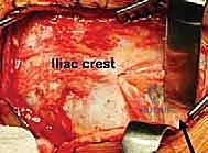
The procedure is typically performed via an anterior Smith-Petersen approach or a modified ilioinguinal approach. The incision begins along the anterior crest of the ilium, extending distally between the tensor fasciae latae and the sartorius.

The superficial plane is developed, and the lateral femoral cutaneous nerve is identified and protected. The abductor musculature (gluteus medius and minimus) is carefully elevated subperiosteally from the outer table of the ilium. This dissection proceeds posteriorly toward the greater sciatic notch. Concurrently, the iliacus muscle is elevated from the inner table of the ilium, exposing the pelvic brim and the medial aspect of the sciatic notch.
The anterior inferior iliac spine (AIIS) is identified. The straight head of the rectus femoris may be detached to improve visualization of the superior joint capsule. It is imperative that the joint capsule remains intact; the capsule is the substrate for the future fibrocartilaginous bearing surface. The superior capsular reflection is meticulously delineated.
Osteotomy Execution and Displacement
Blunt retractors (such as malleable or specific sciatic retractors) are placed subperiosteally within the greater sciatic notch to protect the sciatic nerve and superior gluteal vessels. An additional retractor is placed anteriorly to protect the femoral neurovascular bundle.

The osteotomy line is marked just superior to the capsular reflection. The cut is initiated with an oscillating saw or sharp osteotomes. Many surgeons prefer passing a Gigli saw through the sciatic notch and bringing it anteriorly to ensure a smooth, continuous cut. The osteotomy must be directed 10 to 15 degrees cephalad in the coronal plane. In the axial plane, the cut should follow the curve of the acetabulum to ensure adequate posterior coverage.
Once the osteotomy is complete, the displacement is performed. The distal fragment is displaced medially by abducting the freely draped lower extremity while applying lateral traction to the proximal iliac fragment. The distal fragment hinges on the pubic symphysis. The goal is to achieve medial displacement equal to approximately 50% of the iliac width, or until the lateral edge of the proximal iliac fragment adequately covers the subluxated femoral head and capsule. Fluoroscopy is utilized to confirm the exact degree of medialization and the coverage of the femoral head.
Fixation and Adjunctive Procedures
Once optimal displacement is achieved, the osteotomy is provisionally held with smooth K-wires. Definitive fixation is typically achieved using two or three fully threaded Steinmann pins or large-fragment cannulated screws. These are directed from the proximal iliac fragment, across the osteotomy site, and into the dense bone of the medialized distal fragment (often directed toward the posterior column or the quadrilateral plate).

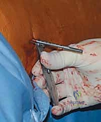
If the anterior or lateral coverage remains deficient despite adequate displacement, structural bone graft (often harvested from the anterior iliac crest) can be wedged between the proximal fragment and the capsule to augment the shelf.
Because the lateralization of the proximal fragment shortens the abductor lever arm, a concomitant distal and lateral advancement of the greater trochanter may be performed. The trochanter is osteotomized, advanced distally to restore abductor tension, and fixed with cerclage wires or screws. This adjunctive step is highly effective in reducing the incidence of postoperative Trendelenburg gait. The wound is then closed in layers over a suction drain.
Complications and Management
The Chiari osteotomy is a technically demanding procedure with a recognized complication profile. Meticulous surgical technique and adherence to anatomical safe zones are paramount.

| Complication | Incidence / Risk Factors | Prevention and Management Strategies |
|---|---|---|
| Sciatic Nerve Injury | 1-3%. Caused by retractor misplacement or over-penetration of the saw into the sciatic notch. | Prevention: Strict subperiosteal dissection; use of blunt, perfectly contoured retractors in the notch. Management: Observation for neurapraxia; AFO for foot drop. Surgical exploration if transection is suspected. |
| Intra-articular Osteotomy | Varies. Occurs when the osteotomy is placed too low, violating the articular cartilage. | Prevention: Precise fluoroscopic templating; identifying the capsular reflection. Management: If recognized intraoperatively, redirect the cut. Postoperatively, may lead to rapid joint degeneration requiring early THA. |
| Inadequate Coverage (High Cut) | Varies. Osteotomy placed too far superior to the capsule. | Prevention: Keep the cut immediately adjacent to the superior capsule. Management: The interposed capsule will stretch, failing to provide stability. May require revision shelf arthroplasty or structural bone grafting. |
| Nonunion / Delayed Union | <5%. Increased risk with inadequate fixation or poor bone stock. | Prevention: Rigid internal fixation (screws/pins); ensuring 10-15° cephalad angulation for compressive forces. Management: Prolonged non-weight bearing; revision fixation with bone grafting if symptomatic nonunion persists. |
| Severe Trendelenburg Gait | Common (up to 30-50% without trochanteric advancement). | Prevention: Concomitant greater trochanteric advancement; minimizing the cephalad angle to reduce abductor shortening. Management: Aggressive physical therapy for abductor strengthening; late trochanteric advancement if refractory. |
| Heterotopic Ossification | 5-10%. Increased risk with extensive muscle stripping or concomitant trochanteric osteotomy. | Prevention: Gentle soft tissue handling; prophylactic NSAIDs (e.g., Indomethacin) or single-dose radiation in high-risk patients. Management: Surgical excision only if mature and mechanically restricting range of motion. |
Post Operative Rehabilitation Protocols
The postoperative rehabilitation protocol must balance the need for rigid immobilization to ensure osteotomy union with the requirement for early motion to stimulate the fibrocartilaginous metaplasia of the interposed capsule.
In pediatric and adolescent populations, particularly those with spasticity, a one and one-half spica cast is often applied for 6 weeks to ensure absolute stability of the osteotomy and control deforming muscle forces.
In older adolescents and adults with reliable compliance, cast immobilization may be avoided. The patient is placed on strict non-weight bearing or toe-touch weight bearing precautions on the operative extremity for 6 to 8 weeks. During this period, continuous passive motion (CPM) or active-assisted range of motion exercises are initiated immediately. Motion is the primary catalyst for the differentiation of the highly vascularized hip capsule into a resilient, weight-bearing fibrocartilaginous surface.
Radiographic evaluation is performed at 2, 6, and 12 weeks postoperatively. Once clinical and radiographic evidence of bony consolidation at the osteotomy site is confirmed (typically around 6 to 8 weeks), progressive partial weight bearing is initiated.
Physical therapy during the weight-bearing phase heavily emphasizes abductor strengthening. Because the biomechanics of the hip have been altered, the gluteus medius and minimus must be retrained to function at their new resting lengths. Aquatic therapy is highly beneficial in the early weight-bearing phases. Full weight bearing without assistive devices is generally achieved by 3 to 4 months postoperatively, contingent upon the resolution of the Trendelenburg lurch.
Summary of Key Literature and Guidelines
The academic literature surrounding the Chiari osteotomy emphasizes its role as a durable, time-buying procedure for the dysplastic hip facing inevitable arthrosis.
Karl Chiari's original long-term follow-up studies established the viability of capsular interposition arthroplasty, demonstrating that the capsule reliably undergoes metaplasia into a tissue histologically indistinguishable from fibrocartilage when subjected to compressive loading and motion.

Subsequent robust survival analyses by authors such as Windhager, Lack, and Kotz have quantified the longevity of the procedure. In patients operated on for DDH with mild to moderate arthrosis (Tönnis grade 1 or 2), 15-year survivorship (with conversion to THA as the endpoint) frequently exceeds 75%. However, survivorship drops precipitously in patients older than 45 years at the time of surgery, or in those with advanced, bone-on-bone osteoarthritis (Tönnis grade 3).
Modern guidelines dictate that while the Bernese periacetabular osteotomy (PAO) is the gold standard for the congruous, dysplastic hip, the Chiari osteotomy remains a critical tool. It is specifically indicated in the algorithm for the incongruous, aspherical hip where reconstructive osteotomies are destined to fail. Furthermore, long-term studies indicate that a prior Chiari osteotomy does not preclude a successful subsequent total hip arthroplasty. While the medialized acetabulum and altered pelvic anatomy present technical challenges during THA (often requiring smaller, medialized cups and careful assessment of the sciatic nerve), the restoration of bone stock provided by the Chiari shelf frequently facilitates robust acetabular component fixation during the ultimate arthroplasty.
Clinical & Radiographic Imaging
You Might Also Like