Bennett's Fracture-Dislocation: Clinical Presentation, Diagnostics & Management
Key Takeaway
Bennett's fracture-dislocation is an intra-articular fracture of the first metacarpal base, with a small volar-ulnar fragment and dorsal-radial-proximal displacement of the main shaft. Diagnosis involves clinical findings (pain, instability) and confirmed by specialized X-rays (Robert's view) and CT scans for precise articular evaluation and surgical planning.
Introduction and Epidemiology
Bennett fracture dislocations represent the most common intra-articular fractures involving the base of the first metacarpal. Originally described by Irish surgeon Edward Hallaran Bennett in 1882, this injury is defined by a characteristic fracture pattern where a small volar ulnar fragment remains anatomically reduced while the larger metacarpal shaft displaces proximally, dorsally, and radially. The injury typically occurs secondary to an axial load applied to a partially flexed and adducted thumb, frequently sustained during altercations, athletic endeavors, or high energy occupational trauma.
Epidemiologically, these fractures predominantly affect young, active males, particularly those in their second to fourth decades of life. The dominant hand is most frequently involved. Because the first carpometacarpal joint is critical for opposition, pinch, and grip strength, failure to recognize and anatomically restore the articular congruity can lead to rapid onset post traumatic osteoarthritis, profound weakness, and significant functional impairment.
Clinical Patient Presentation and History
To illustrate the classic presentation of this injury, we present the case of a 32-year-old male, right-hand dominant, who sustained an injury to his right thumb. The mechanism of injury was a direct axial load to a partially flexed and adducted thumb metacarpal, occurring when he punched a solid object. He reported immediate, severe pain and swelling at the base of his thumb, with an inability to move the digit.
His past medical history is unremarkable, with no significant comorbidities such as diabetes, peripheral vascular disease, or known osteopenia or osteoporosis. He is a non-smoker and takes no regular medications. There is no prior history of trauma or surgery to the affected hand. The patient is employed in a physically demanding role requiring fine motor skills and strong grip strength.
Initial Clinical Examination
Upon initial inspection of the right hand, significant swelling and ecchymosis were noted over the base of the thumb and the thenar eminence. A characteristic thumb-in-palm deformity with noticeable shortening of the thumb metacarpal was observed, along with a prominent metacarpal base palpable dorsoradially.
Palpation elicited exquisite tenderness directly over the first carpometacarpal joint. Axial loading of the thumb metacarpal through the carpometacarpal joint, as well as attempts at passive and active range of motion, reproduced severe pain and demonstrated gross instability.
Active and passive range of motion of the right thumb carpometacarpal joint was severely limited and painful, particularly abduction and opposition. Flexion and extension of the interphalangeal and metacarpophalangeal joints were also restricted due to the proximal instability and associated pain. Grip strength and pinch strength were impossible to assess due to pain.
Neurological examination revealed intact sensation in the median, ulnar, and radial nerve distributions. Capillary refill was brisk in all digits, and radial and ulnar pulses were palpable and strong, indicating no acute neurovascular compromise. Specific attention was paid to the radial sensory nerve distribution, which remained intact throughout the initial assessment.
Surgical Anatomy and Biomechanics
A profound understanding of the complex osteology, ligamentous restraints, and dynamic muscular forces acting upon the first carpometacarpal joint is mandatory for the orthopedic surgeon managing a Bennett fracture.
First Carpometacarpal Joint Osteology
The first carpometacarpal joint is a highly mobile, biconcave saddle joint formed by the articulation between the trapezium and the base of the first metacarpal. The trapezium is concave in the radioulnar plane and convex in the dorsovolar plane, while the first metacarpal base possesses the reciprocal morphology. This unique geometric configuration permits a wide degree of freedom, facilitating flexion, extension, abduction, adduction, and the composite motion of opposition. However, this inherent mobility comes at the expense of intrinsic osseous stability, rendering the joint highly reliant on its ligamentous capsule and dynamic muscle forces.
Ligamentous Stabilizers
The stability of the first carpometacarpal joint is governed by a robust capsuloligamentous complex. The primary ligaments include the anterior oblique ligament, the dorsoradial ligament, the posterior oblique ligament, and the intermetacarpal ligament.
The anterior oblique ligament, often referred to as the beak ligament, is the most critical structure in the context of a Bennett fracture. It originates from the palmar tubercle of the trapezium and inserts onto the volar ulnar aspect of the first metacarpal base. In a Bennett fracture, the anterior oblique ligament remains entirely intact and firmly attached to the small volar ulnar fracture fragment. This tethering effect is why the Bennett fragment remains anatomically located within the articular fossa of the trapezium, while the remainder of the metacarpal shaft dislocates.
Abductor Pollicis Longus Role in Fracture Displacement
The pathognomonic displacement pattern of a Bennett fracture is dictated entirely by the uninhibited pull of the extrinsic and intrinsic musculature acting on the destabilized metacarpal shaft.
The abductor pollicis longus plays the primary role in the displacement of the metacarpal shaft. Originating from the posterior surfaces of the radius and ulna, the abductor pollicis longus tendon inserts onto the dorsal radial base of the first metacarpal. When the structural integrity of the metacarpal base is compromised by the fracture, the resting tone and active contraction of the abductor pollicis longus exert a powerful proximal, dorsal, and radial vector force. This pulls the main metacarpal shaft out of the saddle joint, creating the classic subluxation.
Simultaneously, the adductor pollicis muscle, which inserts onto the ulnar sesamoid and the ulnar base of the proximal phalanx, exerts a strong ulnar pull on the distal aspect of the first metacarpal. This creates a fulcrum effect, driving the metacarpal head ulnarly into the palm and forcing the proximal shaft further radially and dorsally. Furthermore, the adductor pollicis induces a supination torque on the metacarpal shaft. Successful surgical reduction requires reversing these exact vectors through longitudinal traction, palmar abduction, and pronation.
Indications and Contraindications
The primary goal in the management of a Bennett fracture is the anatomical restoration of the articular surface and the establishment of a stable, congruent carpometacarpal joint. Because the thumb carpometacarpal joint is subjected to massive compressive forces during normal pinch and grip activities, even minor articular step-offs can rapidly progress to severe post traumatic osteoarthritis.
Operative Versus Non Operative Criteria
Historically, up to 2 millimeters of articular step-off was considered acceptable. However, contemporary orthopedic consensus strongly favors operative intervention for almost all displaced Bennett fractures, with most hand surgeons accepting no more than 1 millimeter of articular incongruity.
| Management Strategy | Primary Indications | Relative Contraindications |
|---|---|---|
| Non Operative Management | Truly non displaced fractures (< 1mm step off). Anatomically reducible fractures that remain stable in a thumb spica cast. Patients with severe medical comorbidities precluding anesthesia. Low demand or non ambulatory patients. |
Displaced intra articular fractures. Inability to maintain reduction in a cast. High demand functional requirements. Polytrauma or open fractures. |
| Operative Management | Articular step off > 1mm. Subluxation of the carpometacarpal joint. Inability to achieve or maintain closed reduction. Open fractures. Associated neurovascular compromise. Polytrauma necessitating early mobilization. |
Active local soft tissue infection. Severe, life limiting systemic illness. Pre existing, end stage carpometacarpal osteoarthritis (may warrant primary arthrodesis or arthroplasty instead of fixation). |
Pre Operative Planning and Patient Positioning
Meticulous preoperative planning is essential for achieving an optimal surgical outcome. This involves a comprehensive radiographic analysis to understand the fracture morphology, fragment size, and degree of comminution.
Radiographic Imaging and Diagnostics
Initial radiographic evaluation included standard posteroanterior, lateral, and oblique views of the right hand and thumb. Crucially, a Robert's view (a true anteroposterior view of the thumb carpometacarpal joint obtained by rotating the wrist 20 degrees internally and the hand 45 degrees internally) was obtained, providing optimal visualization of the first carpometacarpal joint.
Standard Radiographic Findings
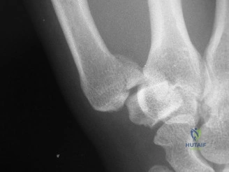
The radiographs demonstrated an intra-articular fracture at the base of the first metacarpal. A small, triangular volar-ulnar fragment (the eponymous Bennett fragment) was clearly visible, remaining anatomically reduced or minimally displaced relative to the trapezium. The main shaft of the first metacarpal was significantly displaced radially, proximally, and dorsally relative to the Bennett fragment and the trapezium. This characteristic pattern of subluxation and fracture indicated a classic Bennett fracture dislocation. There was an estimated articular step-off of approximately 3mm and significant subluxation of the metacarpal shaft.
Figure 1: Typical radiographic appearance of a Bennett fracture dislocation demonstrating the small volar-ulnar fragment and the proximally, radially, and dorsally displaced metacarpal shaft.
Advanced Cross Sectional Imaging
Given the intra-articular nature of the fracture and the significant displacement observed on plain radiographs, a computed tomography scan of the right hand was obtained. This was critical for a precise three-dimensional characterization of the articular step-off, the exact size and morphology of the Bennett fragment, potential comminution, and the overall degree of subluxation. The computed tomography confirmed an intra-articular fracture dislocation with a single, well-defined volar ulnar fragment and no significant secondary comminution, confirming suitability for either percutaneous pinning or open reduction and internal fixation based on fragment size.
Operating Room Setup and Positioning
The patient is positioned supine on the operating table with the affected extremity extended onto a radiolucent hand table. A well padded pneumatic tourniquet is applied to the proximal arm. Intravenous regional anesthesia (Bier block), regional brachial plexus blockade (supraclavicular or axillary), or general anesthesia may be utilized depending on patient and surgeon preference.
The fluoroscopy unit (C-arm) is positioned parallel to the longitudinal axis of the operating table, entering from the distal end of the hand table or perpendicularly from the contralateral side. The surgeon typically sits in the axilla, allowing unimpeded access to the radial aspect of the hand and wrist. A standard hand surgery tray equipped with fine reduction forceps (pointed Weber clamps), a motorized wire driver, Kirschner wires (0.045 inch and 0.062 inch), and a mini fragment or headless compression screw set (1.0 mm to 1.5 mm) should be available.
Detailed Surgical Approach and Technique
The choice between closed reduction and percutaneous pinning versus open reduction and internal fixation depends primarily on the size of the volar ulnar fragment, the degree of comminution, and the ability to achieve a concentric, anatomic closed reduction.
Closed Reduction and Percutaneous Pinning
Closed reduction and percutaneous pinning is indicated when the Bennett fragment is too small to accommodate a screw, or when a perfectly anatomic closed reduction can be achieved and confirmed fluoroscopically.
The closed reduction maneuver must directly counteract the deforming forces of the abductor pollicis longus and adductor pollicis. The surgeon grasps the patient's thumb and applies longitudinal traction. While maintaining traction, the thumb metacarpal is brought into palmar abduction. Finally, a pronation force is applied to the metacarpal shaft while simultaneous direct manual pressure is exerted over the dorsal radial aspect of the metacarpal base, pushing it volarly and ulnarly toward the anatomically reduced Bennett fragment.
Once reduction is achieved and verified on multiple fluoroscopic views (particularly the true anteroposterior and lateral views of the carpometacarpal joint), percutaneous fixation is performed.
1. Metacarpal to Trapezium Pinning: A 0.045 inch Kirschner wire is driven from the dorsal radial aspect of the first metacarpal shaft, across the carpometacarpal joint, and into the trapezium. This pin neutralizes the proximal and dorsal pull of the abductor pollicis longus.
2. Metacarpal to Metacarpal Pinning: A second 0.045 inch or 0.062 inch Kirschner wire is often driven from the first metacarpal shaft transversely into the second metacarpal shaft. This pin acts as a robust strut, preventing radial subluxation and maintaining the first web space.
3. Fragment Pinning: If the Bennett fragment is large enough, a dedicated pin can be passed from the main metacarpal shaft directly into the fragment, though this is technically demanding percutaneously.
Pins are typically cut outside the skin and bent to facilitate removal in the clinic, though they may be buried beneath the skin based on surgeon preference.
Open Reduction and Internal Fixation via Wagner Approach
Open reduction and internal fixation is indicated for fractures with a large Bennett fragment (typically > 20% of the articular surface) where closed reduction is inadequate, or when precise articular restoration is demanded. The classic radiopalmar approach, originally described by Wagner, provides excellent exposure of the first carpometacarpal joint.
Incision and Superficial Dissection:
A curved or lazy-S incision is made along the glabrous border of the thenar eminence, beginning distally at the level of the first metacarpophalangeal joint and extending proximally toward the distal wrist crease, curving ulnarly toward the flexor carpi radialis tendon.
Blunt dissection is utilized in the subcutaneous tissues. The surgeon must meticulously identify and protect the superficial branches of the radial sensory nerve dorsally, and the lateral antebrachial cutaneous nerve volarly. The thenar fascia is incised.
Deep Dissection and Internervous Planes:
The internervous plane lies between the abductor pollicis longus and extensor pollicis brevis tendons (innervated by the radial nerve) dorsally, and the thenar musculature (abductor pollicis brevis and opponens pollicis, innervated by the median nerve) volarly.
The thenar muscles are elevated subperiosteally from the radial and volar aspects of the first metacarpal shaft and retracted ulnarly. This exposes the capsule of the first carpometacarpal joint. A longitudinal or T-shaped capsulotomy is performed. The joint is irrigated to evacuate the fracture hematoma, and small periarticular debris is meticulously debrided to allow direct visualization of the articular surface.
Reduction and Internal Fixation:
The fracture surfaces are identified. The small volar ulnar fragment is visualized attached to the anterior oblique ligament. The main metacarpal shaft is mobilized, and the joint is reduced under direct vision using the traction, abduction, and pronation maneuver described previously.
A pointed reduction forceps (Weber clamp) is carefully applied. One tine is placed on the dorsal radial aspect of the metacarpal shaft, and the other tine is placed delicately on the volar ulnar fragment. Gentle compression is applied to achieve anatomic articular reduction.
Fixation is typically achieved using one or two small screws (1.0 mm, 1.3 mm, or 1.5 mm). The trajectory of the screw must be perpendicular to the fracture plane to achieve maximal interfragmentary compression.
1. A glide hole is drilled in the near cortex (the main metacarpal shaft).
2. A thread hole is drilled in the far cortex (the Bennett fragment).
3. The hole is countersunk to prevent hardware prominence.
4. The length is measured, and an appropriately sized cortical screw is inserted. Alternatively, headless compression screws can be utilized to minimize hardware irritation beneath the capsule.
If the fragment is highly comminuted or too small for screw fixation, multiple fine Kirschner wires (0.028 inch or 0.035 inch) can be utilized for fixation. Following rigid fixation, the joint capsule is meticulously repaired using absorbable sutures to augment stability. The thenar musculature is allowed to fall back into place, and the skin is closed in a standard fashion.
Complications and Management
Despite meticulous surgical technique, complications following the treatment of Bennett fractures can occur. Early recognition and appropriate salvage strategies are critical for preserving hand function.
| Complication | Estimated Incidence | Etiology and Clinical Presentation | Management and Salvage Strategies |
|---|---|---|---|
| Post Traumatic Osteoarthritis | 20% to 50% (Long term) | Results from residual articular incongruity (>1mm), initial cartilage impaction damage, or chronic joint subluxation. Presents as progressive pain, stiffness, and weakness with pinch activities. | Initial management includes NSAIDs, splinting, and intra articular corticosteroid injections. Surgical salvage options include trapeziectomy with ligament reconstruction and tendon interposition, or primary carpometacarpal arthrodesis. |
| Malunion | 10% to 15% | Occurs due to loss of reduction, inadequate initial fixation, or failure to neutralize the abductor pollicis longus. Results in a shortened, adducted thumb with a narrowed first web space. | Corrective intra articular or extra articular closing wedge osteotomies of the first metacarpal base. Soft tissue releases of the first web space (Z-plasty) may be required for severe adduction contractures. |
| Pin Tract Infection | 5% to 10% | Superficial bacterial colonization around percutaneous Kirschner wires. Presents with erythema, localized swelling, and purulent discharge at the pin site. | Oral antibiotics (e.g., cephalexin) and increased local pin care. If the infection tracks deeply or osteomyelitis is suspected, early hardware removal and intravenous antibiotics are mandatory. |
| Sensory Nerve Injury | 2% to 5% | Iatrogenic injury to the superficial radial sensory nerve or lateral antebrachial cutaneous nerve during the surgical approach or percutaneous pin insertion. Presents as numbness, paresthesias, or painful neuromas. | Observation for neuropraxia. Gabapentinoids for neuropathic pain. If a painful neuroma develops, surgical exploration, neuroma excision, and burying the nerve end into deep muscle may be required. |
| Nonunion | < 1% | Extremely rare in the highly vascularized metaphyseal base of the first metacarpal. Usually associated with severe infection or massive bone loss. | Revision open reduction internal fixation with autologous cancellous bone grafting from the distal radius. |
Post Operative Rehabilitation Protocols
The postoperative rehabilitation protocol must balance the need for fracture healing with the prevention of debilitating joint stiffness. The exact timeline is dictated by the stability of the fixation achieved intraoperatively.
Phase 1: Immobilization (Weeks 0 to 4)
Immediately following surgery,
