ABOS Part I Orthopedic Surgery Review: Ankle, Knee, and Foot Arthritis Management | Part 22168

Key Takeaway
This ABOS Part I comprehensive review module covers critical orthopedic topics including ankle arthritis management (arthrodesis, TAR), total knee arthroplasty for valgus deformities (surgical techniques, complications), and hallux valgus surgery (osteotomies, MTP arthrodesis). It provides in-depth explanations of diagnosis, surgical planning, and post-operative considerations for board exam preparation.
ABOS Part I Orthopedic Surgery Review: Ankle, Knee, and Foot Arthritis Management | Part 22168
A 42-year-old manual worker presents with chronic left ankle pain following a fracture 7 years prior, which was managed non-operatively. An AP weightbearing radiograph is obtained.
Based on the provided image and the candidate's initial description, which of the following is the most accurate radiographic interpretation?
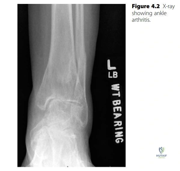
Correct Answer: C
The candidate's description of the radiograph explicitly states: 'This is an AP weightbearing radiograph of a left ankle showing narrowing of the joint space and some subchondral sclerosis consistent with post-traumatic arthritis. There is evidence of a previous fibula fracture superior to the syndesmosis and varus angulation of the ankle.' This directly matches option C.
Option A is incorrect as the description does not mention a trimalleolar fracture malunion or subtalar arthritis. Option B is incorrect as medial clear space widening is not noted; rather, varus angulation is described. Option D is incorrect as avascular necrosis of the talar dome and valgus deformity are not mentioned in the description. Option E is incorrect as the findings are consistent with post-traumatic arthritis, not inflammatory arthritis, and diffuse osteopenia or erosions are not described.
A 55-year-old patient is diagnosed with end-stage ankle arthritis. During counseling, the physician discusses the most common etiologies.
According to the provided case, which of the following is considered the most common cause of end-stage ankle arthritis?
Correct Answer: D
The case explicitly states: 'Primary osteoarthritis is thought to be relatively uncommon and the most common cause of ankle arthritis is probably post-traumatic arthritis. Other causes are inflammatory arthritis and septic arthritis.' Therefore, post-traumatic arthritis is the most common cause.
Option A is incorrect because primary osteoarthritis is noted as relatively uncommon. Options B and C are incorrect as inflammatory and septic arthritis are listed as 'other causes' but not the most common. Option E, avascular necrosis of the talus, is a potential cause of ankle arthritis but is not listed in the case as one of the most common etiologies.
A 68-year-old low-demand patient with ankle arthritis is initiating conservative management. The patient has failed NSAIDs and activity modification.
All of the following conservative management strategies are mentioned in the case as potentially beneficial for ankle arthritis, EXCEPT:
Correct Answer: E
The case discusses several conservative measures: 'optimize the patient’s analgesia adding in NSAIDs, and suggest activity modification. He could try footwear modification with a cushioned sole and rocker-bottom shoe which may improve his symptoms as may use of an ankle brace or AFO. Similarly an injection of intra-articular steroid or viscosupplementation may be of symptomatic benefit. Physiotherapy could be an adjunctive treatment in patients with symptoms of instability or weakness but may aggravate symptoms.' Long-term oral corticosteroids are not mentioned as a conservative management strategy for ankle arthritis in this context.
Options A, B, C, and D are all explicitly mentioned as potential conservative treatments in the case.
A 35-year-old active patient with symptomatic ankle arthritis, not yet ready for definitive surgical treatment, is exploring options to 'buy time' before considering fusion or replacement.
Which of the following surgical options is described in the case as a temporizing measure for ankle arthritis?
Correct Answer: C
The case clearly categorizes surgical options into 'temporizing measures' and 'definitive treatments'. It states: 'The temporizing measures are debridement of the joint which can be performed arthroscopically or open... The other option is distraction arthroplasty.' Therefore, distraction arthroplasty is a temporizing measure.
Options A (Total ankle replacement) and B (Ankle arthrodesis) are described as 'definitive surgical options.' Options D (Supramalleolar osteotomy) and E (Subtalar fusion) are not mentioned in the case as either temporizing or definitive treatments for ankle arthritis.
The 42-year-old manual worker from the case, with post-traumatic ankle arthritis and varus angulation, insists on a total ankle replacement (TAR) despite being advised against it.
Based on the case discussion, which of the following is the primary contraindication for total ankle replacement in this specific patient?
Correct Answer: C
The examiner specifically asks about the 42-year-old patient wanting an ankle replacement, and the candidate responds: 'He is a young patient, in a manual job. He wouldn’t be a candidate for total ankle replacement and I would explain to him that if his symptoms have failed to be controlled by non-operative measures and he requires definitive surgical treatment then an ankle fusion would be a better option for him.' This clearly identifies his young age and high-demand occupation as the primary contraindications.
While varus angulation (Option B) is a contraindication if significant (>10 degrees), the case highlights the patient's age and activity level as the primary reasons for not being a candidate. A previous fibula fracture (Option A) and subchondral sclerosis (Option D) are findings associated with his post-traumatic arthritis but are not listed as primary contraindications for TAR in the same way age and activity level are. Option E is irrelevant to the surgical decision-making for this patient.
A patient with end-stage ankle arthritis is undergoing ankle arthrodesis.
According to the case, what is the ideal position for ankle fusion?
Correct Answer: B
The case explicitly states the ideal position for ankle fusion: 'The foot should be plantigrade with a physiological 5 of hindfoot valgus and 5 of external rotation.'
Options A, C, D, and E describe various other positions that do not match the specific recommendations provided in the case for optimal ankle fusion.
A surgeon is discussing the evolution of total ankle replacement (TAR) designs with a resident.
The case mentions that most modern total ankle replacement designs are characterized by which of the following features?
Correct Answer: B
The case states: 'The earlier designs involved a two-component design such as the Agility total ankle replacement, which required fusion of the distal tibiofibular joint. Most modern designs are three-component uncemented mobile bearing prostheses.' This directly describes option B.
Option A describes older, less common features. Option C describes earlier designs like the Agility, but not 'most modern designs.' Options D and E do not accurately reflect the description of modern TAR designs provided in the case.
A 45-year-old active patient with post-traumatic ankle arthritis, similar to the case patient, desires a total ankle replacement to maintain activity levels and avoid a stiff ankle, despite being counseled against it.
In this scenario, what is the most appropriate advice to give the patient regarding ankle fusion, as per the case?
Correct Answer: C
When the examiner presses the candidate about the patient still wanting a replacement to get back to hill walking and sports, the candidate explains: 'A fusion would provide a stable pain-free ankle that would allow him to return to the majority of activities that he wishes to do. I would explain that many patients return to sports after ankle fusion. I would also explain that an ankle fusion would only sacrifice the residual movement that he has at his ankle joint and that his subtalar, midfoot and forefoot movements would still be present.' This comprehensive explanation aligns perfectly with option C.
Option A is incorrect because the case explicitly states that subtalar, midfoot, and forefoot movements are preserved, and many patients return to sports. Option B is incorrect as the case states fusion is still considered the 'gold standard' and is not outdated for all patients. Option D is incorrect as the case suggests fusion is suitable for active patients like the manual worker. Option E is incorrect; while adjacent joint arthritis is a known complication of fusion, the case does not state it's a higher risk than TAR, and the overall tone is that fusion is a better option for this patient.
A patient is considering ankle fusion and asks about potential complications.
Which of the following is NOT listed in the case as a potential complication of ankle fusion?
Correct Answer: D
The candidate lists the following complications for ankle fusion: 'Non-union, malunion, delayed union, infection, wound-healing problems, nerve or vessel damage, DVT/PE, risk of exacerbating or developing arthritis in other joints.' Stress fracture of the ipsilateral tibia is not mentioned in this list.
Options A, B, C, and E are all explicitly listed as potential complications of ankle fusion in the case.
A 62-year-old patient considering total ankle replacement (TAR) asks about the long-term prognosis.
Based on the information provided in the case, what is the approximate 10-year survival rate for total ankle replacements, and how does this compare to hip and knee replacements?
Correct Answer: B
The candidate's response to the question about TAR longevity is: 'The 10-year survival is approaching 85% but there are fewer data available than for knee and hip replacements. Many series are small.' This directly matches option B.
Options A, C, D, and E provide incorrect survival rates or make inaccurate comparisons regarding the availability of data for ankle replacements versus hip and knee replacements.
A 72-year-old female presents with bilateral knee pain and gradual deformity. Weightbearing anteroposterior radiographs are obtained, as shown below. Based on the provided image and case description, which of the following best describes the primary radiographic findings and the associated arthritic pattern?
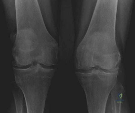
Correct Answer: C
The case explicitly states, and the image demonstrates, 'narrowing of joint spaces with bone-on-bone contact in the lateral compartments of both knees. There is early arthrosis affecting the medial compartments of both knees. There is moderate valgus deformity.' This directly matches option C.
Option A is incorrect because the deformity is valgus, not varus, and the primary bone-on-bone contact is in the lateral compartment, not medial.
Option B is incorrect as the primary finding is tibiofemoral arthritis with valgus deformity, not isolated patellofemoral arthrosis.
Option D is incorrect because while arthritis is present, the description specifies lateral compartment bone-on-bone contact and early medial arthrosis, not diffuse narrowing as the primary descriptor, and the image does not strongly suggest diffuse inflammatory changes over a specific compartment pattern.
Option E is incorrect because the radiographs clearly show significant arthritic changes with bone-on-bone contact, not preserved joint space, and the primary diagnosis is arthritis with deformity, not an isolated meniscal tear.
A 68-year-old female presents with a valgus knee deformity and symptomatic arthritis, similar to the patient in the case. During the preoperative workup, the surgeon considers potential etiologies for this pattern of joint disease. Which of the following conditions is LEAST commonly associated with a valgus deformity of the knee with arthritis?
Correct Answer: D
The case specifically lists conditions commonly associated with valgus deformity of the knee with arthritis: 'inflammatory joint conditions such as rheumatoid arthritis... primary osteoarthritis, overcorrection of high tibial osteotomy (HTO), post-traumatic arthritis following lateral meniscectomy and osteonecrosis.' Post-traumatic arthritis following a *medial* meniscectomy would typically predispose to *varus* deformity and medial compartment arthritis, not valgus deformity.
Option A (Rheumatoid arthritis) is incorrect because the case explicitly states it is a common association.
Option B (Primary osteoarthritis) is incorrect because the case explicitly states it can occur in primary osteoarthritis.
Option C (Overcorrection of a high tibial osteotomy (HTO)) is incorrect because the case explicitly states it can occur due to HTO overcorrection.
Option E (Osteonecrosis of the lateral femoral condyle) is incorrect because the case explicitly states it can be a cause of valgus arthritis.
During total knee arthroplasty for a valgus knee, the surgeon notes significant deficiency of the lateral femoral condyle. This anatomical variation can lead to a specific rotational malalignment if standard referencing techniques are used. To prevent this malalignment, which of the following intraoperative techniques is most appropriate for determining femoral component rotation?
Correct Answer: C
The case states: 'Due to the posterior femoral condyle deficiency, the standard 3 posterior condylar referencing can result in internal rotation of the component. In this situation, AP axis (Whiteside line) is used to prevent malrotation in the form of internal rotation.' The Whiteside line (AP axis) is a reliable anatomical landmark that is less affected by condylar wear, making it crucial for accurate femoral rotation in valgus knees with condylar deficiency.
Option A is incorrect because while 3 degrees of external rotation relative to the posterior condylar axis is a common goal, the posterior condylar deficiency in valgus knees makes this reference unreliable and prone to internal rotation malalignment.
Option B (Transepicondylar axis) is incorrect as it is a valid reference but the case specifically highlights the AP axis (Whiteside line) as the preferred method to address the posterior condylar deficiency issue in valgus knees.
Option D (Aligning the femoral component parallel to the tibial cut) is incorrect as this does not directly address femoral rotation relative to the femur's anatomical axes and could lead to malrotation if the tibial cut is not perfectly aligned or if there are significant soft tissue imbalances.
Option E (Relying solely on the measured resection technique from the posterior condyles) is incorrect because, as stated in the case, posterior condylar deficiency makes this method unreliable and can lead to internal rotation of the component.
A surgeon is performing a TKR on a patient with a severe valgus deformity. During soft tissue balancing, significant release of the lateral and posterior structures is required to achieve adequate correction. What is the most likely immediate consequence of this extensive soft tissue release on the flexion-extension gap, and how might it impact implant selection?
Correct Answer: B
The case states: 'With regards to flexion–extension gap, the release of lateral and posterior structures results in increased extension gap requiring a thicker insert which may elevate the joint line.' This directly describes the consequence of extensive lateral and posterior soft tissue release in a valgus knee.
Option A is incorrect because releasing contracted structures would increase, not decrease, the extension gap.
Option C is incorrect because significant release of contracted structures will almost certainly alter the gap, making it unlikely to be perfectly balanced without specific intervention.
Option D is incorrect because the primary effect of releasing lateral and posterior structures is on the extension gap, not typically an isolated increase in the flexion gap that would necessitate a larger femoral component.
Option E is incorrect because releasing structures would loosen, not tighten, the knee, making a decreased flexion gap unlikely.
A 72-year-old female, similar to the patient in the case, undergoes TKR for a valgus knee. Intraoperatively, after femoral and tibial cuts, the surgeon observes persistent lateral subluxation of the patella despite appropriate component rotation and a mild distal femoral valgus cut. What is the most appropriate next step to address this issue?
Correct Answer: C
The case states: 'Patients with severe valgus deformity usually require lateral retinacular release to achieve proper patella tracking.' The scenario describes persistent lateral subluxation, indicating the need to release the tight lateral structures that are pulling the patella laterally.
Option A is incorrect because while a distal femoral cut of 7 degrees can help in mild valgus, increasing it further is not the primary solution for persistent patellar subluxation after initial cuts and may lead to other issues like over-resection or instability. The case mentions 7 degrees for *mild* valgus to *avoid* release, implying that for persistent issues, a release is needed.
Option B (Medial retinacular release) is incorrect as this would worsen lateral patellar subluxation by releasing the medial restraints.
Option D (Downsizing the femoral component) is incorrect as this would primarily affect flexion gap and overall knee size, not directly address patellar tracking issues caused by tight lateral retinaculum.
Option E (Use a thicker polyethylene insert) is incorrect as this primarily addresses flexion-extension gap balancing and joint line elevation, not patellar tracking.
Following a total knee arthroplasty for a severe valgus deformity, a 72-year-old patient experiences new-onset foot drop and numbness along the dorsum of the foot. Which of the following nerves is most likely affected?
Correct Answer: D
The case explicitly warns: 'With correction of significant valgus deformity, one has to watch for peroneal nerve palsy in the postoperative period.' Foot drop and numbness along the dorsum of the foot are classic symptoms of peroneal nerve palsy, which can occur due to traction injury during significant valgus correction.
Option A (Femoral nerve) is incorrect. Femoral nerve injury typically presents with quadriceps weakness and sensory loss over the anterior thigh and medial leg, not foot drop.
Option B (Saphenous nerve) is incorrect. Saphenous nerve injury causes sensory loss along the medial aspect of the leg and foot, without motor deficits like foot drop.
Option C (Tibial nerve) is incorrect. Tibial nerve injury typically affects plantarflexion and sensation on the sole of the foot, not dorsiflexion or sensation on the dorsum.
Option E (Obturator nerve) is incorrect. Obturator nerve injury affects adduction of the thigh and sensation over the medial thigh, which is unrelated to foot drop.
In a patient presenting with a valgus knee deformity requiring TKR, the case describes specific anatomical and rotational characteristics of the femur and tibia. Which of the following statements accurately describes these characteristic deformities?
Correct Answer: B
The case explicitly states: 'In valgus knees the lateral femoral condyle is deficient, therefore the femur is internally rotated and tibia is externally rotated.' This directly matches option B.
Option A is incorrect because the lateral femoral condyle is deficient, not the medial, and the rotational deformities are reversed.
Option C is incorrect because the lateral femoral condyle is specifically deficient, not both equally, and the rotational deformities are not both external.
Option D is incorrect because the lateral femoral condyle is deficient, not hypertrophied, and the rotational deformities are reversed.
Option E is incorrect because the lateral femoral condyle is deficient, not the medial, and the tibial rotation is external, not internal.
A surgeon is planning a TKR for a patient with a valgus knee. While a medial parapatellar approach is commonly used, the surgeon considers a lateral approach. According to the case, what is the theoretical advantage of utilizing a lateral surgical approach for total knee arthroplasty in a valgus knee?
Correct Answer: C
The case directly addresses this: 'EXAMINER : What is the theoretical advantage of a lateral approach? CANDIDATE : It is a direct approach providing easier access and preserves the neurovascular supply to the extensor mechanism.' This statement directly supports option C.
Option A is incorrect because a lateral approach would make medial compartment access more challenging, not easier.
Option B is incorrect because the case states that a medial parapatellar approach 'gives good access to the whole knee and better soft tissue cover,' implying this is an advantage of the medial approach, not the lateral.
Option D is incorrect because while component rotation is critical, the advantage of the lateral approach is not specifically tied to easier correction of femoral internal rotation compared to other approaches.
Option E is incorrect because the risk of peroneal nerve palsy is related to the degree of valgus correction and traction on the nerve, not inherently reduced by the surgical approach itself.
During a TKR for a valgus knee, the surgeon encounters a situation where excessive posterior cruciate ligament (PCL) release is performed during soft tissue balancing. Based on the principles outlined in the case, what is the most likely consequence of this excessive PCL release and the appropriate implant choice?
Correct Answer: C
The case explicitly states: 'Excessive PCL release usually requires cruciate sacrificing implants in order to balance the knee.' This is a direct consequence of losing the PCL's contribution to stability and balance.
Option A is incorrect because excessive PCL release would lead to a looser, not tighter, flexion gap, and would preclude PCL retention.
Option B is incorrect because excessive PCL release would destabilize the knee, making it unbalanced, and would not allow for PCL-retaining implants.
Option D is incorrect because while soft tissue releases can affect the extension gap, excessive PCL release specifically impacts flexion stability and necessitates a change in implant type, not just insert thickness while retaining the PCL.
Option E is incorrect because PCL release has a significant impact on knee kinematics and balance, directly influencing implant choice.
A 65-year-old patient with a mild valgus deformity (8 degrees) is undergoing TKR. The surgeon is focused on optimizing patellar tracking and avoiding unnecessary lateral retinacular release. According to the case, what specific distal femoral cut angle can be utilized to improve patellar tracking in this scenario?
Correct Answer: C
The case specifically mentions: 'In mild valgus deformity (7–10) a distal femoral cut of 7 can improve patella tracking and avoid the need for lateral retinacular release.' This directly provides the recommended angle for mild valgus deformities to aid patellar tracking.
Option A (3 degrees) is incorrect as this is a common valgus angle for a varus knee or a neutral knee, but not specifically highlighted for improving patellar tracking in mild valgus.
Option B (5 degrees) is incorrect as the case specifies 7 degrees for this particular purpose.
Option D (9 degrees) is incorrect as while it falls within the 7-10 degree range of mild valgus, the specific recommendation for the cut is 7 degrees to improve tracking and avoid release.
Option E (0 degrees) is incorrect as a neutral cut would not address the valgus deformity or specifically aid patellar tracking in a valgus knee.
A 65-year-old female presents with bilateral foot pain. The examiner provides the following clinical image of her feet.
Based on the provided image and the initial candidate's observations, which of the following is the MOST accurate initial observation regarding the patient's right foot?
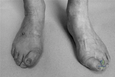
Correct Answer: C
The candidate's initial observation states, 'There is hallux valgus with the hallux over-riding the second toes.' The provided image (Figure 4.7, AP view) clearly depicts hallux valgus on the right foot, where the great toe is deviated laterally and appears to override the second toe. The case mentions the absence of the second toe on the left foot, not the right. Hallux varus is a medial deviation of the great toe, which is contrary to the image. Scars and specific surgical evidence are not definitively identifiable for the right foot from this initial clinical image alone.
A 65-year-old female presents with hallux valgus. The examiner asks about the value of plain radiographs. The candidate states they would measure the intermetatarsal angle (IMA), hallux valgus angle (HVA), and distal metatarsal articular angle (DMAA).
Which of the following represents the correct normal range for these angles as stated in the case?
Correct Answer: B
The case explicitly states the normal ranges for these angles: 'The intermetatarsal angle is normally less than 9. The hallux valgus angle should be less than 15. The distal metatarsal articular angle is normally a maximum of 15 from perpendicular to the axis of the first metatarsal.' Therefore, option B accurately reflects these normal values.
The 65-year-old lady presents with a right foot deformity characterized by an intermetatarsal angle (IMA) of 15 degrees, a hallux valgus angle (HVA) of 35 degrees, and minimal passive correction of the hallux. Her first tarsometatarsal (TMT) joint is assessed as normal.
Based on these findings and the candidate's proposed management, which surgical approach would be MOST appropriate for her right foot?
Correct Answer: C
The examiner presents a specific scenario: 'If this lady had an intermetatarsal angle of 15 on the right with a hallux valgus angle of 35 and minimal passive correction of the hallux, what surgery would you plan?' The candidate responds: 'If the first tarsometatarsal joint is normal, I would plan a scarf osteotomy combined with a lateral release and an Akin osteotomy of the proximal phalanx if necessary.' This directly matches option C. Chevron and Mitchell osteotomies are typically for less severe deformities or have disadvantages like shortening, as discussed later in the case. Arthrodesis is usually reserved for severe arthrosis or recurrent cases, especially with missing toes, and Keller is a salvage procedure.
Following the decision to perform a Scarf osteotomy for the patient's right foot, the examiner asks the candidate to justify this choice over simpler procedures.
Which of the following is a key advantage of the Scarf osteotomy, as described by the candidate, that makes it suitable for this patient's deformity?
Correct Answer: C
When asked 'Why would you choose a scarf osteotomy?', the candidate replies: 'It is a very versatile procedure with stable fixation allowing postoperative mobilization without a cast. It maintains length of the metatarsal but allows translation, angulation and depression of the metatarsal head as necessary.' This statement directly supports option C, highlighting stable fixation, early mobilization, and versatility. Options A and D are incorrect as Scarf is for moderate to severe deformities and is not necessarily 'simpler' than distal osteotomies. Option B is incorrect as Scarf maintains length or can even lengthen, unlike Mitchell. Option E is incorrect as arthrodesis is preferred for significant MTP arthritic change.
When discussing consent for the proposed Scarf osteotomy, the candidate outlines potential complications.
Which of the following complications is explicitly mentioned by the candidate as a possibility following a Scarf osteotomy?
Correct Answer: C
The candidate explicitly states: 'A minority of patients will have significant stiffness of the MTP joint afterwards and there can be sensory loss if the dorsomedial sensory nerve is injured.' While DVT/PE and CRPS are general surgical risks, they are not specifically highlighted by the candidate in this discussion. Avascular necrosis is a known complication of some distal osteotomies (e.g., Chevron) but not specifically emphasized for Scarf in this context. The recurrence risk is mentioned as 'greatest in adolescent cases' and not given a specific high percentage like 50% for this patient.
During the discussion of complications, the examiner specifically asks about the treatment of hallux varus, which is a potential complication of hallux valgus surgery.
According to the candidate, what is a reliable surgical option for hallux varus in the presence of significant stiffness or arthrosis?
Correct Answer: C
The candidate states: 'Whilst soft tissue procedures such as abductor hallucis and medial capsular release or transfer of a slip of EHL are described for flexible deformity, arthrodesis of the first MTP joint is a reliable option in the presence of significant stiffness or arthrosis.' This directly identifies MTP joint arthrodesis as the reliable option for stiff or arthritic hallux varus. Other options are either not mentioned or are inappropriate for this specific indication.
After successfully treating the patient's right foot, the examiner asks if the same procedure would be performed on the left foot, given its presentation. The oblique view of the left foot is provided.
Considering the patient's left foot, as depicted in the image and described in the case (specifically the prior surgery), what is the MOST appropriate surgical plan for the hallux valgus recurrence?
Correct Answer: C
The examiner asks: 'So you have successfully treated this lady’s right foot and she is pleased with the result. Would you go ahead and do the same on the left?' The candidate responds: 'No. The absence of the second toe predisposes to recurrence and I would propose arthrodesis of the hallux MTP joint.' The image (Figure 4.8, oblique view) shows the left foot, which, as noted in the initial clinical description, has only three lesser toes due to the prior removal of the second toe. This unique anatomical situation on the left foot, predisposing to recurrence, makes MTP joint arthrodesis the preferred and most reliable option for long-term stability.
The examiner asks the candidate to describe their further assessment of the patient, beyond initial observations. The candidate outlines a comprehensive physical examination.
Which of the following is NOT a component of the physical examination specifically mentioned by the candidate for this patient?
Correct Answer: C
The candidate details the physical examination: 'I would palpate for areas of tenderness, paying particular attention to the hallux MTP joint and lesser metatarsal heads. I would assess the degree of active and passive correction possible and the range of movement of the involved joints and look for gastrocnemius tightness. I would also perform a grind test to assess pain from loading the MTP joint. Neurovascular status must also be assessed.' While 'Neurovascular status must also be assessed' is mentioned, the specific 'Measurement of ankle-brachial index (ABI)' is not explicitly stated as part of the physical examination components by the candidate. All other options are directly mentioned.
The examiner challenges the candidate on why a simpler procedure like a Chevron or Mitchell osteotomy would not be chosen for the described deformity (IMA 15°, HVA 35°, minimal passive correction).
According to the candidate, what is a significant disadvantage of a Mitchell osteotomy for this patient's specific deformity?
Correct Answer: C
When asked 'Why not use a simpler procedure such as a chevron or Mitchell osteotomy?', the candidate responds: 'For the degree of deformity described, combined with the lack of passive correction of the hallux, I believe the correction that could be achieved with a distal osteotomy would be inadequate. A further disadvantage of a Mitchell osteotomy is that it produces shortening of the first metatarsal, which could lead to transfer metatarsalgia.' This statement directly supports option C as a significant disadvantage of the Mitchell osteotomy.
The examiner presents the clinical photographs (Figures 4.7 and 4.8) and asks the candidate to describe what they see.
Based on the candidate's initial description of the clinical photographs, which of the following observations is made regarding the patient's left foot?
Correct Answer: C
The candidate's initial description of the clinical photographs (Figures 4.7 and 4.8) includes: 'I can only count three lesser toes on the left foot and there is a scar in the webspace lateral to the hallux.' This observation is clearly visible in the images, particularly Figure 4.8 (oblique view of the left foot), which shows the absence of the second toe and a scar. The case later confirms that the left second toe was removed years ago. Options A, B, D, and E are either inaccurate or not explicitly stated as initial observations for the left foot.
Based on the altered biomechanics shown postoperatively, which adjacent joint is at the highest risk of developing progressive symptomatic osteoarthritic changes over the next 10 years?
A 65-year-old female presents with severe, bilateral knee pain limiting her ambulation to a single block. She has failed extensive conservative measures. Assuming the radiograph demonstrates advanced tricompartmental osteoarthritis with bone-on-bone changes, what is the most appropriate definitive management?
None
