Master Orthopedic Board Review: Bone Tumors & Metabolic Diseases - Lymphoma, MPS, CMF | Part 11
Key Takeaway
This orthopedic board review covers key bone pathologies including primary lymphoma of bone, mucopolysaccharidoses (Hurler, Morquio syndromes), chondromyxoid fibroma, and Camurati-Engelmann disease. It details clinical presentations, imaging, histology, and genetics for comprehensive exam preparation in musculoskeletal oncology and metabolic bone disorders.
Question 1
During surgical exploration for a suspected tumor in the proximal humerus of a 68-year-old patient, the surgeon encounters a lesion with poorly defined margins that appears whitish and fleshy.
View Answer & Explanation
Correct Answer: D
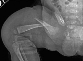
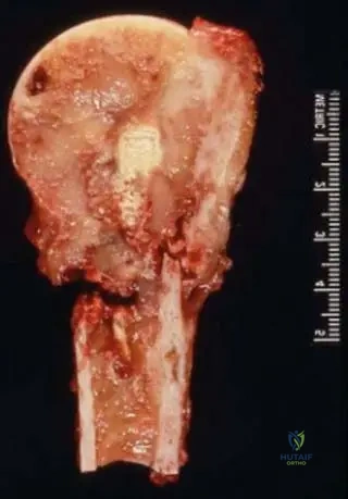
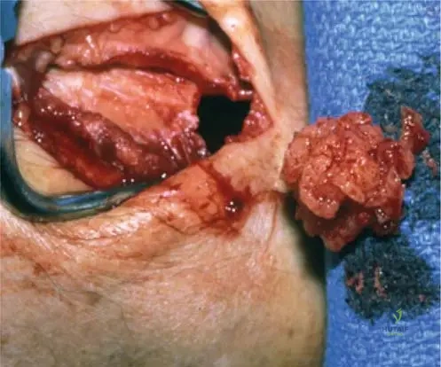
Rationale: Fig. 8.107 and its caption state, "Gross pathologic image of proximal humeral lymphoma. The lesion is permeative with poorly defined margins. Lymphoma often appears whitish and fleshy." Osteosarcoma typically has a gritty, bony appearance due to osteoid production. Chondrosarcoma is cartilaginous. Ewing sarcoma can be soft and fleshy but the description specifically matches lymphoma. Fibrous dysplasia is a benign fibro-osseous lesion.
Question 2
A 72-year-old male presents with new-onset back pain and progressive weakness in his lower extremities. Physical examination reveals bilateral lower extremity motor weakness and sensory deficits. Imaging reveals a destructive lesion involving a lumbar vertebral body.
View Answer & Explanation
Correct Answer: C
Rationale: The clinical text states, "Patients with spine involvement can have neurologic deficits." The patient's symptoms of weakness and sensory deficits directly indicate neurologic compromise. While pathologic fracture is a risk with any destructive bone lesion, neurologic deficits are the most critical and specific complication of spinal involvement. Spontaneous resolution is highly unlikely for a malignant tumor. Hypercalcemia can occur with malignancy but is not the most direct or critical complication of spinal involvement itself.
Question 3
A 50-year-old woman presents with chronic pain in her forearm. Radiographs show a subtle area of cortical thickening and mild lucency in the ulna, but no overt aggressive features. A soft tissue mass is palpable.
View Answer & Explanation
Correct Answer: C
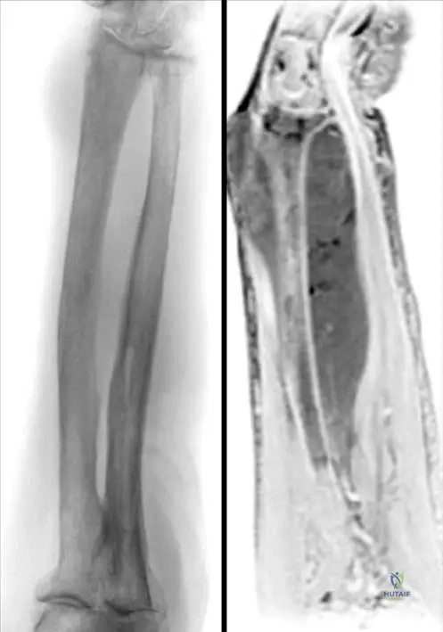
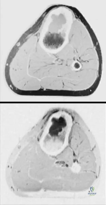
Rationale: Fig. 8.108 and its caption describe "Lymphoma involving left forearm. Characteristically there is a soft tissue mass with minimal roenterographic changes (a). The MR picture (b) shows the large size of the tumor." This description perfectly matches the vignette. Osteoid osteoma typically presents with a nidus and prominent reactive sclerosis, often without a large soft tissue mass. Enchondroma, non-ossifying fibroma, and fibrous cortical defect are typically intraosseous lesions with more characteristic radiographic appearances and do not usually present with a significant soft tissue mass.
Question 4
A pathologist is reviewing a high-power microscopic slide from a bone biopsy. The cells are described as having round to oval vesicular nuclei with margination of chromatin at the nuclear membrane. Nucleoli are prominent, either single and central or multiple and adjacent to the nuclear membrane.

View Answer & Explanation
Correct Answer: C
Rationale: The clinical text accompanying Fig. 8.106b states, "Higher power view (b) of primary lymphoma of bone. Note the round or oval appearing nuclei, which appear vesicular, owing to the margination of chromatin at the nuclear membrane. Nucleoli may be single and central, or may number two or three and be located adjacent to the nuclear membrane." This description is specific to lymphoma cells. Chondroblastic cells, osteoblastic cells, and fibroblasts have distinct nuclear and cytoplasmic features. Giant cell tumor stromal cells have oval nuclei but lack the specific chromatin margination described.
Question 5
A 65-year-old patient is diagnosed with a primary bone tumor that is histologically confirmed as lymphoma. The patient asks about other names for this condition.
View Answer & Explanation
Correct Answer: C
Rationale: The clinical text explicitly lists "Reticulum cell sarcoma" and "non-Hodgkin lymphoma of bone" as synonyms for lymphoma. The other options are distinct types of bone tumors.
Question 6
A 55-year-old male presents with persistent pain in his lower extremity. Radiographs show a subtle lytic lesion in the metadiaphyseal region of a long bone.
View Answer & Explanation
Correct Answer: D
Rationale: The clinical text states, "Patients present at any age and any bone can be affected, but most often the femur is involved." While other bones can be affected, the femur is the most common site.
Question 7
A 48-year-old patient is diagnosed with primary lymphoma of bone after a biopsy of a lytic lesion in the distal femur. Further workup, including PET scan and bone marrow biopsy, reveals no other sites of disease.
View Answer & Explanation
Correct Answer: B
Rationale: The clinical text states, "The majority of bone lymphomas arise in nodal tissues and subsequently metastasize. Rarely, patients will present with a solitary osseous lesion." Therefore, a solitary presentation is rare. It can occur at any age, not just pediatric. Solitary lesions are less likely to have systemic symptoms, and it is a malignant condition, not benign.
Question 7
A 55-year-old male presents with 3 months of progressive left thigh pain and swelling. He reports occasional night sweats and generalized fatigue. Physical examination reveals a tender, ill-defined mass in the distal left femur. Radiographs show subtle cortical irregularity.
View Answer & Explanation
Correct Answer: C
Rationale: The clinical text states, "Patients with multifocal bone involvement may complain of systemic symptoms such as malaise, night sweats, etc." Night sweats are a classic B symptom associated with lymphoma. Weight gain is incorrect; unexplained weight loss is a common systemic symptom of malignancy. Polyuria, diarrhea, and tremors are not typically associated systemic symptoms of lymphoma.
Question 7
A 68-year-old woman presents with chronic right knee pain. Radiographs of the distal femur show subtle lucency with cortical thickening. Subsequent MRI reveals a large, permeative lesion extending significantly beyond the radiographic findings, with an associated small soft tissue mass along the medial femoral condyle.
View Answer & Explanation
Correct Answer: D
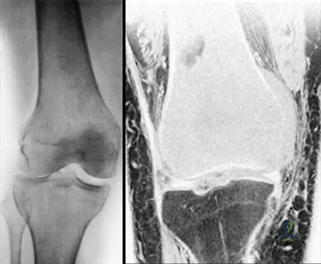
Rationale: The clinical text and Fig. 8.105 highlight that "Radiographic fi ndings may be subtle and easy to overlook. Coronal T1-weighted MRI ( b ) of the same lesion. The size of lesion detected on MRI with lymphoma is often for far greater than suggested on radiographs." This discrepancy between subtle radiographs and extensive MRI findings is characteristic of lymphoma. While other tumors can have soft tissue components, this specific radiographic subtlety combined with extensive MRI findings is a hallmark of lymphoma. Osteosarcoma typically presents with more aggressive and obvious radiographic changes.
Question 7
A 48-year-old patient undergoes biopsy of a lytic lesion in the proximal humerus. Histopathological examination reveals a proliferation of large cells with nuclear pleomorphism and moderately abundant cytoplasm. Immunohistochemistry is pending.
View Answer & Explanation
Correct Answer: C
Rationale: The clinical text explicitly states, "Note the large cell size and the nuclear pleomorphism typically found in diff use large B-cell lymphoma (the most common lymphoma of bone)." This description directly matches the provided histopathological findings. Hodgkin lymphoma is less common as a primary bone lymphoma and has distinct Reed-Sternberg cells.
Question 7
A 62-year-old male presents with a painful lesion in his left forearm. Biopsy confirms a diagnosis of primary lymphoma of bone. Immunohistochemical staining is performed to characterize the tumor.
View Answer & Explanation
Correct Answer: C
Rationale: The clinical text states, "As these are mature B-cell tumors, they generally will stain positive for the immunohistochemical markers CD19 and CD20." CD3 and CD5 are T-cell markers. CD1a and S100 are associated with Langerhans cell histiocytosis. CD30 and CD15 are characteristic of Hodgkin lymphoma. Myogenin and Desmin are markers for rhabdomyosarcoma.
Question 7
During surgical resection of a proximal humeral lesion in a 75-year-old patient, the surgeon notes a permeative tumor with poorly defined margins that appears whitish and fleshy.
View Answer & Explanation
Correct Answer: C
Rationale: Fig. 8.107 and its caption state, "Gross pathologic image of proximal humeral lymphoma. The lesion is permeative with poorly defi ned margins. Lymphoma often appears whitish and fl eshy." This description directly matches the gross appearance of lymphoma. Osteosarcoma typically has a gritty, hemorrhagic, and often calcified appearance. Osteochondromas are cartilaginous exostoses, and enchondromas are cartilaginous lesions within bone, neither matching the "whitish and fleshy" description.
Question 7
A 70-year-old patient is diagnosed with primary lymphoma of bone. While any bone can be affected, understanding the typical distribution is important for clinical suspicion.
View Answer & Explanation
Correct Answer: D
Rationale: The clinical text states, "Patients present at any age and any bone can be aff ected, but most oft en the femur is involved." While other bones like the humerus, vertebrae, and pelvis can be involved, the femur is explicitly mentioned as the most frequent site. The tibia is also a possible site but not the most common.
Question 7
A 30-year-old presents with a painful, enlarging mass in the left forearm. Radiographs show minimal changes, but MRI reveals a large soft tissue mass with underlying bone involvement.
View Answer & Explanation
Correct Answer: C
Rationale: Fig. 8.108 and its caption describe "Lymphoma involving left forearm. Characteristically there is a soft tissue mass with minimal roenterographic changes ( a ). The MR picture ( b ) shows the large size of the tumor a b." This imaging pattern of a large soft tissue mass with subtle radiographic bone changes is highly characteristic of lymphoma. Lipoma is a benign fatty tumor, typically not associated with bone involvement or significant pain. Myositis ossificans involves heterotopic bone formation in soft tissue, which would be visible on radiographs. Ganglion cysts are benign fluid-filled sacs, and osteoid osteoma has a characteristic nidus.
Question 7
A pathology report for a bone lesion in a 65-year-old patient uses the term "reticulum cell sarcoma." This term is considered a synonym for which of the following conditions?
View Answer & Explanation
Correct Answer: D
Rationale: The clinical text explicitly lists "Synonyms:reticulum cell sarcoma, non-Hodgkin lymphoma of bone" under the Lymphoma section. Ewing sarcoma is a distinct small round blue cell tumor, though historically there was some confusion in terminology. The other options are distinct bone tumors.
Question 7
A 60-year-old female presents with isolated pain in her distal femur. Imaging reveals a lytic lesion without evidence of other systemic involvement. Biopsy confirms lymphoma.
View Answer & Explanation
Correct Answer: C
Rationale: The clinical text states, "Th e majority of bone lymphomas arise in nodal tissues and subsequently metastasize. Rarely, patients will present with a solitary osseous lesion." This indicates that primary solitary osseous lymphoma is an uncommon presentation. Options A and B are incorrect as they suggest a common occurrence. Options D and E are incorrect as the text does not specify age or immunodeficiency as exclusive factors for solitary lesions.
Question 7
A 58-year-old male with a known diagnosis of lymphoma presents with new onset lower extremity weakness and paresthesias. Physical examination reveals diminished sensation and motor strength in his left leg.
View Answer & Explanation
Correct Answer: D
Rationale: The clinical text explicitly states, "Patients with spine involvement can have neurologic defi cits." Lower extremity weakness and paresthesias are classic signs of spinal cord or nerve root compression, which would be caused by lymphoma involving the spine. Involvement of the femur, humerus, forearm, or pelvis would not typically cause these specific neurological deficits unless there was a pathological fracture with nerve impingement, but spine involvement is the direct cause mentioned.
Question 7
A pathologist is reviewing a biopsy specimen from a suspected primary bone lymphoma in a 50-year-old patient. On higher power magnification, the nuclei are described as round or oval, vesicular, with margination of chromatin at the nuclear membrane. Nucleoli are single and central, or two to three and located adjacent to the nuclear membrane.
View Answer & Explanation
Correct Answer: C
Rationale: The description provided in the vignette and Fig. 8.106 refers to the characteristic nuclear features of the neoplastic lymphocytes found in lymphoma. The text states, "Note the round or oval appearing nuclei, which appear vesicular, owing to the margination of chromatin at the nuclear membrane. Nucleoli may be single and central, or may number two or three and be located adjacent to the nuclear membrane." These are features of the malignant B-cells. Osteoblasts, chondrocytes, fibroblasts, and adipocytes have distinct morphological characteristics.
Question 7
A 25-year-old male presents with a painful lesion in his tibia. Imaging and biopsy confirm primary lymphoma of bone. Understanding the epidemiology is crucial for diagnosis.
View Answer & Explanation
Correct Answer: C
Rationale: The clinical text explicitly states, "Patients present at any age." This indicates that lymphoma of bone does not have a specific age predilection, making options A, B, D, and E incorrect as they suggest specific age groups or distributions.
Question 7
A 65-year-old woman presents with a tender mass in her distal femur, and radiographs show subtle changes. Given the suspicion for lymphoma, what is the most appropriate next imaging step to assess the true extent of the lesion and potential soft tissue involvement?
View Answer & Explanation
Correct Answer: C
Rationale: The clinical text and Fig. 8.105 emphasize the role of MRI: "The size of lesion detected on MRI with lymphoma is often for far greater than suggested on radiographs. There is a small soft tissue mass along the medial femoral condyle
Question 8
A 55-year-old male presents with 3 months of progressive left thigh pain and swelling. He reports occasional night sweats and generalized fatigue. Physical examination reveals a tender, ill-defined mass in the distal left femur. Radiographs show subtle cortical irregularity.
View Answer & Explanation
Correct Answer: C
Rationale: The clinical text states, "Patients with multifocal bone involvement may complain of systemic symptoms such as malaise, night sweats, etc." Night sweats are a classic B symptom associated with lymphoma. Weight gain is incorrect; unexplained weight loss is a common systemic symptom of malignancy. Polyuria, diarrhea, and tremors are not typically associated systemic symptoms of lymphoma.
Question 9
A 68-year-old woman presents with chronic right knee pain. Radiographs of the distal femur show subtle lucency with cortical thickening. Subsequent MRI reveals a large, permeative lesion extending significantly beyond the radiographic findings, with an associated small soft tissue mass along the medial femoral condyle.
View Answer & Explanation
Correct Answer: D
Rationale: The clinical text and Fig. 8.105 highlight that "Radiographic fi ndings may be subtle and easy to overlook. Coronal T1-weighted MRI ( b ) of the same lesion. The size of lesion detected on MRI with lymphoma is often for far greater than suggested on radiographs." This discrepancy between subtle radiographs and extensive MRI findings is characteristic of lymphoma. While other tumors can have soft tissue components, this specific radiographic subtlety combined with extensive MRI findings is a hallmark of lymphoma. Osteosarcoma typically presents with more aggressive and obvious radiographic changes.
Question 10
A 48-year-old patient undergoes biopsy of a lytic lesion in the proximal humerus. Histopathological examination reveals a proliferation of large cells with nuclear pleomorphism and moderately abundant cytoplasm. Immunohistochemistry is pending.
View Answer & Explanation
Correct Answer: C
Rationale: The clinical text explicitly states, "Note the large cell size and the nuclear pleomorphism typically found in diff use large B-cell lymphoma (the most common lymphoma of bone)." This description directly matches the provided histopathological findings. Hodgkin lymphoma is less common as a primary bone lymphoma and has distinct Reed-Sternberg cells.
Question 11
A 62-year-old male presents with a painful lesion in his left forearm. Biopsy confirms a diagnosis of primary lymphoma of bone. Immunohistochemical staining is performed to characterize the tumor.
View Answer & Explanation
Correct Answer: C
Rationale: The clinical text states, "As these are mature B-cell tumors, they generally will stain positive for the immunohistochemical markers CD19 and CD20." CD3 and CD5 are T-cell markers. CD1a and S100 are associated with Langerhans cell histiocytosis. CD30 and CD15 are characteristic of Hodgkin lymphoma. Myogenin and Desmin are markers for rhabdomyosarcoma.
Question 12
During surgical resection of a proximal humeral lesion in a 75-year-old patient, the surgeon notes a permeative tumor with poorly defined margins that appears whitish and fleshy.
View Answer & Explanation
Correct Answer: C
Rationale: Fig. 8.107 and its caption state, "Gross pathologic image of proximal humeral lymphoma. The lesion is permeative with poorly defi ned margins. Lymphoma often appears whitish and fl eshy." This description directly matches the gross appearance of lymphoma. Osteosarcoma typically has a gritty, hemorrhagic, and often calcified appearance. Osteochondromas are cartilaginous exostoses, and enchondromas are cartilaginous lesions within bone, neither matching the "whitish and fleshy" description.
Question 13
A 70-year-old patient is diagnosed with primary lymphoma of bone. While any bone can be affected, understanding the typical distribution is important for clinical suspicion.
View Answer & Explanation
Correct Answer: D
Rationale: The clinical text states, "Patients present at any age and any bone can be aff ected, but most oft en the femur is involved." While other bones like the humerus, vertebrae, and pelvis can be involved, the femur is explicitly mentioned as the most frequent site. The tibia is also a possible site but not the most common.
Question 14
A 30-year-old presents with a painful, enlarging mass in the left forearm. Radiographs show minimal changes, but MRI reveals a large soft tissue mass with underlying bone involvement.
View Answer & Explanation
Correct Answer: C
Rationale: Fig. 8.108 and its caption describe "Lymphoma involving left forearm. Characteristically there is a soft tissue mass with minimal roenterographic changes ( a ). The MR picture ( b ) shows the large size of the tumor a b." This imaging pattern of a large soft tissue mass with subtle radiographic bone changes is highly characteristic of lymphoma. Lipoma is a benign fatty tumor, typically not associated with bone involvement or significant pain. Myositis ossificans involves heterotopic bone formation in soft tissue, which would be visible on radiographs. Ganglion cysts are benign fluid-filled sacs, and osteoid osteoma has a characteristic nidus.
Question 15
A pathology report for a bone lesion in a 65-year-old patient uses the term "reticulum cell sarcoma." This term is considered a synonym for which of the following conditions?
View Answer & Explanation
Correct Answer: D
Rationale: The clinical text explicitly lists "Synonyms:reticulum cell sarcoma, non-Hodgkin lymphoma of bone" under the Lymphoma section. Ewing sarcoma is a distinct small round blue cell tumor, though historically there was some confusion in terminology. The other options are distinct bone tumors.
Question 16
A 60-year-old female presents with isolated pain in her distal femur. Imaging reveals a lytic lesion without evidence of other systemic involvement. Biopsy confirms lymphoma.
View Answer & Explanation
Correct Answer: C
Rationale: The clinical text states, "Th e majority of bone lymphomas arise in nodal tissues and subsequently metastasize. Rarely, patients will present with a solitary osseous lesion." This indicates that primary solitary osseous lymphoma is an uncommon presentation. Options A and B are incorrect as they suggest a common occurrence. Options D and E are incorrect as the text does not specify age or immunodeficiency as exclusive factors for solitary lesions.
Question 17
A 58-year-old male with a known diagnosis of lymphoma presents with new onset lower extremity weakness and paresthesias. Physical examination reveals diminished sensation and motor strength in his left leg.
View Answer & Explanation
Correct Answer: D
Rationale: The clinical text explicitly states, "Patients with spine involvement can have neurologic defi cits." Lower extremity weakness and paresthesias are classic signs of spinal cord or nerve root compression, which would be caused by lymphoma involving the spine. Involvement of the femur, humerus, forearm, or pelvis would not typically cause these specific neurological deficits unless there was a pathological fracture with nerve impingement, but spine involvement is the direct cause mentioned.
Question 18
A pathologist is reviewing a biopsy specimen from a suspected primary bone lymphoma in a 50-year-old patient. On higher power magnification, the nuclei are described as round or oval, vesicular, with margination of chromatin at the nuclear membrane. Nucleoli are single and central, or two to three and located adjacent to the nuclear membrane.
View Answer & Explanation
Correct Answer: C
Rationale: The description provided in the vignette and Fig. 8.106 refers to the characteristic nuclear features of the neoplastic lymphocytes found in lymphoma. The text states, "Note the round or oval appearing nuclei, which appear vesicular, owing to the margination of chromatin at the nuclear membrane. Nucleoli may be single and central, or may number two or three and be located adjacent to the nuclear membrane." These are features of the malignant B-cells. Osteoblasts, chondrocytes, fibroblasts, and adipocytes have distinct morphological characteristics.
Question 19
A 25-year-old male presents with a painful lesion in his tibia. Imaging and biopsy confirm primary lymphoma of bone. Understanding the epidemiology is crucial for diagnosis.
View Answer & Explanation
Correct Answer: C
Rationale: The clinical text explicitly states, "Patients present at any age." This indicates that lymphoma of bone does not have a specific age predilection, making options A, B, D, and E incorrect as they suggest specific age groups or distributions.
Question 20
A 3-year-old male presents with coarse facial features, restricted elbow motion, and corneal clouding. Initial workup suggests a mucopolysaccharidosis.
View Answer & Explanation
Correct Answer: B
Rationale: The provided text explicitly states that mucopolysaccharidoses (MPS) are lysosomal storage diseases with autosomal recessive inheritance. This is a fundamental characteristic of the disorder. The main distractor, Autosomal dominant, is incorrect as MPS follows a recessive pattern, meaning two copies of the mutated gene are required for the disease to manifest.
Question 21
A 2-year-old female presents with developmental delay, hepatosplenomegaly, and joint stiffness. Mucopolysaccharidosis is suspected.
View Answer & Explanation
Correct Answer: C
Rationale: The clinical text states that diagnosis of MPS is made using urine analysis for glycosaminoglycan, tissue samples, and leukocyte enzyme analysis. Urine analysis for glycosaminoglycan is a primary and common screening method. Muscle biopsy is incorrect as it is not listed as a primary diagnostic method for MPS in the provided text, although it might be used in other neuromuscular disorders.
Question 22
A 2-year-old male is brought to the clinic due to concerns about his facial appearance. His parents describe him as having a "flat face" and "bulging forehead."

View Answer & Explanation
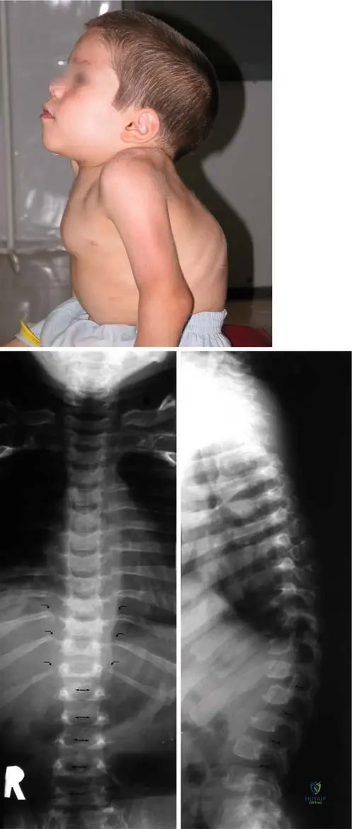
Correct Answer: D
Rationale: The clinical context for Fig. 1.37b explicitly lists "distinct facial features including flat face, depressed nasal bridge, flared nostrils, widely spaced, prominent eyes, thick lips with open mouth and bulging forehead" as characteristic of Hurler syndrome. Prominent chin is incorrect as the description mentions a flat face and thick lips, not a prominent chin.
Question 23
A 4-year-old male presents with a short stature and a noticeably short body trunk. His height is measured at 3 feet, 8 inches.
View Answer & Explanation
Correct Answer: A
Rationale: The clinical context for Fig. 1.37a states, "In Hurler type... of MPS the patient develops a short body trunk and a maximum stature of less than 4 ft." 4-5 ft is incorrect as the text specifies a maximum stature of less than 4 feet.
Question 24
A 5-year-old male with known Hurler syndrome presents for orthopedic evaluation. On physical examination, his lower extremities show a specific angular deformity.
View Answer & Explanation
Correct Answer: A
Rationale: The clinical context for Fig. 1.37a explicitly states, "The valgus knee deformity is not rare in this mucopolysaccharidosis." Varus knee deformity is incorrect; the text specifically mentions valgus deformity.
Question 25
A 3-year-old male with coarse facial features and joint stiffness is noted to have a prominent curvature of his upper back.
View Answer & Explanation
Correct Answer: A
Rationale: The clinical context for Fig. 1.39a states, "Characteristic features for Hurler syndrome are the dorsal kyphosis (a)..." Lumbar lordosis is incorrect; the text specifies dorsal kyphosis.
Question 26
A 4-year-old male with suspected Hurler syndrome undergoes spinal radiographs. The images reveal specific abnormalities in the vertebral bodies.
View Answer & Explanation
Correct Answer: A
Rationale: The clinical context for Fig. 1.39b states, "Characteristic features for Hurler syndrome are... ossification defect on the vertebral bodies (b)..." Sclerosis is incorrect; the text describes an ossification defect, which is a lack of bone formation, rather than increased bone density.
Question 27
A 2-year-old female with developmental delay and coarse features presents with a distinct angular prominence in her lower back.
View Answer & Explanation
Correct Answer: A
Rationale: The clinical context for Fig. 1.39c states, "Characteristic features for Hurler syndrome are... with the very typical dorsolumbar gibbus (c)." Lumbar scoliosis is incorrect; the text specifically identifies a dorsolumbar gibbus as the typical deformity.
Question 28
A 6-year-old male with Hurler syndrome is examined. His hands are noted to have a particular appearance.

View Answer & Explanation
Correct Answer: A
Rationale: The clinical context for Fig. 1.40a states, "The hand is relatively small but wide, the fingers are shortened (a)." Long and slender fingers is incorrect as the description clearly states the fingers are shortened and the hand is wide.
Question 29
A 7-year-old female with Hurler syndrome undergoes hand radiographs. The images show specific changes in the metacarpals and phalanges.
View Answer & Explanation
Correct Answer: A
Rationale: The clinical context for Fig. 1.40b states, "Widening of the proximal part of the phalanges and pointing of the proximal part of the 2–5 metacarpal bones together with claw hand can be observed on radiograph (b) in Hurler disease." Narrowing of the joint spaces is incorrect; the text describes widening of phalanges and pointing of metacarpals, not joint space narrowing.
Question 30
A 5-year-old male with Hurler syndrome presents with difficulty extending his fingers. Radiographs show characteristic changes in the metacarpals and phalanges.
View Answer & Explanation
Correct Answer: A
Rationale: The clinical context for Fig. 1.40b states, "Widening of the proximal part of the phalanges and pointing of the proximal part of the 2–5 metacarpal bones together with claw hand can be observed on radiograph (b) in Hurler disease." Boutonnière deformity is incorrect; the text specifically mentions claw hand in association with the radiographic findings.
Question 31
A 6-year-old female with Morquio disease presents with difficulty walking. On examination, her feet show a specific deformity.

View Answer & Explanation
Correct Answer: A
Rationale: The clinical context for Fig. 1.41 states, "Toe axis deformity and flatfeet can be observed due to generalized ligamentous laxity in Morquio disease." Pes cavus is incorrect; the text describes flatfeet, which is the opposite of pes cavus.
Question 32
A pediatrician suspects a mucopolysaccharidosis in a 1-year-old infant presenting with coarse facial features and hepatosplenomegaly.
View Answer & Explanation
Correct Answer: B
Rationale: The provided text states, "The Hurler syndrome (MPS type I.) is the most common..." Morquio syndrome (MPS type IV) is incorrect as the text identifies it as the most severe, not the most common.
Question 33
A 4-year-old male is diagnosed with a mucopolysaccharidosis, and his parents are inquiring about the prognosis and severity of different types.
View Answer & Explanation
Correct Answer: A
Rationale: The provided text states, "...Morquio syndrome (MPS type IV.) is the most severe MPS." Hurler syndrome (MPS type I) is incorrect as the text identifies it as the most common, not the most severe.
Question 34
A 3-year-old female with suspected mucopolysaccharidosis presents with limited range of motion in her upper extremities.
View Answer & Explanation
Correct Answer: B
Rationale: The general description of MPS states, "...epiphyseal deformation with restricted motion of the joints (particularly in the elbow)..." Shoulder is incorrect; while other joints can be affected, the elbow is specifically highlighted in the text.
Question 35
A 2-year-old male is diagnosed with a mucopolysaccharidosis. Beyond skeletal and facial features, what other systemic involvement is commonly observed?
View Answer & Explanation
Correct Answer: B
Rationale: The general description of MPS lists "corneal clouding" as a characteristic feature. Hyperglycemia is incorrect; it is not mentioned as a characteristic feature of MPS in the provided text.
Question 36
A 4-year-old female with a confirmed diagnosis of mucopolysaccharidosis shows progressive developmental delays.
View Answer & Explanation
Correct Answer: C
Rationale: The general description of MPS lists "mental deterioration" as a characteristic feature. Seizures are incorrect; while some MPS types may have seizures, mental deterioration is explicitly listed as a general characteristic in the provided text.
Question 37
A 5-year-old male with Hurler syndrome is undergoing a comprehensive medical evaluation.
Question 37
A 3-year-old male presents with developmental delay, coarse facial features, and restricted elbow motion. His parents are concerned about the genetic implications for future children. Genetic testing confirms a diagnosis of mucopolysaccharidosis (MPS).
View Answer & Explanation
Correct Answer: D
Rationale: Mucopolysaccharidoses are characterized by autosomal recessive inheritance, meaning both parents must carry a copy of the mutated gene for the child to be affected. X-linked inheritance patterns are not typical for MPS. Main Distractor: Autosomal dominant inheritance would imply only one copy of the mutated gene is needed, which is incorrect for MPS.
Question 38
A 1-year-old female presents with a history of recurrent ear infections, corneal clouding, and a progressively bulging forehead. Physical examination reveals hepatosplenomegaly. To confirm the suspected diagnosis of mucopolysaccharidosis, which of the following is the most appropriate initial diagnostic test?
View Answer & Explanation
Correct Answer: C
Rationale: Diagnosis of mucopolysaccharidoses is typically made using urine analysis for glycosaminoglycan (GAG) levels, which are elevated due to the enzyme deficiency. This is a key initial screening test. Main Distractor: While other tests might be performed later or for complications, a serum electrolyte panel or CBC are non-specific and not primary diagnostic tools for MPS.
Question 39
A 4-year-old male with a known diagnosis of Hurler syndrome (MPS type I) presents for orthopedic evaluation. His parents report increasing difficulty with joint movement. On examination, which joint is most classically noted for restricted motion in MPS patients?
View Answer & Explanation
Correct Answer: E
Rationale: Patients with mucopolysaccharidoses, particularly Hurler syndrome, are characterized by epiphyseal deformation with restricted motion of the joints, with the elbow being particularly affected. Main Distractor: While other joints can be affected, the elbow is specifically highlighted in the text as a site of particular restriction.
Question 40
A 2-year-old female is diagnosed with a mucopolysaccharidosis. Beyond the orthopedic manifestations, which of the following systemic findings is commonly associated with this group of disorders?
View Answer & Explanation
Correct Answer: C
Rationale: Common systemic features of MPS include coarsening of the face, epiphyseal deformation, restricted joint motion, corneal clouding, deafness, mental deterioration, and cardiac disease. Main Distractor: Hyperthyroidism, renal failure, type 1 diabetes, and hemophilia are not characteristic systemic features of MPS.
Question 41
A 2-year-old male presents with a short body trunk and his parents note changes in his facial appearance over the past year. On examination, he exhibits distinct facial features including a flat face, depressed nasal bridge, and thick lips with an open mouth. These findings are characteristic of which specific mucopolysaccharidosis type?
View Answer & Explanation
Correct Answer: D
Rationale: The distinct facial features described (flat face, depressed nasal bridge, flared nostrils, widely spaced, prominent eyes, thick lips with open mouth, and bulging forehead) are characteristic of Hurler syndrome (MPS type I), as shown in Fig. 1.37b. Main Distractor: While other MPS types have facial features, the specific combination described is classic for Hurler syndrome.
Question 42
A 5-year-old male with a confirmed diagnosis of Hurler syndrome (MPS type I) is being evaluated for growth concerns. His parents report he is significantly shorter than his peers. What is the typical maximum stature observed in patients with Hurler syndrome?
View Answer & Explanation
Correct Answer: B
Rationale: In Hurler type MPS, the patient develops a short body trunk and a maximum stature of less than 4 ft, as stated in the clinical context for Fig. 1.37. Main Distractor: While patients are short, "less than 3 ft" is too extreme, and "less than 5 ft" or "less than 6 ft" are not specific to the provided information.
Question 43
A 3-year-old female with Hurler syndrome (MPS type I) presents with gait abnormalities. On physical examination, she is noted to have a significant angular deformity of her lower extremities. Which of the following knee deformities is commonly observed in Hurler syndrome?
View Answer & Explanation
Correct Answer: C
Rationale: The clinical context for Fig. 1.37a explicitly states that "The valgus knee deformity is not rare in this mucopolysaccharidosis (Hurler type)." Main Distractor: Genu varum (bow-leggedness) is the opposite deformity and not specifically mentioned as common in Hurler syndrome.
Question 44
A 4-year-old male with Hurler syndrome (MPS type I) is undergoing spinal imaging due to concerns about posture. Radiographs reveal a characteristic spinal deformity. Which of the following is a typical spinal finding in Hurler syndrome?
View Answer & Explanation
Correct Answer: D
Rationale: Characteristic features for Hurler syndrome include dorsal kyphosis and a very typical dorsolumbar gibbus, as shown in Fig. 1.39a and c. Main Distractor: While cervical instability can occur in some MPS types (e.g., Morquio), dorsal kyphosis is specifically highlighted for Hurler syndrome in the provided text and image.
Question 45
A 3-year-old female with Hurler syndrome (MPS type I) has radiographs of her chest as part of her routine evaluation. What characteristic finding related to the ribs might be observed on these radiographs?
View Answer & Explanation
Correct Answer: C
Rationale: Fig. 1.39b explicitly states that characteristic features for Hurler syndrome include "widening the lateral portion of the ribs." Main Distractor: Rib notching is associated with coarctation of the aorta, and bifid ribs are a congenital anomaly not specifically linked to Hurler syndrome in the text.
Question 46
A 2-year-old male with Hurler syndrome (MPS type I) presents with a prominent dorsolumbar gibbus. Radiographs of the spine are obtained. What specific vertebral body finding is characteristic of Hurler syndrome?
View Answer & Explanation
Correct Answer: C
Rationale: Fig. 1.39b highlights "ossification defect on the vertebral bodies" as a characteristic feature for Hurler syndrome, often contributing to the dorsolumbar gibbus. Main Distractor: While compression fractures can occur in various conditions, an ossification defect is a specific developmental anomaly noted in Hurler syndrome.
Question 47
A 3-year-old female with Hurler syndrome (MPS type I) is noted to have unusual hand morphology. On physical examination, her hands are described as relatively small but wide, with shortened fingers. This appearance is often associated with which of the following radiographic findings?
View Answer & Explanation
Correct Answer: B
Rationale: The text for Fig. 1.40a describes the hand as "relatively small but wide, the fingers are shortened." Fig. 1.40b further states that "Widening of the proximal part of the phalanges... can be observed on radiograph." Main Distractor: Carpal tunnel syndrome is a complication, not a primary radiographic finding of the bone morphology. Polydactyly and syndactyly are congenital anomalies not described here.
Question 48
A 5-year-old male with Hurler syndrome (MPS type I) undergoes hand radiographs due to progressive stiffness and deformity. The radiograph reveals a characteristic appearance of the metacarpal bones. Which of the following is a typical radiographic finding of the metacarpals in Hurler disease?
View Answer & Explanation
Correct Answer: B
Rationale: Fig. 1.40b explicitly states: "pointing of the proximal part of the 2–5 metacarpal bones together with claw hand can be observed on radiograph in Hurler disease." Main Distractor: While metacarpals can be affected, "shortening of all metacarpals" is less specific than the characteristic pointing described.
Question 49
A 6-year-old female with Morquio disease (MPS type IV) presents with difficulty walking and complains of foot pain. On examination, she exhibits a characteristic foot deformity. Which of the following foot deformities is commonly observed in Morquio disease?
View Answer & Explanation
Correct Answer: C
Rationale: Fig. 1.41 states: "Toe axis deformity and flatfeet can be observed due to generalized ligamentous laxity in Morquio disease." Main Distractor: Clubfoot and pes cavus are different foot deformities not specifically mentioned as characteristic of Morquio disease in the provided text.
Question 50
A 7-year-old male with Morquio disease (MPS type IV) presents with progressive toe axis deformity and flatfeet. What is the underlying pathological mechanism contributing to these foot deformities in Morquio disease?
View Answer & Explanation
Correct Answer: C
Rationale: The clinical context for Fig. 1.41 explicitly states that "Toe axis deformity and flatfeet can be observed due to generalized ligamentous laxity in Morquio disease." Main Distractor: While bone dysplasia is part of MPS, the specific cause for the foot deformities in Morquio is attributed to ligamentous laxity, not primarily bone dysplasia or neuromuscular issues.
Question 51
A 1-year-old male presents with coarse facial features, corneal clouding, and hepatosplenomegaly. Genetic testing confirms a diagnosis of mucopolysaccharidosis. Among the various types of MPS, which one is considered the most common?
View Answer & Explanation
Correct Answer: D
Rationale: The introductory text states: "The Hurler syndrome (MPS type I.) is the most common and Morquio syndrome (MPS type IV.) is the most severe MPS." Main Distractor: Morquio syndrome is noted as the most severe, not the most common.
Question 52
A 2-year-old female is diagnosed with a mucopolysaccharidosis, presenting with severe skeletal dysplasia, short stature, and significant orthopedic challenges. Her prognosis is guarded due to the severity of her condition. Which type of MPS is described as the most severe?
View Answer & Explanation
Correct Answer: D
Rationale: The introductory text states: "The Hurler syndrome (MPS type I.) is the most common and Morquio syndrome (MPS type IV.) is the most severe MPS." Main Distractor: Hurler syndrome is the most common, but Morquio is specifically noted as the most severe.
Question 53
A 1-year-old male presents with developmental delay and coarse facial features. Diagnostic workup confirms a mucopolysaccharidosis. This condition is fundamentally caused by a deficiency in which type of enzyme?
View Answer & Explanation
Correct Answer: D
Rationale: The introductory text states: "It is caused when a hydrolase enzyme deficiency creates an accumulation of mucopolysaccharides." Main Distractor: Amylase, lipase, protease, and kinase are different classes of enzymes not primarily implicated in the pathophysiology of MPS.
Question 54
A 6-month-old female is suspected of having a mucopolysaccharidosis based on early clinical signs. The underlying pathophysiology involves the accumulation of which specific substance due to an enzyme deficiency?
View Answer & Explanation
Correct Answer: C
Rationale: The introductory text states: "It is caused when a hydrolase enzyme deficiency creates an accumulation of mucopolysaccharides." Main Distractor: Glycogen and sphingolipids accumulate in other lysosomal storage diseases (e.g., glycogen storage diseases, sphingolipidoses), but mucopolysaccharides (glycosaminoglycans) are specific to MPS.
Question 55
A 9-month-old male is undergoing evaluation for suspected mucopolysaccharidosis due to subtle developmental delays and early signs of facial coarsening. At what age do most patients with MPS typically become symptomatic?
View Answer & Explanation
Correct Answer: B
Rationale: The introductory text states: "Most patients become symptomatic in early childhood." While some signs may appear in infancy, the full symptomatic picture typically emerges in early childhood. Main Distractor: Infancy might show some signs, but "early childhood" is the more accurate general statement from the text.
Question 56
A 22-year-old male presents with 6 months of localized pain and swelling in his proximal tibia. Physical examination reveals mild tenderness over the area with no significant warmth or erythema. Radiographs show a lytic lesion in the metaphysis. Biopsy is planned. Based on the most common presentation of this rare benign cartilaginous tumor, which of the following is the most likely diagnosis?
View Answer & Explanation
Correct Answer: D
Rationale: Chondromyxoid fibroma is a rare benign cartilaginous tumor that commonly presents in males in the first three decades of life with pain and local swelling, most frequently in the tibia. Osteochondroma typically presents as a painless exostosis. Enchondroma is often asymptomatic unless complicated by fracture. Chondroblastoma is also a benign cartilaginous tumor but often affects epiphyses. Low-grade chondrosarcoma is a malignant tumor, and while it can present with pain, the demographics and typical benign presentation described make CMF more likely in this initial assessment.
Question 57
A 19-year-old male presents with chronic pain and swelling in his left ankle. Imaging reveals a lytic lesion in the distal tibia. A biopsy is performed. Given the typical demographic and most common location for chondromyxoid fibroma, which bone is most frequently affected?
View Answer & Explanation
Correct Answer: D
Rationale: The clinical context explicitly states that chondromyxoid fibromas occur most commonly in the tibia. While other options like the femur, humerus, and small bones of the hands and feet are also common sites, the tibia is listed as the most frequent location. The fibula is not specifically mentioned as a common site.
Question 58
A 25-year-old male presents with a several-month history of pain and swelling in his right hand. Radiographs show a lytic lesion in the metacarpal. An intraoperative image of the resected tissue is obtained. Which of the following best describes the gross appearance of a chondromyxoid fibroma?
View Answer & Explanation
Correct Answer: C
Rationale: The clinical context for Fig. 8.49 explicitly states that the tissue of a chondromyxoid fibroma often resembles hyaline cartilage, which is typically described as gelatinous and bluish-white. The image itself shows a glistening, somewhat translucent appearance consistent with cartilage. The other options describe appearances more typical of other bone tumors (e.g., osteosarcoma, lipoma, giant cell tumor).
Question 59
A 17-year-old female presents with pain in her foot. Radiographs reveal a lytic lesion in a metatarsal bone. A biopsy is performed. On low-power microscopic examination, the pathologist notes a distinctive architectural pattern. Which of the following is the characteristic growth pattern of chondromyxoid fibroma seen on low-power histology?
View Answer & Explanation
Correct Answer: C
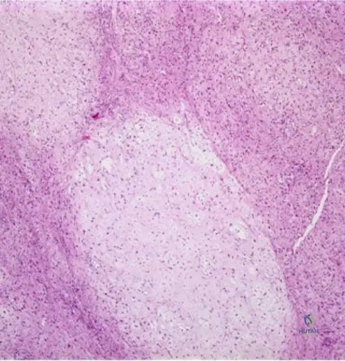
Rationale: The clinical context for Fig. 8.50 explicitly states that a low-power photomicrograph of chondromyxoid fibroma shows a lobular growth pattern. This is a key diagnostic feature. Spiculated or trabecular bone formation is characteristic of osteoid-forming tumors, diffuse infiltrative growth is more typical of malignancy, and a cartilaginous cap is seen in osteochondromas.
Question 60
A 28-year-old male presents with localized swelling and discomfort in his distal femur. Imaging reveals a benign-appearing lytic lesion. A biopsy confirms the diagnosis of chondromyxoid fibroma. On higher power microscopic examination, which of the following cellular and stromal features are characteristic?
View Answer & Explanation
Correct Answer: B
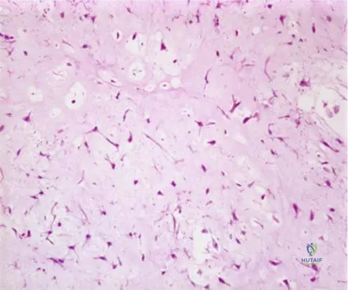
Rationale: The clinical context for Fig. 8.51 explicitly describes the higher power photomicrograph showing "myxoid/chondroid stroma and lobules of spindle shaped or stellate cells with abundant myxoid or chondroid intercellular material." This directly matches option B. The other options describe features of other tumors (e.g., clear cell chondrosarcoma, giant cell tumor, chondrosarcoma, osteoblastoma/osteosarcoma).
Question 61
A 16-year-old male presents with a several-month history of mild pain and swelling in his proximal humerus. Physical examination reveals subtle tenderness. Radiographs show a well-defined lytic lesion. A biopsy is performed. Based on the typical nature of chondromyxoid fibroma, what is its classification?
View Answer & Explanation
Correct Answer: D
Rationale: The introductory text explicitly states, "Chondromyxoid fibromas are rare benign cartilaginous tumors." This directly answers the question. The other options describe different types of tumors.
Question 62
A 20-year-old male presents with pain and swelling in his right foot, specifically involving a small bone. Physical examination reveals localized tenderness. Radiographs show a lytic lesion. Which of the following locations, besides the tibia, is commonly affected by chondromyxoid fibroma?
View Answer & Explanation
Correct Answer: D
Rationale: The clinical context for Fig. 8.48 states, "Lesions occur most commonly in the tibia, followed by small bones of the hands and feet, femur, and humerus." Therefore, small bones of the hands and feet are a common location. The image itself shows a metacarpal lesion, reinforcing this point. The other options are not listed as common sites for CMF.
Question 63
A 15-year-old male presents with a several-month history of mild pain in his distal femur. Physical examination is largely unremarkable, with only subtle tenderness on deep palpation. Radiographs show a well-defined lytic lesion. What are the two most common patient complaints associated with chondromyxoid fibroma?
View Answer & Explanation
Correct Answer: C
Rationale: The text explicitly states, "Pain and local swelling are the most common patient complaints." The other options describe symptoms not typically associated with CMF or are less common.
Question 64
A 21-year-old male presents with chronic pain in his proximal tibia. Physical examination reveals localized tenderness but no significant mass or deformity. Radiographs show a lytic lesion. Which of the following statements best describes the typical physical examination findings for chondromyxoid fibroma?
View Answer & Explanation
Correct Answer: C
Rationale: The text states, "Physical exam may reveal tenderness but many lesions have minimal findings on examination." This aligns with option C. The other options describe more pronounced or different physical findings not typical for CMF.
Question 65
A 14-year-old male presents with pain in his right humerus. Radiographs reveal a lytic lesion. A biopsy is performed, and the pathologist notes a myxoid/chondroid stroma with spindle-shaped cells. Which of the following is a key characteristic of the intercellular material in chondromyxoid fibroma?
View Answer & Explanation
Correct Answer: C
Rationale: The clinical context for Fig. 8.51 explicitly mentions "abundant myxoid or chondroid intercellular material" as a feature of chondromyxoid fibroma. This is a defining histological characteristic. The other options describe features of other bone or soft tissue lesions.
Question 66
A 23-year-old male presents with chronic pain and swelling in his distal tibia. Radiographs show a lytic lesion. Given the typical demographics of chondromyxoid fibroma, which age group is most commonly affected?
View Answer & Explanation
Correct Answer: C
Rationale: The text states that "most patients present in the first 3 decades." This corresponds to ages 0-30 years. The other age groups are less common for this tumor.
Question 67
A 17-year-old male presents with localized pain and swelling in his proximal tibia. Radiographs show a lytic lesion. A biopsy is performed. Which of the following cell types is characteristic of chondromyxoid fibroma on high-power microscopy?
View Answer & Explanation
Correct Answer: C
Rationale: The clinical context for Fig. 8.51 explicitly mentions "lobules of spindle shaped or stellate cells" as a feature of chondromyxoid fibroma. This is a key cellular characteristic. The other options describe cell types associated with other tumors (e.g., epithelioid sarcoma, Ewing's sarcoma, osteochondroma, giant cell tumor).
Question 68
A 26-year-old male presents with pain in his right femur. Radiographs show a lytic lesion. Given the typical gender predisposition for chondromyxoid fibroma, which statement is true?
View Answer & Explanation
Correct Answer: C
Rationale: The introductory text states, "They more commonly occur in males." This directly answers the question. While it's more common in males, it's not exclusively found in them, making option E incorrect.
Question 69
A 19-year-old male presents with localized pain and swelling in his left metacarpal. Radiographs confirm a lytic lesion. An intraoperative image of the resected specimen is shown. What type of tissue does the gross appearance of chondromyxoid fibroma typically resemble?
View Answer & Explanation
Correct Answer: C
Rationale: The clinical context for Fig. 8.49 explicitly states, "The tissue often resembles hyaline cartilage." This is a direct factual recall from the provided text. The image itself shows a glistening, somewhat translucent appearance consistent with cartilage.
Question 70
A 24-year-old male presents with pain in his right humerus. Radiographs show a lytic lesion. A biopsy is performed. On low-power microscopy, the pathologist observes a distinct lobular architecture. What is the significance of this lobular pattern in the diagnosis of chondromyxoid fibroma?
View Answer & Explanation
Correct Answer: B
Rationale: The clinical context for Fig. 8.50 highlights the "lobular growth pattern" as a feature of chondromyxoid fibroma. For a benign tumor, this is a characteristic architectural feature aiding in diagnosis. It does not indicate malignancy, vascular origin, or a non-specific finding. The lobular pattern is a key diagnostic clue for CMF.
Question 71
A 16-year-old male presents with pain and swelling in his right foot. Radiographs show a lytic lesion in a tarsal bone. A biopsy is performed. High-power microscopy reveals spindle-shaped cells within a myxoid stroma. Which of the following terms best describes the overall rarity of chondromyxoid fibroma?
View Answer & Explanation
Correct Answer: D
Rationale: The introductory text explicitly states, "Chondromyxoid fibromas are rare benign cartilaginous tumors." This directly answers the question regarding its prevalence. The other options are incorrect descriptions of its rarity.
Question 72
A 29-year-old male presents with localized pain and swelling in his distal tibia. Physical examination reveals mild tenderness. Radiographs show a lytic lesion. Considering the common locations for chondromyxoid fibroma, which of the following bones is *least* likely to be affected among the choices provided?
View Answer & Explanation
Correct Answer: E
Rationale: The clinical context for Fig. 8.48 lists the tibia (most common), small bones of the hands and feet (e.g., metacarpal), femur, and humerus as common locations. The vertebrae are not mentioned as a common site for chondromyxoid fibroma, making it the least likely among the given options based on the provided text.
Question 73
A 17-year-old male presents with a 4-month history of pain and swelling in his proximal tibia. Physical examination reveals mild tenderness. Radiographs show a lytic lesion. A biopsy is performed, and the pathologist notes a lobular growth pattern with spindle-shaped cells in a myxoid matrix. Which of the following is the primary tissue component from which chondromyxoid fibroma is derived?
View Answer & Explanation
Correct Answer: C
Rationale: The introductory text explicitly states, "Chondromyxoid fibromas are rare benign cartilaginous tumors." This indicates their derivation from cartilaginous tissue. The histological features (myxoid/chondroid stroma) further support this classification.
Question 74
A 20-year-old male presents with localized pain and swelling in his right distal femur. Physical examination reveals mild tenderness. Radiographs show a lytic lesion. A biopsy is performed. The pathologist notes a lobular pattern with stellate cells in a myxoid background. What is the typical age range of presentation for most patients with chondromyxoid fibroma?
View Answer & Explanation
Correct Answer: B
Rationale: The text states that "most patients present in the first 3 decades." This corresponds to adolescence and young adulthood, which is typically considered up to around 30 years of age. The other options represent different age ranges that are less common for CMF.
Question 75
A 27-year-old male presents with chronic pain and swelling in his left proximal humerus. Physical examination reveals mild tenderness. Radiographs show a lytic lesion. A biopsy is performed. On high-power microscopy, the pathologist identifies lobules of spindle-shaped cells within an abundant myxoid and chondroid intercellular matrix. Which of the following is a characteristic feature of the stromal component of chondromyxoid fibroma?
View Answer & Explanation
Correct Answer: C
Rationale: The clinical context for Fig. 8.51 explicitly describes "myxoid/chondroid stroma" as a key feature of chondromyxoid fibroma. This is a defining characteristic of the tumor's intercellular material. The other options describe stromal components of different types of tumors.
Question 76
A 22-year-old male presents with chronic pain and swelling in his distal tibia. Radiographs show a lytic lesion in the metaphysis. A biopsy is performed. On high-power photomicrography, which of the following features is most characteristic of chondromyxoid fibroma?
View Answer & Explanation
Correct Answer: B
Rationale: The higher power photomicrograph (Fig. 8.51) of chondromyxoid fibroma characteristically shows lobules of spindle-shaped or stellate cells with abundant myxoid or chondroid intercellular material. This is a defining histological feature. Option C describes Giant Cell Tumor, while Option A describes epithelioid sarcoma or similar lesions not typical for CMF. Option D describes normal bone marrow or lipoma. Option E describes osteochondroma. The main distractor, C, describes a Giant Cell Tumor, which can also present as a lytic lesion in a young adult, but has distinct histological features.
Question 77
A 19-year-old male presents with localized tenderness and swelling in his foot. Imaging reveals a benign-appearing lesion in a metatarsal. A biopsy is performed. On low-power photomicrography, which growth pattern is most indicative of a chondromyxoid fibroma?
View Answer & Explanation
Correct Answer: C
Rationale: Low power photomicrographs of chondromyxoid fibroma (Fig. 8.50) typically demonstrate a distinct lobular growth pattern. This is a key diagnostic feature distinguishing it from other bone tumors. Option B describes osteosarcoma or osteoblastoma. Option D describes fibrosarcoma or fibrous dysplasia. The main distractor, D, describes a fibrous lesion, which has a different architectural pattern.
Question 78
During an intraoperative procedure for a benign bone tumor in a 25-year-old male, the surgeon notes the gross appearance of the resected tissue. Which description is most consistent with a chondromyxoid fibroma?
View Answer & Explanation
Correct Answer: C
Rationale: As noted in the clinical context for Fig. 8.49, the intraoperative gross appearance of chondromyxoid fibroma often shows tissue that resembles hyaline cartilage, typically described as gelatinous and glistening due to its myxoid and chondroid components. Option A describes a more aggressive or malignant lesion. Option B describes osteoma or osteochondroma. Option D describes a lipoma. Option E describes a fibroma or desmoid tumor. The main distractor, A, describes features more commonly associated with malignant or highly aggressive benign lesions.
Question 79
A 17-year-old male presents with localized pain and swelling in his lower extremity. Radiographs reveal a lytic lesion. Based on the typical epidemiology of chondromyxoid fibroma, which of the following bones is the most common site for this tumor?
View Answer & Explanation
Correct Answer: C
Rationale: The clinical context for Fig. 8.48 explicitly states that chondromyxoid fibromas occur most commonly in the tibia. While other locations listed are possible, the tibia is the most frequent site. The main distractor, A (Femur), is a common site for many bone tumors but is listed as less common than the tibia for CMF.
Question 80
A 28-year-old female presents with persistent pain and swelling in her left hand. Radiographs show a well-defined lytic lesion in the proximal phalanx. Considering the typical locations for chondromyxoid fibroma, which category of bones is the second most common site for this tumor?
View Answer & Explanation
Correct Answer: D
Rationale: The clinical context for Fig. 8.48 states that lesions occur most commonly in the tibia, followed by small bones of the hands and feet. The image itself shows a metacarpal lesion, reinforcing the prevalence in small bones. The main distractor, A (Femur), is listed as a common site but less common than small bones of the hands and feet.
Question 81
A 45-year-old male presents with a new onset of pain and swelling in his distal femur. Radiographs show a lytic lesion. While chondromyxoid fibroma is a possibility, which demographic factor makes this presentation less typical for this specific tumor?
View Answer & Explanation
Correct Answer: C
Rationale: The teaching case states that most patients with chondromyxoid fibroma present in the first 3 decades (i.e., under 30 years old). A 45-year-old patient is outside this typical age range, making age the demographic factor that makes this presentation less typical. Gender (male) and symptoms (pain and swelling) are consistent. Femur is a listed location, though less common than tibia or small bones. The main distractor, A (Gender), is incorrect because males are more commonly affected, making the patient's gender consistent with CMF.
Question 82
A 20-year-old patient presents with a painful lesion in the proximal humerus. Imaging suggests a benign cartilaginous tumor. When considering chondromyxoid fibroma, which gender is more commonly affected?
View Answer & Explanation
Correct Answer: A
Rationale: The teaching case explicitly states that chondromyxoid fibromas more commonly occur in males. The main distractor, B (Females), is incorrect as males are more commonly affected.
Question 83
A 16-year-old male presents to the clinic with concerns about his left ankle. He reports a gradual onset of discomfort and a noticeable enlargement over the past few months. On examination, there is mild tenderness and palpable swelling. Which of the following are the most common patient complaints for chondromyxoid fibroma?
View Answer & Explanation
Correct Answer: C
Rationale: The teaching case states that "Pain and local swelling are the most common patient complaints" for chondromyxoid fibroma. The vignette describes these exact symptoms. The main distractor, A (Pathologic fracture and fever), describes symptoms more typical of aggressive or malignant lesions, or infection, which are not the most common for benign CMF.
Question 84
A 23-year-old female presents with mild, intermittent pain in her right foot. She points to the dorsal aspect of her navicular bone. On physical examination, the orthopaedic surgeon notes minimal findings, with only slight tenderness upon deep palpation. This presentation is consistent with chondromyxoid fibroma because:
View Answer & Explanation
Correct Answer: B
Rationale: The teaching case explicitly states that "Physical exam may reveal tenderness but many lesions have minimal findings on examination." This aligns perfectly with the vignette's description of minimal findings and slight tenderness. The main distractor, C (Chondromyxoid fibromas are usually asymptomatic), is incorrect because pain and local swelling are the most common complaints, indicating they are often symptomatic, even if physical exam findings are minimal.
Question 85
A 21-year-old male presents with a several-month history of pain and swelling in his proximal tibia. Radiographs show a well-defined, eccentric lytic lesion. A biopsy confirms the diagnosis of chondromyxoid fibroma. Based on the nature of this tumor, it is classified as a:
View Answer & Explanation
Correct Answer: B
Rationale: The very first sentence of the teaching case states, "Chondromyxoid fibromas are rare benign cartilaginous tumors." The main distractor, A (Malignant fibrous tumor), is incorrect as CMF is benign and cartilaginous, not fibrous or malignant.
Question 86
A 15-year-old male presents with pain in his right hand. Radiographs reveal a well-defined, expansile lytic lesion involving the metacarpal bone, as depicted in the image. This radiographic appearance is characteristic of chondromyxoid fibroma in this location.
View Answer & Explanation
Correct Answer: C
Rationale: The provided image (Fig. 8.48) clearly shows a well-defined, expansile lytic lesion in the metacarpal, which implies a narrow zone of transition, typical of a benign process like chondromyxoid fibroma. While the text doesn't explicitly describe the radiographic appearance, the image provides this context. The main distractor, B (Lytic lesion with internal calcifications), can be seen in other cartilaginous lesions like enchondromas, but the image primarily shows a lytic defect without obvious calcifications.
Question 87
A 24-year-old male presents with pain and swelling in his proximal phalanx of the great toe. Radiographs show a lytic lesion. Given the location in a small bone of the foot and the patient's age, which of the following benign tumors would be a primary differential diagnosis, alongside chondromyxoid fibroma?
View Answer & Explanation
Correct Answer: C
Rationale: Chondromyxoid fibroma commonly occurs in the small bones of the hands and feet. Enchondroma is another very common benign cartilaginous tumor in these locations and often presents in a similar age group with lytic lesions. Osteosarcoma, Ewing Sarcoma, Chondrosarcoma, and Multiple Myeloma are either malignant or typically present in different age groups/locations or with different radiographic features. The main distractor, A (Osteosarcoma), is a malignant tumor and while it can occur in young adults, it is less common in the small bones of the hands and feet and would typically have more aggressive radiographic features.
Question 88
A 14-year-old male presents with a painful lesion in his distal femur. A biopsy confirms chondromyxoid fibroma. The combination of the patient's age, gender, and the location in the femur is consistent with the typical presentation of this tumor. Which of the following statements about its demographics is true?
View Answer & Explanation
Correct Answer: C
Rationale: The teaching case states that chondromyxoid fibromas "more commonly occur in males and most patients present in the first 3 decades." This directly supports option C. The main distractor, A (It is more common in females over 40), is incorrect as CMF is more common in younger males.
Question 89
A 20-year-old male presents with a painful swelling in his right hand. Radiographs reveal a lytic lesion in the metacarpal bone, as shown in the image. This specific location (metacarpal) for chondromyxoid fibroma is categorized as:
View Answer & Explanation
Correct Answer: C
Rationale: The clinical context for Fig. 8.48 states that lesions occur most commonly in the tibia, followed by small bones of the hands and feet. The metacarpal is a small bone of the hand, fitting into this second most common category. The main distractor, A (The most common site overall), is incorrect because the tibia is the most common site.
Question 90
A 26-year-old male presents with a lytic lesion identified in his cervical spine. While chondromyxoid fibroma is a consideration for benign bone tumors in young adults, based on the provided information, which of the following locations is LEAST commonly affected by chondromyxoid fibroma?
View Answer & Explanation
Correct Answer: E
Rationale: The teaching case lists the most common locations as tibia, small bones of the hands and feet, femur, and humerus. The spine is not mentioned in this list, implying it is a less common or rare site compared to the options provided. The main distractor, B (Femur), is incorrect because the femur is explicitly listed as a common location, albeit less common than the tibia or small bones.
Question 91
A 65-year-old female presents with a lytic lesion in her distal tibia, causing mild pain. A biopsy is planned. Considering the typical demographics of chondromyxoid fibroma, which aspect of this patient's presentation makes it the LEAST typical for this diagnosis?
View Answer & Explanation
Correct Answer: D
Rationale: Chondromyxoid fibromas most commonly occur in patients in the first 3 decades of life (under 30 years old) and are more common in males. A 65-year-old female is significantly outside the typical age range and gender predilection. While the tibia is a common location and pain is a common symptom, the age is the most striking atypical feature. The main distractor, C (Patient's gender), is also atypical (more common in males), but the age (65 vs. first 3 decades) is a more significant deviation from the typical demographic profile.
Question 92
A 20-year-old male undergoes a biopsy for a lytic lesion in his proximal humerus. The pathologist examines the tissue under high power. Which specific histological characteristic, as depicted in the image, is most crucial for diagnosing chondromyxoid fibroma?
View Answer & Explanation
Correct Answer: B
Rationale: The higher power photomicrograph (Fig. 8.51) and its context describe "myxoid/chondroid stroma and lobules of spindle shaped or stellate cells with abundant myxoid or chondroid intercellular material." This is the most characteristic high-power histological feature. The main distractor, A (Presence of numerous osteoclast-like giant cells), describes a Giant Cell Tumor, which can be a differential but has distinct stromal cells.
Question 93
A 17-year-old female presents with a painful lesion in her distal fibula. A biopsy is performed. On low-power microscopic examination, the pathologist observes a distinct architectural arrangement of the tumor cells. Which low-power feature, as shown in the image, is most characteristic of chondromyxoid fibroma?
View Answer & Explanation
Correct Answer: C
Rationale: The low power photomicrograph (Fig. 8.50) and its context specifically highlight a "lobular growth pattern" as a characteristic feature of chondromyxoid fibroma. The main distractor, A (A diffuse, infiltrative pattern), is incorrect as CMF typically has a more circumscribed, lobular architecture, even if it can be locally aggressive.
Question 94
During surgical excision of a benign bone tumor from the proximal tibia of a 28-year-old male, the surgeon observes the gross appearance of the tumor tissue. Which description of the tissue's appearance, as shown in the image, would strongly suggest a diagnosis of chondromyxoid fibroma?
View Answer & Explanation
Correct Answer: C
Rationale: The clinical context for Fig. 8.49 explicitly states that the gross appearance of chondromyxoid fibroma tissue often resembles hyaline cartilage, which is typically glistening and translucent. The main distractor, D (Reddish-brown, friable, and hemorrhagic), describes features more commonly associated with aneurysmal bone cysts or giant cell tumors, or more aggressive lesions.
Question 95
A 24-year-old male presents with a history of progressive limb pain and muscle weakness, leading to a waddling gait. Physical examination reveals a thin habitus with columnar shaped extremities and decreased muscle mass. Radiographs of the long bones demonstrate periosteal and endosteal sclerosis and thickening of the diaphyses. Skull radiographs show sclerosis of the skull base. Which of the following is the most likely diagnosis?

View Answer & Explanation
Correct Answer: C
Rationale: The clinical presentation of progressive limb pain, muscle weakness, waddling gait, thin habitus, and characteristic radiographic findings of periosteal and endosteal sclerosis and thickening of the diaphyses of long bones, along with skull base involvement, are pathognomonic for Progressive Diaphyseal Dysplasia, also known as Camurati–Engelmann Disease. Osteopetrosis (D) also involves bone sclerosis but typically presents with diffuse skeletal sclerosis, bone fragility, and often hematological issues, rather than isolated diaphyseal thickening and the specific clinical habitus described.
Question 96
A 15-year-old male presents with chronic limb pain and a family history of similar symptoms in his father. Radiographs show diffuse diaphyseal cortical thickening and sclerosis of the long tubular bones. Genetic testing is being considered. What is the most likely inheritance pattern for this condition?
View Answer & Explanation
Correct Answer: C
Rationale: Progressive diaphyseal dysplasia (Camurati–Engelmann disease) is explicitly stated to be an autosomal dominant disorder. The family history of similar symptoms in the father further supports an autosomal dominant inheritance pattern. Autosomal recessive (A) would typically require both parents to be carriers, and X-linked patterns (B, D) would show different inheritance patterns related to sex chromosomes.
Question 97
A 30-year-old female presents with a long-standing history of limb pain and progressive muscle weakness. Radiographs of her humerus, femur, tibia, and fibula reveal significant cortical thickening and sclerosis primarily affecting the diaphyses of these long bones. Which of the following best describes the primary radiographic characteristic of Camurati–Engelmann disease?
View Answer & Explanation
Correct Answer: C
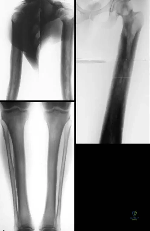
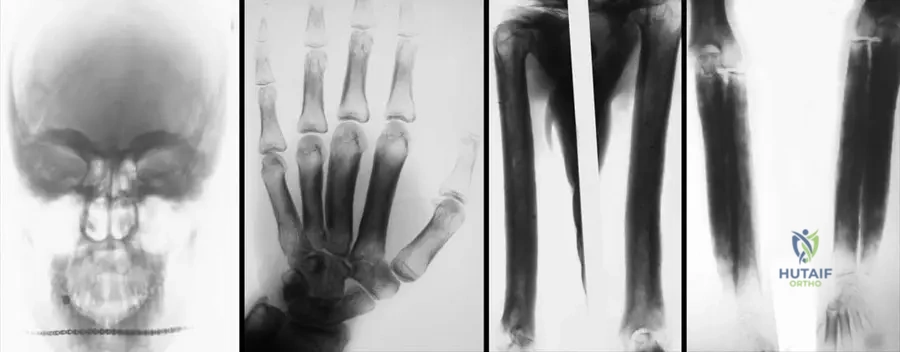
Rationale: The clinical text and accompanying images (Fig. 1.120) clearly state that "Diaphyseal cortical thickening and sclerosis of long tubular bones are the main characteristics of Camurati-Engelmann disease." Options A, B, D, and E describe features of other bone dysplasias or conditions not consistent with the provided information.
Question 98
A 10-year-old boy is brought to the clinic by his parents due to increasing difficulty walking and frequent complaints of leg pain, especially after activity. His gait is described as a "waddle." Physical examination confirms muscle weakness in the lower extremities. Radiographs show early signs of diaphyseal sclerosis in the long bones. Which of the following is a frequent symptom associated with this condition?
View Answer & Explanation
Correct Answer: D
Rationale: The text explicitly lists "limb pain, muscle weakness, waddling gait, and sometimes deafness" as the most frequent symptoms. Limb pain (D) directly matches this description. The other options are not mentioned as frequent symptoms of Camurati–Engelmann disease.
Question 99
A 45-year-old male with a known diagnosis of Progressive Diaphyseal Dysplasia presents with new-onset headaches and hearing loss. Radiographs of his skull are obtained. Based on the typical presentation of this disease, which area of the skull is most likely to show involvement?
View Answer & Explanation
Correct Answer: B
Rationale: The teaching case states that Camurati–Engelmann disease involves the "skull base." The image Fig. 1.122a also shows sclerosis of the skull. Sclerosis of the skull base (B) is consistent with the disease and can explain symptoms like hearing loss due to cranial nerve impingement. The other options are not characteristic features of this condition.
Question 100
A 7-year-old girl presents with progressive leg pain and a waddling gait. Radiographs show bilateral symmetrical diaphyseal cortical thickening and sclerosis of the femurs and tibias. Her parents are concerned about other bone disorders. Which of the following conditions is most important to differentiate from Camurati–Engelmann disease, given its similar radiographic feature of increased bone density?
View Answer & Explanation
Correct Answer: C
Rationale: Osteopetrosis (C) is a group of disorders characterized by increased bone density (sclerosis) due to defective osteoclast function, making it a key differential for conditions presenting with sclerotic bones. However, osteopetrosis typically involves diffuse skeletal sclerosis, often with bone fragility and hematological complications, whereas Camurati–Engelmann disease primarily affects the diaphyses of long bones, skull base, and clavicles with cortical thickening. Rickets (A) involves decreased bone mineralization, Achondroplasia (B) is a dwarfism with short limbs, and Marfan (D) and Ehlers-Danlos (E) syndromes are connective tissue disorders, none of which primarily present with diaphyseal sclerosis.
You Might Also Like