Complex Environmental and Ballistic Trauma to the Hand: Shotgun Injuries and Frostbite
Key Takeaway
Shotgun wounds to the upper extremity present complex reconstructive challenges due to clustered shot destruction and wadding contamination. Conversely, frostbite injuries require extreme patience, relying on advanced imaging like Tc-99m bone scans to delineate tissue necrosis before delayed amputation. Both mechanisms demand meticulous, staged surgical protocols, ranging from aggressive early debridement in ballistic trauma to delayed reconstructive interventions for cold-induced physeal arrest in pediatric patients.
Comprehensive Introduction and Patho-Epidemiology
The management of severe upper extremity trauma requires a profound understanding of the underlying mechanism of injury, as the energy transfer and physiological insult dictate both the immediate surgical approach and the long-term reconstructive strategy. Within the broad spectrum of orthopedic traumatology, environmental and ballistic injuries represent two of the most challenging and physiologically distinct paradigms. This chapter comprehensively addresses two highly destructive mechanisms of hand trauma: close-range shotgun injuries and severe frostbite. While shotgun wounds demand aggressive, immediate surgical intervention to mitigate gross contamination and stabilize massive tissue disruption, frostbite requires a counterintuitive paradigm of extreme patience, delayed surgical intervention, and meticulous physiological preservation.
Shotgun injuries represent a unique and devastating subset of ballistic trauma. Unlike standard handgun injuries, which typically involve a single projectile generating a predictable wound tract, shotgun blasts deliver multiple projectiles (pellets) simultaneously, often accompanied by the introduction of highly contaminated foreign bodies. The severity of a shotgun wound is inversely proportional to the distance between the weapon and the victim. While shotguns are generally classified as low-velocity weapons—with muzzle velocities typically ranging between 1,000 and 1,500 feet per second—the clustered shot mass at close range (less than 3 meters) acts as a single, highly destructive projectile. The kinetic energy transfer at this proximity is massive, resulting in a wound profile characterized by severe cavitation, tissue avulsion, and a zone of thermal coagulation that mimics or exceeds the destruction seen with high-velocity military rifles. Epidemiologically, these injuries are frequently encountered in civilian trauma centers as a result of hunting accidents, close-quarters altercations, and suicide attempts, with the upper extremity often sustaining defensive or accidental point-blank trauma.
In stark contrast to the acute, kinetic destruction of ballistic trauma, frostbite is an insidious environmental insult characterized by localized tissue freezing and subsequent microvascular collapse. The pathophysiology of frostbite is biphasic, involving both direct cellular toxicity and delayed microvascular thrombosis. As tissue temperatures drop below freezing, extracellular ice crystals form, creating an osmotic gradient that draws water out of the cells. This leads to profound intracellular dehydration, protein denaturation, and ultimately, cell death. However, the indirect, and often more devastating, injury occurs during the thawing phase. Endothelial damage triggers an aggressive inflammatory cascade, releasing mediators such as thromboxane A2 and prostaglandins. This results in severe vasoconstriction, platelet aggregation, and progressive microvascular thrombosis. Epidemiologically, frostbite of the upper extremity is predominantly seen in unhoused populations, winter sports enthusiasts, high-altitude mountaineers, and individuals with psychiatric or substance use disorders who suffer prolonged environmental exposure.
Understanding the diametrically opposed pathophysiological mechanisms of these two entities is paramount for the treating orthopedic surgeon. The acute, chaotic destruction of a close-range shotgun blast necessitates a reliance on the foundational principles of trauma surgery: hemorrhage control, aggressive serial debridement, skeletal stabilization, and delayed reconstruction. Conversely, the progressive necrosis of severe frostbite requires the surgeon to resist the urge for early intervention, relying instead on advanced perfusion imaging, targeted medical therapy, and a delayed approach to amputation. In both scenarios, the ultimate goal remains the preservation of maximal functional anatomy and the meticulous restoration of the complex biomechanics of the human hand.
Detailed Surgical Anatomy and Biomechanics
The human hand and upper extremity represent a marvel of biomechanical engineering, characterized by a dense concentration of critical structures within a highly compact anatomical space. When managing complex environmental and ballistic trauma, a granular understanding of this anatomy is essential for both navigating the zone of injury and planning functional reconstruction. The vascular architecture of the hand is of particular importance in both shotgun and frostbite injuries. Blood supply is derived primarily from the radial and ulnar arteries, which anastomose to form the superficial and deep palmar arches. These arches give rise to the common digital arteries, which subsequently bifurcate into the proper digital arteries. In frostbite, this terminal microvascular network is the primary site of endothelial damage and thrombosis. The digital arteries are highly susceptible to cold-induced vasospasm due to the dense concentration of sympathetic nerve fibers and arteriovenous anastomoses (glomus bodies) in the fingertips, which physiologically shunt blood away from the digits to preserve core body temperature during cold exposure.
The fascial compartments of the forearm and hand play a critical role in the pathophysiology and management of shotgun injuries. The forearm contains three primary compartments (volar, dorsal, and mobile wad), while the hand is divided into ten distinct fascial compartments: four dorsal interosseous, three volar interosseous, the thenar, the hypothenar, and the adductor compartment. The massive kinetic energy transfer and subsequent tissue edema associated with close-range shotgun blasts frequently result in acute compartment syndrome. The inelastic nature of these fascial envelopes means that any significant increase in intracompartmental pressure will rapidly exceed capillary perfusion pressure, leading to irreversible ischemic necrosis of the intrinsic musculature. The surgeon must maintain a low threshold for prophylactic or therapeutic fasciotomies, ensuring all involved compartments are thoroughly released to preserve the viability of the remaining soft tissues.
The skeletal architecture and tendon gliding mechanisms of the hand are uniquely vulnerable to the indiscriminate destruction of a shotgun blast. The metacarpals and phalanges provide the rigid structural framework necessary for prehension, while the intricate balance of the extrinsic flexor and extensor tendons, coupled with the intrinsic musculature, allows for highly coordinated, fine motor movements. A close-range shotgun wound frequently results in massive segmental bone loss and the avulsion of multiple tendon units. The loss of skeletal stability immediately compromises the tension of the surviving tendons, leading to profound biomechanical dysfunction. Furthermore, the introduction of shotgun wadding and necrotic debris into the complex synovial sheaths of the flexor tendons creates a high-risk environment for rapidly spreading tenosynovitis and deep space infections, which can rapidly ascend into the Parona's space in the distal forearm.
In the pediatric population, the anatomy of the growing hand introduces a unique vulnerability to environmental trauma, specifically regarding the epiphyseal growth plates (physes). The physis is exquisitely sensitive to cold injury, far more so than the surrounding mature bone, skin, or muscle. The chondrocytes within the resting and proliferative zones of the physis are highly susceptible to direct thermal necrosis and microvascular ischemia. This anatomical vulnerability leads to the devastating complication of cold-induced premature physeal arrest. The distinct anatomical predilection for this injury—most frequently involving the index and little fingers, less frequently the middle and ring fingers, and rarely the thumb—is thought to be related to the protective positioning of the digits when clenched in a fist during cold exposure. Understanding this unique pediatric pathoanatomy is crucial for anticipating long-term growth disturbances and planning delayed reconstructive interventions.
Exhaustive Indications and Contraindications
The surgical decision-making process in complex hand trauma is heavily influenced by the mechanism of injury, the physiological status of the tissue, and the overall stability of the patient. Clear indications and absolute contraindications must be meticulously observed to avoid catastrophic iatrogenic complications.
Surgical Indications and Contraindications in Shotgun and Frostbite Trauma
| Clinical Scenario | Mechanism | Primary Indications | Absolute Contraindications |
|---|---|---|---|
| Acute Presentation (<24 hours) | Shotgun Wound (Type III) | Emergent I&D, hemorrhage control, fasciotomy for compartment syndrome, extraction of wadding, skeletal stabilization (ex-fix/K-wires). | Primary wound closure, blind clamping of bleeding vessels, definitive internal fixation (plates/screws) in grossly contaminated fields. |
| Acute Presentation (<24 hours) | Severe Frostbite | Rapid rewarming (37-39°C water bath), systemic ibuprofen, topical aloe vera, consideration of intra-arterial tPA if no contraindications. | Early surgical debridement or amputation, rewarming with dry heat, allowing tissue to refreeze after initial thaw. |
| Subacute Phase (2-7 days) | Shotgun Wound (Type III) | Scheduled second-look debridement, targeted antibiotic therapy, NPWT application, delayed primary closure if wound bed is pristine. | Premature soft tissue coverage over questionable muscle viability, ignoring evolving signs of deep space infection. |
| Subacute Phase (2-7 days) | Severe Frostbite | Tc-99m bone scan to map perfusion, continuous wound care, whirlpool therapy, active ROM of unaffected joints. | Surgical excision of eschar, amputation based on early visual appearance of necrosis ("Freeze in January, amputate in July"). |
| Reconstructive Phase (Weeks-Months) | Shotgun Wound (Type III) | Definitive skeletal reconstruction (bone grafting), pedicled or free tissue transfer for soft tissue coverage, tendon transfers. | Reconstruction in the presence of active osteomyelitis or inadequate soft tissue envelope. |
| Reconstructive Phase (Weeks-Months) | Severe Frostbite | Delayed amputation at the demarcated line of viable tissue, preservation of maximal functional length, coverage of exposed bone/tendon. | Sacrificing functional length to achieve primary closure, early corrective osteotomies in pediatric physeal arrest. |
In the management of close-range shotgun injuries, the absolute indication is immediate, aggressive surgical exploration and debridement. The presence of a Type III wound (sustained at less than 3 meters) guarantees massive tissue destruction and gross contamination. The surgeon must aggressively excise all devitalized tissue, applying the "four Cs" of muscle viability (color, consistency, contractility, and capacity to bleed) with zero compromise. A critical indication in these injuries is the meticulous exploration for and extraction of shotgun wadding. This wadding is heavily contaminated and entirely radiolucent; failure to locate and remove it is an absolute guarantee of catastrophic deep space infection. Conversely, primary closure of a close-range shotgun wound is an absolute contraindication. The zone of injury extends far beyond the macroscopic wound margins, and the risk of clostridial myonecrosis or severe polymicrobial infection demands that these wounds be left open, managed with negative pressure wound therapy (NPWT) or sterile packing, and subjected to planned second-look surgeries.
For severe frostbite, the indications and contraindications represent a complete paradigm shift. The primary indication in the acute setting is rapid rewarming in a circulating water bath, coupled with targeted medical therapy (ibuprofen, aloe vera, and potentially thrombolytics) to halt the progressive microvascular thrombosis. The absolute contraindication in frostbite is early surgical excision or amputation. The initial clinical appearance of frostbitten tissue is notoriously deceptive; tissues that appear completely necrotic in the first week may demonstrate remarkable recovery due to collateral revascularization, while seemingly healthy tissues may succumb to delayed thrombosis. The classic surgical adage, "Freeze in January, amputate in July," remains paramount. Amputation must be strictly delayed until there is a definitive, undeniable line of demarcation between viable and mummified necrotic tissue, a process that frequently takes several weeks to months.
Pediatric frostbite introduces specific indications regarding the management of physeal arrest. While the acute management mirrors that of adults, the long-term indications focus on addressing angular deformities and functional shortening. An absolute contraindication in pediatric cold-induced physeal arrest is early corrective osteotomy. Operating on these deformities before the child approaches skeletal maturity carries an unacceptably high risk of recurrence, as the surrounding normal tissues will continue to grow asymmetrically. Surgical interventions, such as distraction osteogenesis or corrective osteotomies, are indicated only when the deformity severely compromises hand mechanics and the child is nearing skeletal maturity, ensuring a definitive and lasting correction.
Pre-Operative Planning, Templating, and Patient Positioning
Thorough pre-operative planning is the cornerstone of successful outcomes in complex upper extremity trauma. The initial evaluation of a shotgun injury must strictly adhere to Advanced Trauma Life Support (ATLS) protocols, as these high-energy mechanisms are frequently associated with concomitant life-threatening injuries to the torso or head. Once life-threatening hemorrhage is controlled—strictly via direct pressure, as blind clamping is absolutely contraindicated due to the high risk of iatrogenic neurovascular injury—the focus shifts to the extremity. Standard orthogonal radiographs are mandatory to assess fracture patterns, joint involvement, and the distribution of radiopaque pellets. However, the surgeon must remain acutely aware that shotgun wadding, clothing fragments, and plastic debris are radiolucent. In cases of suspected major vascular disruption or absent distal pulses, a CT angiogram of the upper extremity is highly indicated to map the vascular tree and plan for potential reversed saphenous vein grafting or immediate shunting.
For severe frostbite, pre-operative planning relies heavily on advanced imaging modalities rather than immediate surgical exploration. While clinical observation remains the gold standard for determining the final level of amputation, a Technetium-99m (Tc-99m) bone scan performed 48 to 72 hours post-injury is highly sensitive for assessing microvascular perfusion. Areas devoid of radioisotope uptake correlate strongly with eventual tissue necrosis, allowing the surgeon to predict the eventual level of amputation and begin counseling the patient early in the clinical course. Furthermore, Magnetic Resonance Imaging (MRI) and Magnetic Resonance Angiography (MRA) provide high-resolution mapping of the vascular tree and soft tissue viability, aiding the reconstructive microsurgeon in identifying the exact level of demarcation and planning for potential pedicled or free tissue transfer if complex soft tissue coverage will be required following delayed amputation.
Patient positioning and operating room setup must be meticulously planned to facilitate extensile exposures and complex reconstruction. For shotgun injuries to the upper extremity, the patient is positioned supine with the affected arm extended on a radiolucent hand table. A sterile pneumatic tourniquet is applied to the proximal arm; however, a critical nuance in trauma surgery is that the tourniquet should be inflated only when absolutely necessary to identify critical structures or control severe hemorrhage. Operating with the tourniquet deflated is strongly preferred during the debridement phase, as it allows the surgeon to accurately assess tissue perfusion and active bleeding, which are the primary indicators of tissue viability. The surgical field must be prepped widely, often including the ipsilateral lower extremity, to allow for the immediate harvesting of split-thickness skin grafts, structural bone grafts (e.g., iliac crest), or saphenous vein grafts should the intraoperative findings dictate emergent reconstruction.
In the setting of delayed amputation for frostbite, pre-operative planning involves careful templating of the remaining viable skeletal architecture. The goal is always the preservation of maximal functional length. The surgeon must plan local soft tissue flaps (such as cross-finger flaps, thenar flaps, or V-Y advancement flaps) to provide durable, sensate coverage over the bony stumps. If multiple digits are involved, the pre-operative plan must prioritize the preservation of the thumb and at least two opposing digits to maintain basic prehension and pinch mechanics. The positioning remains supine with a hand table, but the tourniquet can generally be utilized more conventionally during the reconstructive phase, provided the limb is exsanguinated gently by elevation rather than aggressive Esmarch wrapping, which could theoretically dislodge organized microthrombi in the adjacent, marginally viable tissues.
Step-by-Step Surgical Approach and Fixation Technique
The surgical execution for close-range shotgun injuries follows a strict, staged orthopedic trauma protocol designed to convert a grossly contaminated, necrotic wound into a clean, stable physiological environment. The procedure begins with the patient under general anesthesia and the limb prepped and draped on a radiolucent table.
Management of Shotgun Trauma
The first critical step is wound extension. The traumatic wound must be extended using standard extensile surgical incisions—such as Bruner zig-zag incisions in the digits and volar or dorsal longitudinal incisions in the forearm—to fully visualize the entire zone of injury. The surgeon must then embark on a radical, uncompromising debridement. All devitalized skin, subcutaneous tissue, and necrotic muscle must be aggressively excised. The "four Cs" of muscle viability must be rigorously applied; any muscle that is dark, friable, non-contractile, or fails to bleed when cut must be removed.
A paramount step during this debridement is the meticulous exploration for foreign bodies. The surgeon must manually explore the depths of the wound tract to locate and extract the radiolucent shotgun wadding, clothing fragments, and environmental debris. It is a common misconception that all lead pellets must be removed; in reality, it is not necessary to extract every single lead pellet, especially those embedded deeply within vital structures (e.g., nerve plexuses or joint capsules), as the collateral iatrogenic damage of extraction often vastly outweighs the minimal risk of lead retention or toxicity.
Once the soft tissue bed is adequately debrided, skeletal stability must be immediately restored. This protects repaired neurovascular structures, prevents further soft tissue compromise, and provides a stable framework for future reconstruction. Depending on the degree of bone loss, stabilization is typically achieved via percutaneous Kirschner wires (K-wires) for the phalanges and metacarpals, or spanning external fixation for massive segmental bone loss in the forearm or wrist. Internal plate osteosynthesis is generally avoided in the acute, contaminated setting due to the high risk of deep implant infection. Following stabilization, the wound is thoroughly irrigated with liters of normal saline. The wound is never closed primarily; it is either packed with sterile dressings or managed with Negative Pressure Wound Therapy (NPWT) at -125 mmHg, and the patient is scheduled for a mandatory second-look debridement within 48 to 72 hours.
Management of Frostbite and Physeal Arrest
The surgical approach to frostbite is fundamentally different, characterized by delayed intervention and tissue preservation. Surgery is deferred until complete mummification and a clear line of demarcation have formed, often weeks after the initial insult. When amputation is finally performed, the incision is planned precisely at the line of demarcation. The necrotic tissue is excised, and the underlying bone is resected just proximal to the soft tissue margin to allow for tension-free closure. The articular cartilage of any exposed joints must be meticulously removed to prevent chronic pain and promote a stable soft tissue envelope over the stump.
If the soft tissue loss exceeds the level of viable bone, and that bone is deemed critical for hand function (e.g., the proximal phalanx of the thumb), complex coverage techniques are employed. This may involve the mobilization of local pedicled flaps, such as a cross-finger flap from an adjacent healthy digit, or a thenar flap for volar distal amputations. In cases of massive tissue loss, free tissue transfer, such as an anterolateral thigh (ALT) flap or a radial forearm free flap, may be required to salvage the functional length of the hand. The nerves are carefully identified, drawn distally, transected sharply, and allowed to retract deep into the proximal soft tissues to minimize the risk of painful terminal neuroma formation, a frequent complication in frostbite amputees.
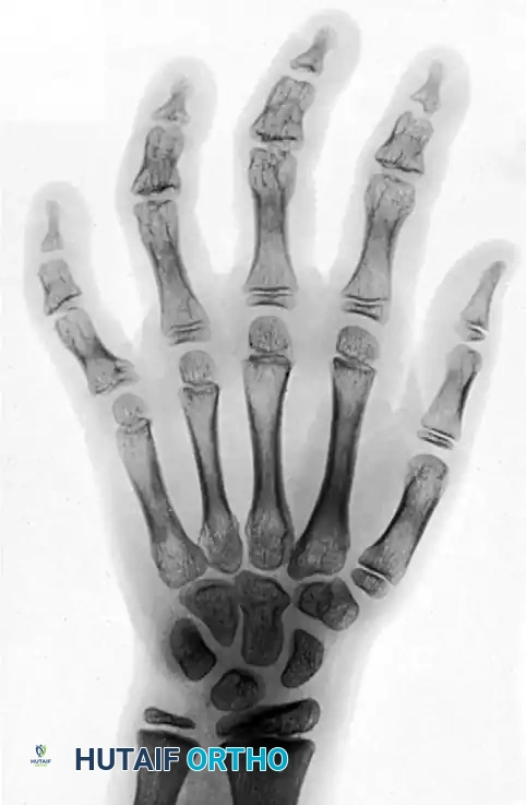
Clinical Radiograph: Deformities of the fingers in a 12-year-old girl caused by severe frostbite incurred at age 2 years. Note the profound destruction of the epiphyses of the middle and distal phalanges across all fingers, and the severe angular deformity of the epiphysis of the proximal phalanx of the little finger. The osseous changes demonstrate the classic delayed manifestation of cold-induced physeal arrest.
In the pediatric population presenting with late-stage cold-induced physeal arrest, the surgical approach shifts to complex deformity correction. Procedures such as opening wedge or closing wedge osteotomies are utilized to realign severe angular deformities (clinodactyly) and restore the mechanical axis of the digit. In cases of profound shortening, distraction osteogenesis is employed. Miniature external fixators are applied to the affected phalanges, a mid-diaphyseal corticotomy is performed, and after a latency period of 5 to 7 days, gradual distraction is initiated at a rate of 0.5 to 1.0 mm per day. This technique not only restores the functional span of the hand but also significantly improves cosmetic appearance, though it requires meticulous pin site care and prolonged patient compliance.
Complications, Incidence Rates, and Salvage Management
The management of complex environmental and ballistic trauma is fraught with severe, often limb-threatening complications. The massive energy transfer of a shotgun blast and the insidious microvascular destruction of frostbite both prime the upper extremity for a cascade of adverse events, requiring the surgeon to remain vigilant and proactive.
Common Complications and Salvage Strategies
| Complication | Associated Mechanism | Estimated Incidence | Pathophysiology & Clinical Presentation | Salvage Management & Treatment Strategy |
|---|---|---|---|---|
| Deep Space Infection / Osteomyelitis | Shotgun (Type III) | 15% - 30% | Retained radiolucent wadding, devitalized bone, polymicrobial contamination. Presents as purulent drainage, systemic sepsis, hardware loosening. | Radical repeat debridement, hardware removal, extraction of retained wadding, placement of antibiotic cement spacers, targeted IV antibiotics, eventual bone grafting. |
| Acute Compartment Syndrome | Shotgun (Type III) | 10% - 20% | Massive kinetic energy transfer causing tissue edema and hemorrhage within rigid fascial envelopes. Pain out of proportion, pain with passive stretch. | Emergent volar and dorsal fasciotomies of the forearm, release of all 10 intrinsic compartments of the hand. Delayed closure or skin grafting. |
| Premature Physeal Arrest | Pediatric Frostbite | 40% - 60% (in severe cases) | Direct cold-induced necrosis of chondrocytes in the resting/proliferative zones. Presents as progressive shortening and angular deformity (clinodactyly) over years. | Delayed intervention until near skeletal maturity. Corrective osteotomies, distraction osteogenesis with mini-ex-fix, or rare epiphysiodesis of contralateral digits. |
| Complex Regional Pain Syndrome (CRPS) | Frostbite / Shotgun | 5% - 15% | Aberrant sympathetic nervous system response following severe trauma or nerve injury. Presents as allodynia, hyperalgesia, sudomotor changes, stiffness. | Aggressive multimodal pain management, early intensive hand therapy, gabapentinoids, stellate ganglion blocks, regional sympathectomy in refractory cases. |
| Terminal Neuroma | Frostbite Amputation | 10% - 25% | Disorganized proliferation of nerve fascicles at the amputation stump due to lack of distal target. Presents as exquisite point tenderness and Tinel's sign. | Surgical exploration, resection of the neuroma, and deep proximal transposition of the nerve end into healthy muscle or bone (TMR or RPNI techniques). |
| Lead Toxicity (Plumbism) | Shotgun (Retained Pellets) | < 1% | Systemic absorption of lead from retained pellets, specifically those located within synovial joints or bursae where synovial fluid dissolves the lead. | Surgical extraction of intra-articular pellets. Chelation therapy (e.g., EDTA) if systemic blood lead levels are dangerously elevated and symptomatic. |
In shotgun injuries, the most catastrophic acute complication is deep space infection, frequently leading to chronic osteomyelitis. This is almost exclusively the result of inadequate initial debridement or the failure to identify and extract the radiolucent shotgun wadding. Once established, osteomyelitis requires a radical salvage approach: all infected bone must be resected back to bleeding margins, existing hardware must be removed, and the defect must be filled with antibiotic-impregnated polymethylmethacrylate (PMMA) spacers (the Masquelet technique). Only after the infection is definitively cleared—often requiring 6 weeks of targeted intravenous antibiotics and negative inflammatory markers—can the surgeon attempt definitive reconstruction with structural bone grafts or vascularized fibular transfers. Lead toxicity, while frequently feared by patients, is exceedingly rare. It typically only occurs if lead pellets are retained within a synovial joint or pseudarthrosis, where the synovial fluid acts as a solvent, slowly leaching lead into the systemic circulation. Intra-articular pellets should therefore be extracted, while extra-articular pellets embedded in muscle are generally left in situ.
In frostbite injuries, the complications are largely related to the long-term sequelae of microvascular damage and nerve injury. Complex Regional Pain Syndrome (CRPS) is a devastating complication characterized by severe allodynia, hyperalgesia, and autonomic dysfunction. The exact etiology is poorly understood but is linked to the profound sympathetic dysregulation inherent in severe cold injury. Salvage management requires a multidisciplinary approach, including early and aggressive hand therapy, pharmacological intervention with gabapentinoids and tricyclic antidepressants, and often interventional pain procedures such as stellate ganglion blocks. Furthermore, frostbite amputees frequently suffer from severe cold intolerance and terminal neuromas. Cold intolerance can persist for years and is managed primarily with behavioral modification and thermal protection. Terminal neuromas, resulting from the disorganized regeneration of transected nerves at the amputation stump, cause exquisite point tenderness that precludes the use of prosthetics or normal hand function. Salvage requires surgical excision of the neuroma and targeted muscle reinnervation (TMR) or regenerative peripheral nerve interface (RPNI) techniques to provide the regenerating nerve with a physiological target, thereby mitigating neuropathic pain.
Phased Post-Operative Rehabilitation Protocols
The functional outcome of complex upper extremity trauma is heavily dependent on a rigorous, phased post-operative rehabilitation protocol. The orthopedic surgeon must work in close concert with specialized hand therapists to navigate the delicate balance between protecting repaired tissues and preventing devastating joint stiffness. The rehabilitation protocols for shotgun injuries and frostbite differ significantly in their early phases due to the distinct nature of the surgical interventions.
Phase 1: Acute Protection and Edema Control (Weeks 0-3)
Following the definitive skeletal stabilization and soft tissue coverage of a shotgun wound, the primary goal is the protection of the reconstruction and the control of massive post-traumatic edema. The hand is strictly immobilized in a custom-molded orthosis in the "intrinsic-plus" or "safe" position: the wrist extended 20 to 30 degrees, the metacarpophalangeal (MCP) joints flexed 70 to 90 degrees, and the interphalangeal (IP) joints in full extension. This position maintains the collateral ligaments of the MCP joints at their maximal length, preventing disabling flexion contractures. Edema is managed with strict elevation, compressive dressings, and retrograde massage. If skeletal stability permits, early passive range of motion (PROM) of the uninvolved digits is initiated immediately to prevent tendon adhesions.
For frostbite patients awaiting delayed amputation, Phase 1 focuses on tissue preservation and infection prevention. The mummifying digits are managed with frequent, gentle washings, often utilizing whirlpool therapy with mild antibacterial solutions (e.g., chlorhexidine or dilute povidone-iodine) to mechanically debride superficial slough and promote a clean demarcation line. The uninjured proximal joints (wrist, elbow, shoulder) must undergo aggressive active range of motion (AROM) exercises to prevent proximal stiffness during the prolonged waiting period.
Phase 2: Intermediate Mobilization and Tendon Gliding (Weeks 3-8)
Once clinical and radiographic signs of early bone healing are evident in shotgun injuries, and soft tissue flaps or grafts have fully incorporated, the protocol transitions to active mobilization. The static splint is converted to a dynamic or hinged orthosis that allows controlled motion while protecting the healing fractures. Intensive tendon gliding exercises are initiated to establish differential movement between the flexor digitorum superficialis (FDS) and flexor digitorum profundus (FDP) tendons, which are highly prone to scarring within the zone of ballistic injury. Scar management techniques, including silicone gel sheeting and deep friction massage, are employed to mobilize the soft tissue envelope and prevent contractures.
In the frostbite population, Phase 2 typically begins immediately following the delayed amputation. The focus is on stump desensitization and the rapid restoration of mobility in the remaining functional joints. Desensitization techniques utilize progressive tactile stimulation—starting with soft materials like silk and progressing to rougher textures like Velcro or rice bins—to help the central nervous system habituate to the altered sensory input of the amputation stump. Active and passive ROM of the remaining digits is pushed aggressively, as the prolonged period of pre-operative immobilization often results in significant capsular stiffness.
Phase 3: Late Strengthening and Functional Restoration (Weeks 8+)
The final phase of rehabilitation for both mechanisms of injury focuses on strengthening, work hardening, and the restoration of complex prehension biomechanics. Progressive resistive exercises using putty, hand grippers, and weighted tools are introduced. For patients with significant skeletal loss or nerve injury from a shotgun blast, this phase may involve training with specialized orthotics or preparing the patient for secondary reconstructive procedures, such as tendon transfers (e.g., transferring the extensor indicis proprius to restore thumb extension).
For frostbite amputees, Phase 3 involves functional integration of the altered hand anatomy. Occupational therapists work extensively with the patient to develop compensatory pinch and grip strategies. If the amputation level is proximal, discussions regarding aesthetic or functional prosthetics are initiated. In the pediatric population suffering from cold-induced physeal arrest, this late phase is essentially continuous, involving longitudinal monitoring of growth and function, with hand therapy utilized intermittently to maintain joint suppleness until the child reaches skeletal maturity and is eligible for definitive deformity correction.
Summary of Landmark Literature and Clinical Guidelines
The modern management of complex environmental and ballistic trauma is deeply rooted in decades of clinical observation, wartime surgical experience, and evolving advanced imaging modalities. A thorough command of
