Operative Spine Surgery: Applied Anatomy, Biomechanics, and Surgical Approaches
Key Takeaway
Mastering spinal surgery requires a profound understanding of vertebral biomechanics, pedicle morphometry, and regional neurovascular anatomy. This comprehensive guide details essential anterior and posterior surgical approaches across the cervical, thoracic, and lumbar spine. It provides orthopedic surgeons and neurosurgeons with evidence-based techniques, step-by-step procedural insights, and critical strategies for avoiding catastrophic complications such as recurrent laryngeal nerve palsy, vascular injury, and spinal cord ischemia.
Comprehensive Introduction and Patho-Epidemiology
The evolution of operative orthopaedics and spine surgery is deeply rooted in a rigorous, uncompromising understanding of spinal anatomy, biomechanics, and the meticulous execution of surgical approaches. The human spine functions as an intricate, dynamic, viscoelastic arch, tasked with the dual, often competing, mandates of protecting the delicate neural elements of the central and peripheral nervous systems while simultaneously facilitating complex, multi-planar motion. Surgical intervention—whether necessitated by high-energy trauma, the relentless progression of degenerative disease, complex triplanar deformity, or aggressive oncologic resection—demands an absolute command of regional neurovascular anatomy, precise pedicle morphometry, and the physiologic limits of spinal cord blood flow.
The patho-epidemiology of spinal disorders is largely defined by the degenerative cascade, a concept classically described by Kirkaldy-Willis. This cascade progresses through phases of dysfunction, instability, and ultimately restabilization via osteophyte formation and hypertrophy. As the global population ages, the incidence of cervical spondylotic myelopathy, lumbar spinal stenosis, and adult spinal deformity has surged, transforming spine surgery into one of the most rapidly advancing and highly scrutinized subspecialties in orthopaedic surgery. The modern spine surgeon must navigate not only the mechanical compression of neural elements but also the microvascular ischemia that accompanies chronic stenosis, leading to irreversible axonal loss if left untreated.
At the intersection of biomechanics and biology lies the fundamental philosophy of spinal reconstruction: the restoration of sagittal and coronal balance, the decompression of neural elements, and the achievement of a solid, enduring arthrodesis or the strategic preservation of motion. The historical reliance on uninstrumented, in-situ fusions has been entirely supplanted by the advent of rigid internal fixation. The principles of the "tension band," load-sharing, and cantilever biomechanics dictate that the success of any construct is inherently dependent on the structural integrity of the anterior column and the robust fixation of the posterior elements. Failure to respect these biomechanical imperatives inevitably leads to construct subsidence, hardware failure, and catastrophic pseudoarthrosis.
This masterclass is meticulously engineered to synthesize foundational biomechanical principles and advanced surgical approaches to the cervical, thoracic, and lumbosacral spine. Designed specifically for orthopedic residents, spine surgery fellows, and practicing consultant surgeons, this exhaustive guide details the precise indications, nuanced patient positioning, step-by-step surgical techniques, and advanced complication avoidance strategies essential for the safe and efficacious practice of modern operative spine surgery.
Detailed Surgical Anatomy and Biomechanics
Osteology and Pedicle Morphometry
The vertebral pedicle serves as the primary and most biomechanically robust conduit for rigid spinal fixation. Transpedicular fixation, the universal workhorse of posterior spinal instrumentation, relies entirely on a precise, three-dimensional understanding of age-related and regional variations in pedicle dimensions. In the cervical spine (C3-C7), the lower cervical pedicles exhibit significant morphometric variability. The pedicle diameter is typically narrowest at C3 and gradually widens toward C7. The medial angulation of cervical pedicles ranges steeply from 30 to 45 degrees, necessitating precise preoperative computed tomography (CT) templating to avoid lateral mass breach and devastating vertebral artery injury.
The thoracic spine (T1-T12) presents an entirely different geometric challenge. The thoracic pedicles are highly complex and notoriously unforgiving. The narrowest pedicles are consistently found in the mid-thoracic apex between T4 and T6, often measuring less than 5 mm in their transverse diameter, which frequently precludes the safe placement of standard pedicle screws and necessitates extrapedicular or in-out-in trajectories. The medial angulation decreases steadily from T1 to T12, while the sagittal trajectory must respect the natural thoracic kyphosis to avoid superior endplate breach.
Conversely, the lumbar pedicles (L1-L5) are robust and voluminous, with the transverse diameter increasing progressively from L1 to L5. The medial angulation also increases caudally, starting at approximately 10 degrees at L1 and reaching up to 30 degrees at L5. The sacrum (S1) presents a massive target, but requires meticulous trajectory planning. The S1 pedicle screw should ideally be directed medially toward the sacral promontory to achieve bicortical purchase; however, an overzealous anterior cortical breach directly endangers the common iliac vessels, the middle sacral artery, and the L5 nerve root traversing the sacral ala.
Ligamentous Anatomy and the Posterior Tension Band
The stability of the spinal column is heavily dependent on its ligamentous restraints, collectively functioning as a sophisticated tension band. The anterior longitudinal ligament (ALL) is a broad, strong band that resists hyperextension and provides a critical tether during anterior column realignment maneuvers, such as anterior lumbar interbody fusion (ALIF) with hyperlordotic cages. The posterior longitudinal ligament (PLL) is narrower, weaker, and provides less resistance to disc herniation, particularly in the posterolateral corners of the spinal canal.
The posterior ligamentous complex (PLC)—comprising the supraspinous ligament, interspinous ligament, ligamentum flavum, and facet joint capsules—acts as the primary restraint against hyperflexion and rotational translation. The ligamentum flavum is highly elastic, composed largely of elastin fibers, but undergoes hypertrophic, fibrotic changes in the setting of degenerative instability, buckling into the spinal canal and contributing significantly to central and lateral recess stenosis. Preservation of the PLC is paramount in motion-preserving surgeries and non-fusion decompressions to prevent iatrogenic instability and progressive kyphotic deformity.
Neurovascular Topography and Spinal Cord Perfusion
The vascular supply to the spinal cord is segmental, tenuous, and highly vulnerable during both anterior and posterior surgical exposures. The critical vascular zone, often referred to as the "watershed area," is located in the mid-thoracic spine (T4-T9), where collateral circulation is notoriously sparse. The anterior two-thirds of the spinal cord is supplied by the anterior spinal artery, while the posterior one-third is supplied by paired posterior spinal arteries.
The Artery of Adamkiewicz (Arteria Radicularis Magna) is the dominant radiculomedullary feeding vessel to the lower thoracic and lumbar spinal cord. It typically arises between T8 and L1 on the left side in approximately 75% to 80% of individuals. Injury to this vessel during anterior thoracolumbar approaches, corpectomies, or aggressive lateral retropleural dissections can result in catastrophic anterior spinal artery syndrome, characterized by profound paraplegia with loss of pain and temperature sensation, but preserved dorsal column function (proprioception and vibratory sense). Furthermore, prolonged spinal cord compression alters local viscoelastic blood flow. Decompression must be executed meticulously, as rapid reperfusion can occasionally lead to reperfusion injury, cytotoxic edema, and transient or permanent neurologic deterioration (e.g., "white cord syndrome").
Biomechanics of Instrumentation
The pullout strength of a pedicle screw is directly proportional to the outer (major) diameter of the screw, the thread depth (the difference between the major and minor diameters), the pitch of the threads, and, most critically, the bone mineral density (BMD) of the pedicle and vertebral body. Maximizing screw diameter without breaching the pedicle cortex is the cornerstone of biomechanically sound fixation. Tapping the pedicle tract under-sized by 1 mm compared to the planned screw diameter increases insertional torque and pullout strength in osteoporotic bone.
Construct rigidity is further dictated by the principles of cantilever bending. A pedicle screw functions as a cantilever beam; the bending moment is highest at the hub-shank junction. Therefore, utilizing screws with a tapered inner minor diameter increases the fatigue strength at the neck of the screw. When bridging long segments, particularly across the cervicothoracic or thoracolumbar junctions, the construct must adequately share the load with the anterior column. Failure to provide anterior column support in the setting of a massive structural defect (e.g., post-corpectomy) places excessive cyclic loading on the posterior instrumentation, inevitably leading to hardware fracture or screw plow.
Exhaustive Indications and Contraindications
Surgical decision-making in spine surgery requires a delicate balance between the patient's patho-anatomy, their physiologic reserve, and the biomechanical goals of the intervention. The fundamental indications for operative intervention generally fall into four categories: progressive neurologic deficit, intractable pain refractory to exhaustive conservative management, structural instability (traumatic or degenerative), and progressive deformity.
The choice of surgical approach—anterior, posterior, lateral, or combined—is dictated by the location of the compressive pathology, the need for sagittal or coronal plane correction, and the patient's specific anatomical constraints. For instance, rigid, fixed kyphotic deformities frequently require anterior column release or three-column posterior osteotomies (e.g., Pedicle Subtraction Osteotomy) to achieve adequate regional alignment. Conversely, pure radiculopathy from a soft, posterolateral disc herniation in a stable spine is elegantly managed with a minimally invasive posterior microdiscectomy.
Contraindications to spine surgery are rarely absolute but require rigorous preoperative optimization. Severe osteoporosis (T-score < -2.5) is a profound relative contraindication to traditional standalone short-segment posterior fixation without adjunctive measures such as cement augmentation, under-tapping, or the use of expandable/fenestrated screws. Medical comorbidities, particularly severe cardiopulmonary disease, may preclude prolonged prone positioning or extensive anterior retroperitoneal exposures.
| Pathology / Condition | Primary Surgical Indication | Relative / Absolute Contraindications | Preferred Surgical Approach |
|---|---|---|---|
| Cervical Spondylotic Myelopathy | Progressive upper motor neuron signs, gait dysfunction, intrinsic hand wasting. | Active anterior neck infection, severe dysphagia (relative for anterior). | ACDF (1-3 levels) or Posterior Laminectomy/Fusion (>3 levels, lordotic spine). |
| Lumbar Spinal Stenosis | Neurogenic claudication refractory to epidural injections, progressive motor deficit. | Pain without neurologic claudication, isolated axial back pain. | Posterior Lumbar Decompression (Laminectomy) +/- Fusion if unstable. |
| Degenerative Spondylolisthesis | Mechanical back pain with radiculopathy, dynamic instability on flexion/extension films. | Severe osteoporosis (requires modified fixation strategies). | TLIF / PLIF or ALIF with posterior percutaneous fixation. |
| Thoracolumbar Burst Fracture | Neurologic deficit, >50% loss of vertebral body height, >20 degrees regional kyphosis. | Hemodynamic instability precluding prolonged surgery (Damage control first). | Posterior Short-Segment Fixation +/- Anterior Corpectomy (if anterior column destroyed). |
| High-Grade Dysplastic Spondylolisthesis | Progressive slip, cauda equina syndrome, severe lumbosacral kyphosis (slip angle >45°). | Poor soft tissue envelope, severe malnutrition. | Combined Anterior/Posterior in-situ fusion or partial reduction with robust pelvic fixation. |
Pre-Operative Planning, Templating, and Patient Positioning
Advanced Imaging and Templating
The foundation of any successful spinal reconstruction is laid long before the incision is made, beginning with exhaustive preoperative imaging and templating. Standard orthogonal radiographs, including standing full-length 36-inch cassettes, are mandatory for assessing global sagittal and coronal balance (e.g., Sagittal Vertical Axis, Pelvic Incidence-Lumbar Lordosis mismatch). Dynamic flexion-extension views evaluate for occult segmental instability. Magnetic Resonance Imaging (MRI) remains the gold standard for evaluating the neural elements, disc hydration, and ligamentous integrity.
However, for complex instrumentation, a fine-cut, non-contrast CT scan is indispensable. CT templating allows the surgeon to measure pedicle diameters, calculate optimal screw trajectories, and assess bone quality using Hounsfield Units (HU). An HU value of less than 110 at the L1 vertebral body is highly predictive of osteoporosis and subsequent hardware loosening, prompting the surgeon to plan for cement-augmented screws or extended construct lengths.
Neuromonitoring Protocols
Multimodal intraoperative neuromonitoring (IONM) has become the standard of care for complex spinal reconstructive procedures. Somatosensory Evoked Potentials (SSEPs) monitor the integrity of the dorsal columns, while Motor Evoked Potentials (MEPs) assess the anterior and lateral corticospinal tracts. Spontaneous and triggered Electromyography (sEMG, tEMG) are utilized to monitor individual nerve roots, particularly during pedicle screw placement and deformity correction maneuvers.
The efficacy of IONM is heavily dependent on the anesthetic regimen. Total Intravenous Anesthesia (TIVA), utilizing propofol and a narcotic infusion (e.g., remifentanil) without the use of volatile halogenated inhalational agents or long-acting paralytics, is mandatory to prevent the suppression of MEP signals. A baseline signal must be obtained prior to positioning, as the mere act of extending the cervical spine or positioning a myelopathic patient prone can precipitate acute spinal cord compression and signal loss.
Patient Positioning and Optimization
Positioning in spine surgery is as critical as the surgical technique itself. For posterior approaches, the patient is typically positioned prone on a specialized radiolucent frame (e.g., Jackson spinal table or Wilson frame). The abdomen must hang completely free; any compression of the abdomen increases intra-abdominal pressure, which is transmitted directly to the epidural venous plexus via the ascending lumbar veins, resulting in massive, uncontrollable intraoperative epidural hemorrhage. All bony prominences must be meticulously padded to prevent pressure necrosis, and the arms should be positioned with the shoulders abducted less than 90 degrees to prevent brachial plexus traction injuries.
For anterior cervical approaches, the patient is positioned supine with a gel roll placed vertically between the scapulae. This induces mild cervical extension, naturally opening the anterior disc spaces and facilitating access. The head is secured with tape or a Mayfield radiolucent skull clamp. For anterior lumbar approaches (ALIF), the patient is positioned supine with the lumbar spine positioned directly over the table break. Breaking the table extends the lumbar spine, maximizing the L5-S1 disc space access and aiding in the recreation of segmental lordosis upon graft insertion.
Step-by-Step Surgical Approach and Fixation Technique
Anterior Cervical Approaches (Smith-Robinson and Transoral)
The transoral approach provides unparalleled direct access to anterior midline compressive pathology at the craniocervical junction (C1-C2), such as rheumatoid pannus, basilar invagination, or complex odontoid fractures. The patient is placed supine with the head secured in a Mayfield clamp. A specialized transoral retractor (e.g., Crockard) is deployed to depress the tongue and elevate the soft palate. A midline vertical incision is made through the posterior pharyngeal wall, extending from the lower clivus to the C2-C3 disc space. The longus colli muscles are elevated subperiosteally and retracted laterally. High-speed, diamond-tipped burrs are utilized for odontoidectomy. The surgeon must remain acutely aware that lateral dissection beyond 15 mm from the midline at the C1-C2 level places the vertebral arteries at imminent risk; preoperative CT angiography is absolutely mandatory to map the vertebral artery groove.
The Smith-Robinson approach to the subaxial cervical spine is the cornerstone of anterior cervical surgery. A transverse incision is made within a natural skin crease (for 1-2 level pathology). The platysma is divided in line with the incision, and the superficial cervical fascia is incised anterior to the sternocleidomastoid (SCM). The critical blunt dissection plane is developed between the carotid sheath (containing the carotid artery, internal jugular vein, and vagus nerve) laterally, and the visceral axis (trachea and esophagus) medially. The prevertebral fascia is incised longitudinally, and the longus colli muscles are elevated bilaterally to accommodate self-retaining retractors. Meticulous discectomy, endplate preparation, and interbody graft placement are performed, followed by anterior plating. To minimize recurrent laryngeal nerve (RLN) injury, retractor tension must be periodically released, and the endotracheal tube cuff should be temporarily deflated and reinflated after retractor placement.
Anterior and Lateral Thoracolumbar Approaches
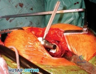
The Anterior Retroperitoneal Approach to the Lumbar Spine (ALIF) provides massive access to the L4-L5 and L5-S1 disc spaces for comprehensive anterior column reconstruction. Following a paramedian fascial incision, the rectus abdominis is mobilized laterally, and the retroperitoneal space is entered. The peritoneal sac is swept medially. At L4-L5, the great vessels bifurcate. The left common iliac vein must be mobilized aggressively to the right. The iliolumbar vein, a critical tethering structure draining into the left common iliac vein, must be identified, double-ligated, and divided. Failure to do so will result in avulsion of the vein from the common iliac vein, leading to catastrophic hemorrhage. At L5-S1, access is achieved within the "vascular window" below the bifurcation, requiring mobilization of the middle sacral artery and vein.
The Lateral Retropleural and Transpsoas approaches allow for minimally invasive access to the anterior thoracic and lumbar spine. In the retropleural approach, a rib is resected, and the parietal pleura is bluntly dissected away from the vertebral body, avoiding the morbidity of a formal thoracotomy. In the transpsoas approach (XLIF/LLIF), the retroperitoneal space is accessed laterally, and sequential dilators are passed directly through the psoas major muscle. Continuous directional EMG monitoring is absolutely critical during this maneuver to map and avoid the lumbar plexus, which lies within the posterior third of the psoas muscle.
Posterior and Posterolateral Approaches
The traditional midline posterior approach requires extensive, meticulous subperiosteal stripping of the multifidus and longissimus muscles off the spinous processes, laminae, and transverse processes. While providing excellent global exposure, this denervates and devascularizes the paraspinal musculature, leading to postoperative atrophy and "fusion disease."
Alternatively, the Paraspinal Sacrospinalis-Splitting Approach (Wiltse) is utilized for far lateral disc herniations and minimally invasive transforaminal lumbar interbody fusions (TLIF). The lumbodorsal fascia is incised 3 to 4 cm lateral to the midline. Blunt dissection is used to separate the multifidus muscle (medially) from the longissimus muscle (laterally). This avascular, intermuscular plane leads directly to the facet joint, the pars interarticularis, and the transverse process—the exact anatomic landmarks required for precise pedicle screw starting points, drastically reducing blood loss and preserving muscle viability.
Pedicle Screw Insertion Techniques
The freehand technique for lumbar pedicle screw insertion relies on precise anatomic landmarks. The starting point is the intersection of the mid-transverse process, the pars interarticularis, and the lateral border of the superior articular facet. The cortex is breached with a high-speed burr or awl. A curved pedicle probe (gearshift) is advanced through the cancellous core of the pedicle. Initially, the curve points laterally to avoid the medial pedicle wall and the spinal canal. Once the probe passes the base of the pedicle (typically at a depth of 20-25 mm), it is rotated 180 degrees so the curve points medially, directing the probe into the voluminous vertebral body. The tract is meticulously palpated with a flexible ball-tip probe to ensure five distinct bony walls (medial, lateral, superior, inferior, and anterior). The tract is tapped, and the appropriate diameter and length screw is inserted.
Complications, Incidence Rates, and Salvage Management
The inherent risks of complex spine surgery are profound. The margin for error is measured in millimeters, and complications can result in devastating, life-altering morbidity. The modern spine surgeon must maintain absolute vigilance, possessing not only the technical acumen to execute the procedure but the foresight to anticipate, recognize, and aggressively manage complications when they arise.
Neurologic deficits remain the most feared complication. C5 palsy is a well-documented phenomenon following extensive cervical decompression, occurring in up to 5-10% of multilevel procedures. It manifests as profound deltoid and biceps weakness, typically presenting 24 to 48 hours postoperatively. The etiology is multi-factorial, attributed to nerve root tethering as the spinal cord shifts posteriorly, or microvascular reperfusion injury. Direct spinal cord injury resulting in paraplegia or quadriplegia is rare (<1% in elective degenerative cases) but requires immediate, protocol-driven intervention.
Vascular and visceral injuries demand immediate, decisive surgical salvage. Vertebral artery injury during cervical spine surgery can result in massive hemorrhage and posterior circulation stroke. Iliac vessel laceration during ALIF or aggressive posterior discectomy requires immediate packing, vascular surgery consultation, and primary repair. Visceral complications, such as chylothorax from thoracic duct injury or retrograde ejaculation from superior hypogastric plexus injury during ALIF, require specialized, multidisciplinary management.
| Complication | Estimated Incidence | Prevention Strategy | Salvage Management / Treatment |
|---|---|---|---|
| C5 Nerve Root Palsy | 5% - 10% (Multilevel cervical decompression) | Prophylactic bilateral C5 foraminotomies, limiting excessive lordotic correction. | Rule out hematoma/graft migration via MRI. Physical therapy; mostly self-limiting (recovers in 6-12 months). |
| Incidental Durotomy (CSF Leak) | 3% - 10% (Higher in revisions and severe stenosis) | Meticulous use of Kerrison rongeurs, avoiding blind dissection under hypertrophic facets. | Primary water-tight repair (6-0 Prolene), fibrin glue, Valsalva maneuver to confirm seal, flat bedrest for 24-48 hours. |
| Vertebral Artery Injury | 0.3% - 0.5% (Cervical instrumentation) | Preoperative CT angiography, precise templating, respecting the 15mm lateral limit at C1-C2. | Direct tamponade with hemostatic agents, immediate intraoperative angiography, endovascular coiling/embolization. |
| Pedicle Screw Breach (Medial) | 1% - 5% (Thoracic > Lumbar) | Anatomic landmark recognition, sequential tract palpation, intraoperative fluoroscopy/navigation. | Immediate screw removal and redirection. If radiculopathy persists post-op, urgent return to OR for revision. |
| Chylothorax | < 1% (Anterior thoracic / cervicothoracic approaches) | Careful dissection of the left posterolateral mediastinum, identifying the thoracic duct. | Closed tube thoracostomy, Medium-Chain Triglyceride (MCT) diet, TPN. Surgical ligation for refractory cases. |
| Retrograde Ejaculation | 1% - 5% (ALIF at L5-S1) | Strict use of blunt dissection and bipolar electrocautery only over the L5-S1 disc space. | Preoperative patient counseling is mandatory. Medical management with sympathomimetics (e.g., pseudoephedrine). |
Phased Post-Operative Rehabilitation Protocols
The success of complex spinal surgery extends far beyond the technical execution in the operating room. Rigorous, phased postoperative protocols are absolutely essential for optimizing bone fusion rates, restoring functional biomechanics, and mitigating systemic and local complications.
Acute Inpatient Phase (Days 0-3)
The immediate postoperative phase prioritizes hemodynamic stability, neurologic monitoring, and pain management. For patients who have undergone extensive decompressions for myelopathy, maintaining a Mean Arterial Pressure (MAP) greater than 85-90 mmHg for the first 72 hours is critical to ensure adequate spinal cord perfusion and mitigate ischemic reperfusion injury. If intraoperative neuromonitoring signals were lost or degraded, the patient must be assessed immediately upon awakening. Unless strictly contraindicated by an unrepaired dural tear or a highly unstable construct, patients should be mobilized on postoperative day one. Early, aggressive mobilization is the most effective strategy to prevent deep vein thrombosis (DVT), pulmonary embolism (PE), and atelectasis. Chemical DVT prophylaxis is typically initiated 24 to 48 hours postoperatively, balancing the risk of epidural hematoma against thromboembolic events.
Subacute Phase and Orthotic Management (Weeks 1-6)
The subacute phase focuses on soft tissue healing and the protection of the surgical construct while the initial stages of osteoinduction and osteoconduction occur. The use of postoperative orthoses (e.g., rigid cervical collars, Thoracolumbosacral Orthoses [TLSO]) depends heavily on the patient's bone quality, the rigidity of the internal construct, and patient compliance. Modern, multi-point rigid pedicle screw fixation has significantly reduced the reliance on postoperative bracing; however, in osteoporotic patients or following complex deformity corrections, a TLSO may be utilized for 6 to 12 weeks to limit cantilever bending forces. Patients are strictly instructed in "BLT" precautions—No Bending, Lifting (greater than 10 lbs), or Twisting. Physical therapy during this phase is limited to walking and gentle isometric core activation.
Long-Term Rehabilitation and Fusion Assessment (Months 3-12)
At the three-month mark, the focus shifts toward dynamic stabilization, core strengthening, and the restoration of normal kinematic function. Patients initiate structured physical therapy focusing on the multifidus, transversus abdominis, and pelvic floor musculature to provide a dynamic muscular tension band that unloads the surgical construct. Radiographic assessment of arthrodesis is performed at 3, 6, and 12 months. True radiographic fusion is defined by the presence of continuous, bridging trabecular bone across the interbody space or posterior elements, the absence of a radiolucent halo around the pedicle screws, and less than 3 degrees of angular motion on dynamic flexion-extension radiographs.
Summary of Landmark Literature and Clinical Guidelines
The practice of operative spine surgery has undergone a paradigm shift from eminence-based to strictly evidence-based medicine, driven by large-scale, multi-center randomized controlled trials and comprehensive clinical guidelines. A profound understanding of this landmark literature is requisite for the academic orthopedic surgeon.
The Spine Patient Outcomes Research Trial (SPORT) remains the most influential body of literature regarding the management of lumbar spine pathology. The SPORT trials definitively demonstrated that for patients with symptomatic lumbar disc herniation, spinal stenosis, and degenerative spondylolisthesis, surgical intervention provides significantly greater and more sustained improvements in pain, function, and quality of life compared to exhaustive non-operative management, provided the clinical presentation precisely matches the radiographic pathology.
In the cervical spine, the AOSpine North America clinical practice guidelines for the management of Cervical Spondylotic Myelopathy (CSM) dictate that surgical intervention is recommended for patients with moderate to severe myelopathy (modified Japanese Orthopaedic Association [mJOA] score < 14) to halt disease progression and improve neurologic status. For mild CSM, surgery or closely monitored conservative management are both viable, though surgery is favored if there is clinical deterioration or inherently unstable patho-anatomy.
Finally, the surgical management of adult spinal deformity is heavily guided by the Schwab-SRS (Scoliosis Research Society) criteria for sagittal spinopelvic alignment. Landmark literature has established that clinical outcomes and health-related quality of life (HRQOL) scores are directly correlated with the restoration of a Sagittal Vertical Axis (SVA) less than 5 cm, a Pelvic Tilt (PT) less than 20 degrees, and a Pelvic Incidence to Lumbar Lordosis (PI-LL) mismatch of less than 10 degrees. Achieving these rigid biomechanical parameters is the ultimate goal of complex spinal reconstruction, ensuring long-term construct survivorship and optimal patient function.
