Central Cord Syndrome: Pathophysiology, Epidemiology, & Clinical Presentation
Key Takeaway
Central Cord Syndrome (CCS) is the most common incomplete spinal cord injury, marked by disproportionately greater upper extremity weakness. It typically occurs from hyperextension injury in older adults with cervical spondylosis, leading to spinal cord compression and ischemia, particularly affecting central gray matter and adjacent motor tracts.
A 72-year-old male presents to the Emergency Department following a mechanical fall at home. He struck his chin on the floor. He complains of burning pain in his hands and significant weakness in his upper limbs, with relatively preserved motor function in his legs. On examination, he is conscious, hemodynamically stable, and has no other obvious trauma. What is your initial assessment and differential diagnosis?
Candidate: The patient has a central cord syndrome, likely secondary to a hyperextension injury in the setting of cervical spondylosis. The primary differential is central cord syndrome, but I must also consider acute cervical disc herniation, traumatic spondylolisthesis, or an underlying spinal cord pathology such as a syrinx or tumor, though the trauma history makes cord syndrome most likely.
Jumping directly to "Central Cord Syndrome" without mentioning a systematic trauma survey (ATLS protocol). Failing to acknowledge that in an elderly patient, you must rule out occult bony injury (fractures) even in the absence of obvious instability.
I would approach this via an ATLS protocol: C-spine immobilization, ABCDE survey. Based on the clinical presentation—disproportionate upper limb motor deficit versus lower limb—I suspect Central Cord Syndrome. This is caused by the "pincer" mechanism of hyperextension on a pre-existing stenotic canal. My differential includes this as the primary diagnosis, but I must exclude unstable fracture-dislocations and acute traumatic disc herniation via urgent CT and MRI.
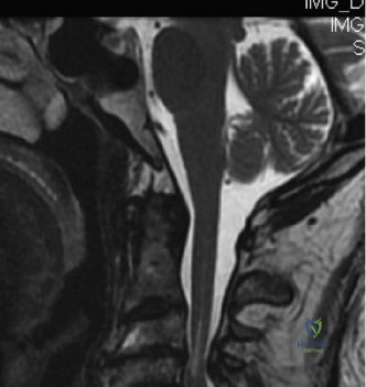
The patient undergoes an MRI, which reveals significant cervical canal stenosis from C3-C6 with evidence of intramedullary T2 signal change. You are presented with the following image:
How do you explain the anatomical basis for his clinical symptoms (upper extremity weakness > lower extremity)?
Candidate: The clinical pattern is explained by the somatotopic organization of the lateral corticospinal tracts. The fibers controlling the upper extremities are located more medially (centrally) within the spinal cord, whereas the fibers for the lower extremities are situated more laterally (peripherally). The hyperextension injury causes central compression and ischemia, affecting the medial fibers disproportionately.
Providing a vague answer about "nerve damage" or failing to use the correct terminology regarding the "Lateral Corticospinal Tract." Examiners look for specific knowledge of the somatotopic map of the cord.
The presentation is secondary to the somatotopic organization of the lateral corticospinal tracts. Upper limb motor fibers are medial (central) and lower limb fibers are lateral (peripheral). The injury produces a central gray matter and white matter insult—specifically to the anterior spinal artery vascular territory—leading to disproportionate upper limb weakness. I would also note that the sensory fibers for the sacral region are located most peripherally, which is why sacral sparing is often observed in incomplete injuries like this.
The patient is currently stable, and his motor function is starting to show minor, early recovery in the lower limbs. What are your indications for surgical intervention versus conservative management in this specific case?
Candidate: I would initially favor conservative management with a rigid cervical collar and close neurological monitoring, given the early sign of recovery and lack of frank instability. Surgery is indicated if he shows a plateau or worsening of his deficit, or if MRI suggests persistent, high-grade compression that is unlikely to resolve with non-operative management.
Stating that surgery is always required, or conversely, stating it is never required. Failing to mention that "early decompression" is an evolving standard of care for patients with persistent compression on MRI, regardless of initial stability.
Decision-making is balanced between neurologic status and radiographic findings. Conservative management (immobilization, BP optimization) is appropriate if there is evidence of neurological recovery. However, surgical decompression is indicated if there is a progressive deficit, documented spinal instability, or persistent, severe cord compression on imaging. Current guidelines lean toward early decompression (24-72 hours) in patients with persistent compression to optimize the potential for neurological recovery, even in the absence of instability.
