Don't Miss Cauda Equina Syndrome: Red Flags in Disc Prolapse
Key Takeaway
In this comprehensive guide, we discuss everything you need to know about Don't Miss Cauda Equina Syndrome: Red Flags in Disc Prolapse. Cauda equina syndrome is a severe condition resulting from compression of the cauda equina nerves, often due to a large acute disc prolapse. It is characterized by urinary retention, faecal incontinence, saddle area numbness, loss of anal tone, and widespread neurological signs. Prompt detection of cauda equina syndrome is vital to prevent permanent neurological deficits.
A 37-year-old man presents with back and left lower limb pain. He reports the leg pain is worse than his back pain, is burning in quality, and associated with paresthesia. What are your clinical priorities?
Candidate: I would take a focused history looking for red flags to rule out Cauda Equina Syndrome (CES) and serious pathology. On examination, I would check for nerve root tension signs (SLR), dermatomal sensory deficits, myotomal weakness, and reflex changes to localize the level of the suspected disc prolapse.
Failing to mention red flags immediately or diving straight into physical examination maneuvers without clarifying the "Red Flag" checklist (bladder/bowel dysfunction, saddle anesthesia, gait disturbance).
Systematically address: 1. Rule out CES (red flags). 2. Characterize the radiculopathy (dermatomal distribution, nature of pain). 3. Neurological exam (SLR, reflexes, power, sensation). 4. Functional impact.
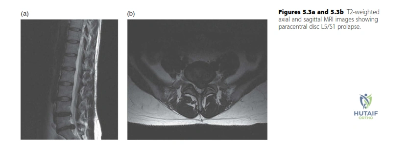
You obtain an MRI of the lumbar spine. Please describe the findings in this imaging.
Candidate: This is a T2-weighted MRI of the lumbar spine. It demonstrates a paracentral disc prolapse at the L5/S1 level, causing indentation of the thecal sac and potential compression of the traversing S1 nerve root.
Confusing the traversing vs. exiting nerve root at the L5/S1 level, or failing to specify if the disc material is central, paracentral, or far-lateral.
State the modality (T2 MRI), the level (L5/S1), the type of pathology (disc herniation/prolapse), and explicitly define which nerve root is at risk (the traversing S1 root).
How does the anatomy of the L5/S1 disc differ from the cervical spine in terms of nerve root nomenclature, and what is your management plan?
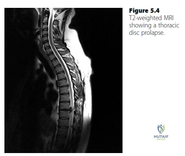
Candidate: In the lumbar spine, a paracentral disc affects the traversing root (L5/S1 affects S1). In the cervical spine, the roots exit above their corresponding pedicles (C5/C6 affects C6). Management is primarily conservative—analgesia, physiotherapy, and reassurance—as natural history is favourable. If failure after 6–12 weeks of non-operative management, I would discuss microdiscectomy.
Stating that surgical intervention is always the answer, or forgetting that cervical root naming follows the pedicle below (C6 exits at C5/C6).
Clearly distinguish between lumbar traversing roots and cervical exiting roots. Emphasize "Conservative first" while acknowledging the timing for surgical consideration (failure of 6–12 weeks of conservative care).
