Mastering the Proximal First Metatarsal Osteotomy: Principles and Techniques
Key Takeaway
Proximal first metatarsal osteotomy is a powerful surgical intervention indicated for severe hallux valgus deformities characterized by an intermetatarsal angle exceeding 10 degrees and a hallux valgus angle greater than 35 degrees. By correcting the metatarsus primus varus near its apex, surgeons achieve substantial distal correction. This comprehensive guide details the biomechanical rationale, step-by-step surgical techniques, stabilization protocols, and postoperative management required to optimize patient outcomes and prevent complications like transfer metatarsalgia.
Comprehensive Introduction and Patho-Epidemiology
Hallux valgus is a remarkably complex, multi-planar, and three-dimensional deformity characterized not merely by the lateral deviation of the great toe, but fundamentally by the medial deviation and pronation of the first metatarsal, clinically termed metatarsus primus varus. Historically viewed as a simple transverse plane deformity, modern orthopedic understanding recognizes that the hallux valgus complex involves significant sagittal and coronal plane abnormalities. When primary or secondary varus of the first metatarsal significantly contributes to the hallux valgus complex, surgical correction near the origin of the deformity—the proximal metatarsal—is biomechanically sound, anatomically logical, and clinically highly effective. The proximal first metatarsal osteotomy represents a cornerstone in the surgical armamentarium for addressing moderate to severe deformities that defy the corrective limits of distal capsulorrhaphies or distal metaphyseal osteotomies.
The epidemiology of hallux valgus reveals a strong predilection for female patients and populations accustomed to wearing constrictive, narrow-toebox footwear. However, severe metatarsus primus varus often has a profound genetic or familial component, frequently presenting in juvenile or adolescent patients and progressing relentlessly into adulthood. In these severe presentations, the deforming forces of the extrinsic musculature, particularly the adductor hallucis and the lateral displacement of the sesamoid apparatus, exacerbate the medial drift of the first metatarsal head. As the intermetatarsal angle widens, the first ray loses its mechanical advantage during the terminal stance phase of gait, leading to load transfer to the lesser metatarsals and the development of intractable plantar keratoses.
A proximal first metatarsal osteotomy, combined with a meticulously executed distal soft tissue procedure at the first metatarsophalangeal (MTP) joint, allows for profound and enduring correction of the intermetatarsal angle (IMA). Because the osteotomy is performed at the base of the metatarsal, leveraging the principles of the center of rotation of angulation (CORA), even a few degrees of angular shift translates into a marked geometric and spatial improvement at the distal articular end. This effectively narrows the forefoot, restores the mechanical axis of the first ray, relocates the sesamoid complex beneath the metatarsal head, and dramatically reduces the chance of recurrent pressure symptoms over the medial eminence.
Despite its tremendous corrective power, the proximal osteotomy is inherently technically demanding. The surgeon must balance the need for multi-planar correction against the risks of destabilizing the first ray. Dorsal elevation of the capital fragment—a catastrophic complication—must be meticulously avoided. As surgical techniques have evolved from traditional open crescentic and Ludloff osteotomies to modern minimally invasive and percutaneous approaches, the fundamental goal remains unchanged: to achieve a stable, anatomic realignment of the first ray that restores physiologic load-bearing to the medial column of the foot.
Detailed Surgical Anatomy and Biomechanics
A profound mastery of the surgical anatomy and biomechanics of the first ray is the absolute prerequisite for executing a successful proximal osteotomy. The first metatarsal is unique among its peers; it is the shortest, thickest, and most robust metatarsal, designed to bear approximately one-third of the body's weight during the push-off phase of normal gait. The proximal metaphysis flares into a broad base that articulates with the medial cuneiform, forming the first tarsometatarsal (TMT) joint. This joint is stabilized by a complex network of ligaments, most notably the Lisfranc ligament complex, and the tendinous insertions of the tibialis anterior and peroneus longus. The tibialis anterior inserts at the plantar-medial aspect of the first metatarsal base and medial cuneiform, while the peroneus longus inserts on the plantar-lateral aspect. Together, these tendons act as a dynamic stirrup, stabilizing the medial column.
The vascular anatomy of the first metatarsal is of paramount importance, particularly when performing osteotomies that threaten the intraosseous and extraosseous blood supply. The first metatarsal receives its primary arterial supply from the first dorsal metatarsal artery, the first plantar metatarsal artery, and the superficial branch of the medial plantar artery. The nutrient artery typically enters the lateral aspect of the middle third of the metatarsal shaft. Proximal osteotomies, by virtue of their location, generally spare the primary nutrient artery, but extensive stripping of the periosteum or aggressive lateral capsular releases at the MTP joint can compromise the epiphyseal and metaphyseal vessels supplying the metatarsal head. Consequently, while avascular necrosis (AVN) is less common with proximal osteotomies compared to distal chevron osteotomies, meticulous soft tissue handling remains critical.
Biomechanically, the rationale for the proximal osteotomy is rooted in the geometric amplification of angular correction. According to the principles of CORA, placing the osteotomy at the apex of the deformity (the metatarsal base) allows for maximal translation of the metatarsal head with minimal angular change. This is critical because it preserves the articular congruity of the first MTP joint while effectively reducing the intermetatarsal angle. The goal is to translate the metatarsal head laterally, reduce the sesamoid complex into their respective grooves on the plantar aspect of the metatarsal head, and maintain or slightly plantarflex the first ray to ensure appropriate load-bearing.
However, the biomechanical advantages of the proximal approach are counterbalanced by significant destabilizing forces. The distal fragment is subjected to ground reaction forces that push it dorsally, while the pull of the extrinsic tendons can cause medial migration or supination. If the osteotomy is not rigidly fixed, these forces will inevitably lead to dorsal malunion. A dorsally elevated first metatarsal fails to engage the ground during terminal stance, neutralizing the windlass mechanism and transferring massive, pathological loads to the second and third metatarsal heads. Therefore, the architectural design of the osteotomy (whether crescentic, chevron, or scarf) and the application of rigid internal fixation are non-negotiable elements of the procedure.
Exhaustive Indications and Contraindications
Patient selection is paramount and requires a judicious synthesis of clinical examination findings, radiographic parameters, and patient expectations. The proximal osteotomy is a technically demanding procedure reserved for moderate to severe deformities where distal procedures alone would fail to adequately reduce the intermetatarsal angle or where the magnitude of correction required would result in lateral cortical overhang and instability if attempted distally.
The primary indication for a proximal metatarsal osteotomy is a symptomatic, moderate-to-severe hallux valgus deformity characterized by significant metatarsus primus varus. Clinically, these patients present with a painful medial eminence, difficulty accommodating standard footwear, and often, impending or established transfer metatarsalgia. Radiographically, the surgeon must carefully assess the Hallux Valgus Angle (HVA), the Intermetatarsal Angle (IMA), and the Distal Metatarsal Articular Angle (DMAA). A patient may benefit from a proximal metatarsal osteotomy and a concomitant distal soft tissue procedure if they present with an HVA greater than 35 degrees and an IMA greater than 13 degrees.
Conversely, the procedure is strictly contraindicated in the presence of severe degenerative joint disease of the first MTP joint (hallux rigidus), as realigning an arthritic joint will only exacerbate pain and stiffness. Furthermore, gross hypermobility of the first TMT joint is a critical contraindication. If the first ray exhibits excessive sagittal or transverse plane instability at the TMT joint, a proximal osteotomy will fail to provide lasting correction, and the deformity will recur. In such cases, a Lapidus arthrodesis (first TMT fusion) is the biomechanically superior choice.
Indications and Contraindications Matrix
| Parameter | Indications for Proximal Osteotomy | Contraindications for Proximal Osteotomy |
|---|---|---|
| Hallux Valgus Angle (HVA) | > 35 degrees (Moderate to Severe) | < 30 degrees (Better suited for distal osteotomy) |
| Intermetatarsal Angle (IMA) | > 13 degrees (or 1st-to-5th IMA ≥ 30 degrees) | < 13 degrees (Mild metatarsus primus varus) |
| First MTP Joint Status | Congruent or subluxated, but viable cartilage | Advanced osteoarthritis (Hallux Rigidus) |
| First TMT Joint Mobility | Stable, normal physiologic motion | Gross hypermobility or TMT arthritis (Requires Lapidus) |
| Bone Quality | Good bone stock for internal fixation | Severe osteoporosis or active osteomyelitis |
| Vascular Status | Intact pedal pulses, good capillary refill | Severe peripheral arterial disease (PAD) |
| Neurological Status | Intact sensation and motor function | Charcot neuroarthropathy or severe neuropathy |
Clinical Pearl: Deformities with intermetatarsal angles of 13 degrees or less and hallux valgus angles of 30 degrees or less can typically be corrected by less technically demanding distal procedures (e.g., distal chevron osteotomy). Reserve the proximal osteotomy for severe metatarsus primus varus where the geometric leverage of a basilar cut is absolutely required.
Pre-Operative Planning, Templating, and Patient Positioning
Thorough pre-operative planning is the foundation of a successful proximal first metatarsal osteotomy. The planning phase begins with the acquisition of high-quality, weight-bearing radiographs of the foot, including anteroposterior (AP), lateral, and sesamoid axial views. Non-weight-bearing films are virtually useless for evaluating the true magnitude of the deformity, as the architectural collapse and splaying of the forefoot only become apparent under physiologic load.
On the AP radiograph, the surgeon must meticulously measure the HVA, IMA, and DMAA. The DMAA is particularly critical; if the articular surface of the metatarsal head is severely laterally deviated (increased DMAA), a simple proximal closing wedge or crescentic osteotomy may correct the IMA but leave the joint incongruent, necessitating a biplanar correction or a double osteotomy. The lateral radiograph is scrutinized to assess the sagittal alignment of the first ray, looking for pre-existing dorsal elevation or plantarflexion, and to evaluate the integrity of the TMT and naviculocuneiform joints. The sesamoid axial view is essential for grading the degree of sesamoid subluxation, which must be anatomically reduced during the procedure to ensure long-term success.
Digital templating software or traditional acetate templates should be utilized to simulate the osteotomy. The surgeon must calculate the exact degree of angular correction required and determine the optimal site for the osteotomy to maximize bone contact and fixation stability. Templating also assists in anticipating the size and trajectory of the internal fixation devices, whether utilizing standard cortical screws, locking plates, or intramedullary Kirschner wires. The surgeon must plan the sequence of the operation, typically beginning with the distal soft tissue release (lateral release) before proceeding to the proximal bone cut.
Patient positioning and anesthesia are critical logistical components. The procedure is typically performed under general anesthesia or a robust regional block (such as a popliteal sciatic nerve block combined with a saphenous nerve block), as the extensive osseous and soft tissue work is poorly tolerated under local infiltration alone. The patient is positioned supine on a radiolucent operating table. A bump is placed under the ipsilateral hip to internally rotate the leg, bringing the foot into a neutral, straight-up position, which is crucial for assessing rotational alignment intraoperatively. A well-padded pneumatic thigh or calf tourniquet is applied to ensure a bloodless surgical field, facilitating precise identification of neurovascular structures and meticulous execution of the osteotomy.
Step-by-Step Surgical Approach and Fixation Technique
Currently, the most frequently utilized open proximal metatarsal osteotomies include the crescentic, chevron, Ludloff, and scarf osteotomies. However, surgical warnings dictate that the specific technique or geometry of the proximal osteotomy is not as important as meticulous attention to surgical detail. Any proximal osteotomy that allows the first metatarsal to deviate laterally and remain rigidly stable in that position—with absolutely no dorsal tilt to the distal fragment—will accomplish the goal of narrowing the IMA. Overcorrection of the IMA is possible but should be avoided through careful intraoperative fluoroscopic assessment.
While traditional open proximal osteotomies are highly effective, advancements in minimally invasive surgery (MIS) have introduced percutaneous techniques that achieve powerful correction with intramedullary stabilization extending to the proximal metatarsal base. The percutaneous distal metatarsal osteotomy (e.g., the Magnan technique) utilizes a distal-to-proximal intramedullary Kirschner wire to achieve the biomechanical stability traditionally reserved for proximal basilar osteotomies. The following details this specific percutaneous approach where the osteotomy is performed distally, but stabilization relies entirely on anchoring the fixation deep into the proximal metatarsal base.
1. Osteotomy Execution
A percutaneous transverse or slightly oblique osteotomy is performed at the metatarsal neck using a specialized Shannon burr under continuous fluoroscopic guidance. The incision is typically no larger than 3 millimeters. The burr is introduced extra-articularly, and the osteotomy is completed with copious cold saline irrigation to prevent thermal necrosis of the bone. Once the osteotomy is complete, the distal capital fragment is translated laterally to correct the IMA. Slight overcorrection is advisable in this specific technique to allow for the early removal of the K-wire without loss of alignment. The lateral translation effectively decompresses the first MTP joint and realigns the soft tissue envelope without the need for a formal open lateral release.
2. Intramedullary Stabilization
Rigid stabilization is the most critical step of this procedure. The fixation must bridge the osteotomy and anchor securely into the proximal metaphysis to resist the deforming forces of weight-bearing and tendon pull. Use a mallet to drive a stout Kirschner wire (typically 2.0 mm) from distal to proximal. The wire enters the medial aspect of the distal fragment, crosses the osteotomy site, and passes into the medullary canal of the first metatarsal. To ensure absolute biomechanical stability, the surgeon must drive the wire as far as the base of the first metatarsal, engaging the dense cancellous bone proximally.
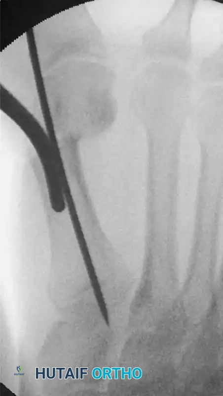
FIGURE 81-36 Percutaneous distal metatarsal osteotomy. The Kirschner wire is driven firmly as far as the base of the first metatarsal to improve stabilization.
3. Final Adjustments and Fluoroscopic Verification
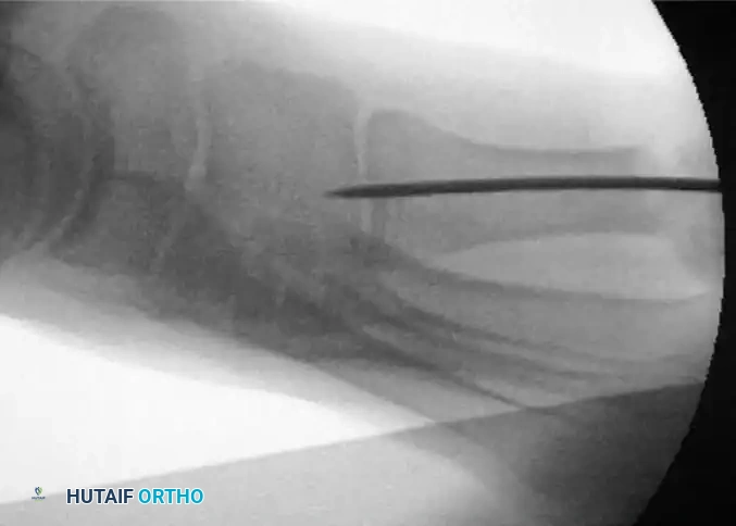
Once the wire is seated proximally, the alignment and hardware placement must be rigorously verified. The surgeon must carefully remove any grooved instruments or tissue protectors used during wire insertion to avoid displacing the osteotomy. A true lateral fluoroscopic image is mandatory to check the trajectory and final position of the K-wire. The surgeon must ensure the wire has not breached the dorsal or plantar cortices and is firmly seated in the proximal base without causing dorsal elevation of the capital fragment.
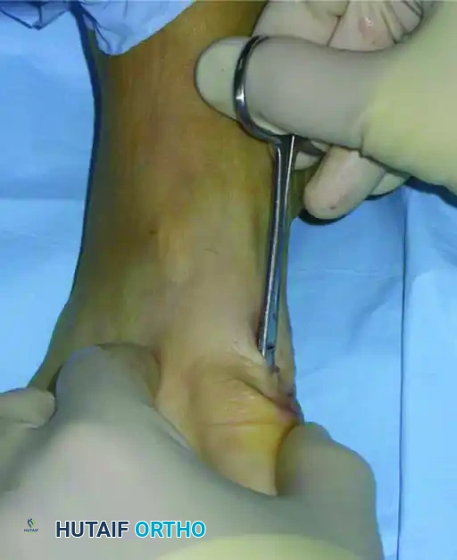
FIGURE 81-37 Percutaneous distal metatarsal osteotomy. Lateral fluoroscopic view shows the position of the Kirschner wire.
4. Soft Tissue Management
Because the capital fragment has been translated laterally by a significant margin (often up to 80% of the metatarsal width), the medial skin and soft tissues may become tethered, blanched, or invaginated around the K-wire insertion site. Left unaddressed, this tension can lead to devastating skin necrosis or deep infection. Using a small hemostat or tenotomy scissors, the surgeon must gently release the soft tissue at the level of the skin incision to free the dermal layers from the underlying capsule and periosteum, thereby alleviating tension.
FIGURE 81-38A Percutaneous distal metatarsal osteotomy. The soft tissue is released at the level of the skin incision.
5. Wire Modification and Closure
The protruding K-wire must be managed to prevent proximal migration into the foot and to assist in the postoperative dressing application. The surgeon bends the Kirschner wire approximately 90 degrees outside the skin before cutting it. This bent tip serves as a crucial anchor point for the postoperative taping protocol and ensures the wire can be easily retrieved in the clinic. The small percutaneous incision is then closed with a single non-absorbable suture, such as a 4-0 nylon.
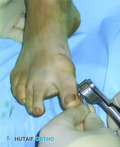
FIGURE 81-38B Percutaneous distal metatarsal osteotomy. The tip of the Kirschner wire is bent before cutting.
Complications, Incidence Rates, and Salvage Management
Surgeons performing proximal metatarsal osteotomies must remain hyper-vigilant regarding several severe complications. The margin for error at the base of the first metatarsal is exceedingly small, and technical failures often lead to profound functional impairment. The most devastating complication is dorsal malunion. Even a few millimeters of dorsal elevation at the proximal osteotomy site will completely unload the first ray during the terminal stance phase of gait. This shifts the biomechanical burden laterally, leading to severe, intractable transfer metatarsalgia beneath the second and third metatarsal heads, often accompanied by lesser MTP joint synovitis or plantar plate ruptures.
Nonunion and delayed union are also significant concerns. While the proximal metaphysis possesses excellent healing potential due to its rich cancellous bone, thermal necrosis from aggressive saw or burr use, inadequate rigid fixation, or premature weight-bearing can lead to a failure of osteogenesis. Smoking, diabetes, and peripheral vascular disease exponentially increase this risk. If a nonunion occurs, it typically presents with persistent pain, swelling, and radiographic lucency at the osteotomy site beyond 12 weeks.
Hallux varus is a distressing iatrogenic complication resulting from overcorrection of the IMA, excessive lateral release, over-resection of the medial eminence, or over-tightening of the medial capsule. A patient with an iatrogenic hallux varus is often more symptomatic and dissatisfied than they were with the original valgus deformity. The toe deviates medially, causing pain in footwear and a cosmetically unacceptable appearance. Finally, avascular necrosis (AVN) of the metatarsal head, while less common with basilar osteotomies than with distal chevron osteotomies, can occur if extensive stripping of the lateral capsular structures disrupts the delicate extraosseous blood supply.
Complications and Salvage Strategies
| Complication | Estimated Incidence | Primary Etiology | Salvage Management Strategy |
|---|---|---|---|
| Dorsal Malunion | 5% - 10% | Inadequate fixation, premature weight-bearing, ground reaction forces | Plantarflexing opening wedge osteotomy of the 1st metatarsal base; Weil osteotomies of lesser rays for transfer metatarsalgia. |
| Nonunion / Delayed Union | 2% - 5% | Thermal necrosis, instability, smoking, poor bone stock | Revision with rigid internal fixation (locking plate) and autologous bone grafting. |
| Iatrogenic Hallux Varus | 3% - 8% | Overcorrection of IMA, excessive lateral release, over-plication of medial capsule | Soft tissue reconstruction (EHL transfer); Medial capsular release; Arthrodesis of 1st MTP joint if arthritic. |
| Avascular Necrosis (AVN) | < 2% | Excessive lateral soft tissue stripping compromising metaphyseal blood supply | Conservative management initially; Core decompression; 1st MTP Arthrodesis for late-stage collapse. |
| Hardware Failure / Migration | 3% - 6% | Poor bone quality, non-compliance with weight-bearing restrictions | Immediate removal of hardware and revision fixation; conversion to Lapidus if TMT instability is noted. |
Phased Post-Operative Rehabilitation Protocols
The success of a proximal metatarsal osteotomy relies as much on meticulous postoperative care as it does on intraoperative execution. The rehabilitation protocol is designed to protect the osteotomy from catastrophic shear and bending forces, prevent dorsal malunion, and ultimately restore normal first MTP joint kinematics. Patient compliance is absolutely critical during this phase, and the surgeon must thoroughly educate the patient on the rationale behind the restrictions.
Phase 1: Immediate Post-Operative Period (Weeks 0-2)
In the immediate postoperative phase, the primary goals are to control edema, manage pain, and protect the surgical construct. The foot is placed in a bulky, sterile compressive dressing. If a percutaneous technique with a protruding K-wire was utilized, the taping and padding protocol is initiated immediately. A plantar kidney-shaped pad is strictly utilized. The concavity of the pad should surround the plantar aspect of the first metatarsal head. This serves a dual purpose: it reduces local weight-bearing pressure beneath the capital fragment and physically prevents dorsiflexion (elevation) of the fragment during the early healing phase. Patients are generally allowed to walk on the day after surgery, provided they strictly use a postoperative shoe with a flat, rigid sole. The rigid sole acts as an external splint, preventing bending moments across the osteotomy site. Elevation of the limb above the level of the heart is strongly encouraged to mitigate swelling.
Phase 2: Intermediate Healing and Taping (Weeks 2-6)
During this phase, the patient returns to the clinic weekly or bi-weekly for dressing changes. The hallux is taped in the corrected position for a minimum of 6 weeks. The tape must be replaced regularly by the surgical team to ensure tension is maintained as postoperative swelling subsides. The taping should maintain a slight hyper-correction of the hallux (slight varus) to counter the natural soft-tissue tendency toward recurrence of the valgus deformity. Weight-bearing in the rigid postoperative shoe continues. The surgeon should obtain interval radiographs (typically at 2 and 6 weeks) to monitor the maintenance of alignment and assess for the early formation of bridging callus.
Phase 3: Hardware Removal and Mobilization (Weeks 6-12)
At 6 weeks, clinical and radiographic union is rigorously assessed. If bridging callus is present across the osteotomy site and the construct is clinically stable (no pain on palpation), the K-wire (if used) is removed in the clinic, and the rigid taping is discontinued. Patients are then transitioned into a wide-toebox, supportive athletic shoe. After tape removal, patients are strongly encouraged to begin active and passive range of motion (ROM) exercises of the first MTP joint. Particular care and formal physical therapy should be directed toward obtaining full dorsiflexion within 4 to 6 weeks following immobilization removal, as MTP joint stiffness is a pervasive complication. Strengthening of the intrinsic foot musculature and gait retraining are essential to restore the windlass mechanism and normalize plantar pressures.
Summary of Landmark Literature and Clinical Guidelines
The evolution of the proximal first metatarsal osteotomy is deeply rooted in landmark orthopedic literature. Historically, the crescentic osteotomy, popularized by Mann and Coughlin, was considered the gold standard for severe hallux valgus. Their long-term follow-up studies demonstrated excellent correction of the IMA and high patient satisfaction, provided that dorsal elevation of the metatarsal head was avoided. They emphasized the necessity of a concomitant distal soft tissue release to achieve a balanced joint.
The Ludloff osteotomy, an oblique diaphyseal-metaphyseal cut, gained resurgence due to its inherent geometric stability and broad surface area for screw fixation. Biomechanical studies by Trnka et al. highlighted that the Ludloff osteotomy, when fixed with multiple cortical screws, provided superior resistance to dorsal displacement compared to single-screw crescentic osteotomies. Similarly, the scarf osteotomy, popularized in Europe by Barouk, demonstrated immense versatility, allowing for lateral translation, shortening, or lengthening, though it carries a steep learning curve and a risk of troughing.
More recently, the shift towards minimally invasive surgery has been championed by pioneers like Magnan, whose percutaneous techniques challenged the dogma that massive open exposures were required for severe deformities. Magnan's prospective studies on the percutaneous distal metatarsal osteotomy with proximal intramedullary stabilization demonstrated comparable radiographic outcomes to open procedures, with significantly reduced soft tissue morbidity, lower infection rates, and faster return to footwear.
Current clinical guidelines from the American Orthopaedic Foot & Ankle Society (AOFAS) and the American Academy of Orthopaedic Surgeons (AAOS) reiterate that surgical decision-making must be tailored to the severity of the deformity and the presence of hypermobility. They strongly advocate for proximal osteotomies or Lapidus fusions in cases of severe metatarsus primus varus (IMA > 13 degrees), warning that distal procedures in this cohort have an unacceptably high rate of recurrence. By adhering to these evidence-based principles, ensuring rigid internal fixation, and executing a flawless postoperative protocol, the proximal first metatarsal osteotomy remains one of the most powerful tools in the orthopedic surgeon's armamentarium.
