Orthopedic Surgery Board Review MCQs: Hand & Wrist, Spinal Nerve & Grafting Part 35
Key Takeaway
This page delivers an interactive MCQ quiz for orthopedic surgeons and residents targeting AAOS/ABOS board certification. It features 50 high-yield questions, formatted like OITE/AAOS exams, on topics such as Graft, Nerve, and Wrist. This resource provides detailed explanations and two learning modes for focused, effective exam preparation.
Orthopedic Surgery Board Review MCQs: Hand & Wrist, Spinal Nerve & Grafting Part 35
Comprehensive 100-Question Exam
00:00
Start Quiz
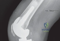
Question 1
A 28-year-old male presents with persistent wrist pain 18 months after a scaphoid waist fracture. Imaging reveals a proximal pole nonunion with avascular necrosis (AVN) of the proximal fragment, which measures 4 mm. A previous attempt at fixation with a Herbert screw and non-vascularized bone graft failed. There is no evidence of radiocarpal arthritis. Which of the following is the most appropriate surgical treatment to maximize the chance of union?
Explanation
Question 2
A 24-year-old motorcyclist sustains a severe closed traction injury to his right brachial plexus. Examination reveals a flail, insensate right upper extremity and a right-sided ptosis and miosis. An MRI of the cervical spine is performed. Which of the following MRI findings is most consistent with the clinical examination and indicates an irreparable lesion at the root level?
Explanation
Question 3
In a two-stage flexor tendon reconstruction utilizing a silicone tendon implant (Hunter rod) for a chronic Zone 2 flexor digitorum profundus (FDP) laceration, what is the primary biological objective of the first stage of the procedure?
Explanation
Question 4
A 50-year-old patient presents with right-sided neck pain radiating down the arm, weakness in elbow extension and wrist flexion, and a diminished triceps reflex. Sensation is decreased over the palmar aspect of the middle finger. MRI demonstrates a paracentral disc herniation at the C6-C7 level. Which spinal nerve root is compressed, and where does it normally exit?
Explanation
Question 5
During autologous nerve grafting for a 4 cm post-traumatic peripheral nerve gap, what is the primary cellular/structural contribution of the interposed donor nerve graft?
Explanation
Question 6
An avid cyclist presents with a 3-month history of right hand weakness. Examination reveals marked atrophy of the dorsal interossei and a positive Froment sign. Sensation is completely intact over the volar and dorsal aspects of the small finger and the ulnar half of the ring finger. Hypothenar muscle bulk is normal. Where is the most likely site of ulnar nerve compression?
Explanation
Question 7
Which of the following physical examination findings is highly specific for distinguishing true neurogenic thoracic outlet syndrome (TOS) from a severe compressive ulnar neuropathy at the elbow (cubital tunnel syndrome)?
Explanation
Question 8
A 35-year-old carpenter sustains a saw injury resulting in a 3.5 cm defect in the proper digital nerve of the index finger in Zone 2. Which of the following reconstructive options represents the gold standard with the most predictable sensory recovery for this specific defect?
Explanation
Question 9
A 42-year-old patient presents with sudden-onset bilateral and symmetric saddle anesthesia, early bowel and bladder incontinence, and impotence. Lower extremity examination reveals hyperreflexia at the knees but absent Achilles reflexes. Motor weakness is mild and symmetric. Where is the most likely neuroanatomic location of the primary lesion?
Explanation
Question 10
In the predictable progression of Scapholunate Advanced Collapse (SLAC) wrist arthritis, which specific radiocarpal or midcarpal articulation is characteristically spared, allowing for surgical salvage via proximal row carpectomy (PRC) in earlier stages?
Explanation
Question 11
When harvesting an autologous corticocancellous bone graft from the anterior iliac crest for the treatment of a structural atrophic nonunion, what is the primary biological property provided by the cancellous portion of the graft?
Explanation
Question 12
A patient is diagnosed with a peripheral, foveal avulsion of the Triangular Fibrocartilage Complex (TFCC), classified as a Palmer 1B lesion. To restore stability to the distal radioulnar joint (DRUJ), which specific anatomical structures within the TFCC must be anatomically repaired to the fovea?
Explanation
Question 13
During a right-sided anterior cervical discectomy and fusion (ACDF) at C6-C7, the patient develops a unilateral vocal cord paralysis. The vulnerability of the right recurrent laryngeal nerve during this approach is anatomically explained by its course looping under which of the following structures?
Explanation
Question 14
In the classic Oberlin transfer used to restore elbow flexion following an upper trunk (C5-C6) brachial plexus avulsion injury, which specific nerve fascicles are transferred to the motor branch of the biceps?
Explanation
Question 15
The off-label use of recombinant human bone morphogenetic protein-2 (rhBMP-2) in Anterior Cervical Discectomy and Fusion (ACDF) procedures has been strongly associated with which of the following serious postoperative complications?
Explanation
Question 16
The tenuous blood supply to the proximal pole of the scaphoid is a primary factor in its high rate of nonunion following fracture. The predominant blood supply to the scaphoid enters at which anatomical location?
Explanation
Question 17
A patient presents with hand weakness and numbness in the ring and small fingers. The examiner is trying to differentiate between a C8 radiculopathy and a severe ulnar neuropathy at the elbow. Weakness in which of the following muscles firmly points to a C8 radiculopathy rather than an ulnar neuropathy?
Explanation
Question 18
In the Masquelet technique for reconstructing a 3 cm segmental bone defect of a metacarpal, a PMMA cement spacer is temporarily placed. During the second stage (typically 6-8 weeks later), a thick membrane surrounds the spacer. What is the primary biological characteristic of this induced membrane?
Explanation
Question 19
A 55-year-old male presents with severe, burning anterior thigh pain, weakness in right knee extension, and a diminished right patellar reflex. Sensation is decreased over the medial aspect of the lower leg. An MRI of the lumbar spine reveals an extraforaminal (far lateral) disc herniation at the L4-L5 level. Which nerve root is primarily compressed?
Explanation
Question 20
A 60-year-old patient with severe, chronic carpal tunnel syndrome presents with profound atrophy of the thenar eminence and an inability to palmar abduct the thumb, though thumb interphalangeal joint flexion is strong. To restore thumb palmar abduction with a transfer that requires minimal motor re-education, which of the following is the most commonly indicated procedure?
Explanation
Question 21
A 55-year-old man presents with chronic wrist pain and stiffness. Radiographs demonstrate advanced joint space narrowing at the radioscaphoid and capitolunate articulations. The radiolunate joint space is completely preserved. He has failed conservative management. Which of the following surgical interventions is most appropriate?
Explanation
Question 22
When performing an autogenous cable nerve graft to bridge a 4-cm defect in the median nerve, standard surgical technique involves reversing the orientation of the harvested nerve graft. What is the primary biological rationale for this step?
Explanation
Question 23
A 48-year-old woman complains of severe neck pain radiating into her medial right forearm and hand. On physical examination, she demonstrates 3/5 strength in the flexor digitorum profundus of her right ring and small fingers, and 3/5 strength in finger abduction. She has diminished sensation over the ulnar border of her right hand and small finger. Her triceps strength and reflex are intact. Which cervical nerve root is most likely compressed?
Explanation
Question 24
A 32-year-old snowboarder sustains a high-energy fall onto an outstretched hand and presents with a volar Barton's fracture of the distal radius. The carpus is subluxated volarly along with the marginal fracture fragment. Which of the following ligamentous structures remains attached to the volar marginal fragment and acts as the primary deforming force tethering the carpus to it?
Explanation
Question 25
A surgeon is considering the use of a synthetic bone graft substitute to fill a metaphyseal void following elevation of a depressed tibial plateau fracture. Which of the following best describes the properties of calcium phosphate cement compared to calcium sulfate?
Explanation
Question 26
A 28-year-old manual laborer is diagnosed with Stage IIIA Kienbock's disease. Radiographs show lunate sclerosis and fragmentation with lunate collapse, but no fixed scaphoid rotation or carpal collapse. His ulnar variance is minus 3 mm. Which of the following is the most appropriate surgical treatment?
Explanation
Question 27
A 45-year-old man presents with acute onset of low back pain radiating down his right leg following heavy lifting. MRI reveals a large paracentral disc herniation at the L4-L5 level on the right side. Which nerve root is most likely compressed, and what clinical finding is expected?
Explanation
Question 28
Recombinant human bone morphogenetic proteins (rhBMPs) are utilized in orthopedics to promote bone healing. At the cellular level, through which type of cell surface receptor do BMPs primarily initiate their intracellular signaling cascade?
Explanation
Question 29
A 25-year-old professional golfer complains of painful snapping on the ulnar side of his wrist during the downswing. Examination reveals subluxation of the extensor carpi ulnaris (ECU) tendon over the ulnar styloid during forearm supination, ulnar deviation, and wrist flexion. This pathology is primarily due to a tear or attenuation of which of the following structures?
Explanation
Question 30
A 60-year-old man undergoes a C4-C6 posterior laminectomy and instrumented fusion for cervical spondylotic myelopathy. On postoperative day 3, he suddenly develops profound unilateral weakness in shoulder abduction and elbow flexion, without a change in his leg strength or bowel/bladder function. What is the most widely accepted pathophysiologic mechanism for this complication?
Explanation
Question 31
When utilizing structural cortical allografts for reconstructive procedures, how does the biological incorporation process critically differ from that of cortical autografts?
Explanation
Question 32
A 21-year-old man sustains a fracture through the proximal pole of the scaphoid. The vulnerability of this specific fracture to avascular necrosis and nonunion is primarily determined by the unique intraosseous retrograde blood supply of the scaphoid. The major blood supply to the scaphoid enters the bone predominantly at the dorsal ridge and is derived from a branch of which artery?
Explanation
Question 33
A 16-year-old gymnast presents with progressive lower back pain and left leg pain. Imaging demonstrates a Grade II L5-S1 isthmic spondylolisthesis with bilateral pars interarticularis defects. If the patient has isolated left lower extremity radicular symptoms, which nerve root is most likely affected by the primary pathoanatomy of this condition?
Explanation
Question 34
A 22-year-old rugby player presents 3 days after injuring his right ring finger when grabbing an opponent's jersey. He is unable to actively flex the distal interphalangeal (DIP) joint. Physical examination reveals tenderness in the palm, and radiographs are negative for a fracture. Based on the Leddy-Packer classification, what is the pathophysiology and recommended timing for surgical repair?
Explanation
Question 35
When evaluating the regenerative properties of various bone grafting materials, the term 'osteoinduction' refers specifically to which of the following processes?
Explanation
Question 36
A 35-year-old man sustained a mid-shaft humerus fracture resulting in a high radial nerve palsy that has shown no clinical or electromyographic signs of recovery at 12 months. In a standard set of tendon transfers (such as the Jones transfer) designed to restore hand and wrist function, which donor tendon is classically transferred to restore wrist extension?
Explanation
Question 37
A 42-year-old woman with a history of chronic low back pain presents to the emergency department with acute worsening of back pain, bilateral sciatica, and perineal numbness. Which of the following urologic findings is the earliest and most reliable indicator of cauda equina syndrome?
Explanation
Question 38
Cancellous bone autograft is considered the 'gold standard' for filling cavitary bone defects due to its rapid incorporation and optimal biological properties. Compared to cortical autograft, what is the primary histological mechanism that accounts for the faster revascularization and incorporation of cancellous bone graft?
Explanation
Question 39
A 65-year-old woman with neglected, severe carpal tunnel syndrome presents with profound thenar atrophy and complete inability to oppose her thumb. The surgeon plans a carpal tunnel release combined with a Bunnell (or Royle-Thompson) opponensplasty. Which of the following describes the most common tendon transfer and pulley utilized in this specific technique to restore true thumb opposition?
Explanation
Question 40
An 8-month-old infant presents with an unresolved Erb-Duchenne palsy (C5-C6 injury) following a difficult vertex delivery with shoulder dystocia. The affected arm rests in internal rotation and adduction at the shoulder, with the elbow extended and forearm pronated. The internal rotation contracture of the shoulder is primarily driven by the unopposed action of which of the following muscles?
Explanation
Question 41
A 26-year-old male presents with a persistent scaphoid nonunion and avascular necrosis of the proximal pole, featuring a humpback deformity and a 6 mm bone defect. Which of the following graft options provides both the structural integrity to correct the deformity and the robust blood supply necessary for this specific scenario?
Explanation
Question 42
To restore elbow flexion in a patient with a traumatic C5-C6 brachial plexus root avulsion, an Oberlin transfer is planned. Which of the following describes the classic donor and recipient nerves in this procedure?
Explanation
Question 43
During an Adams-Berger anatomic reconstruction of the distal radioulnar joint (DRUJ) for chronic instability, a tendon graft is utilized to recreate the palmar and dorsal radioulnar ligaments. Where are the graft ends passed through the radius?
Explanation
Question 44
Following peripheral nerve injury and subsequent grafting, what is the primary role of Schwann cells during the process of Wallerian degeneration?
Explanation
Question 45
A 45-year-old female presents with acute onset of severe unilateral shoulder pain, which subsides after one week, leaving profound weakness in shoulder abduction and external rotation. MRI of the cervical spine is unremarkable. EMG shows active denervation in the supraspinatus and infraspinatus. What is the most likely diagnosis?
Explanation
Question 46
In the standard flexor carpi radialis (FCR) tendon transfer utilized for a high radial nerve palsy, which muscle is typically transferred to the extensor pollicis longus (EPL) to restore thumb extension?
Explanation
Question 47
Demineralized bone matrix (DBM) is widely used in hand and upper extremity osseous reconstruction. Which of the following best describes the biologic properties of DBM?
Explanation
Question 48
A 42-year-old carpenter presents with cold intolerance and a pulsatile mass in the hypothenar eminence of his dominant hand. Angiography reveals occlusion and aneurysmal dilation of the superficial palmar branch of the ulnar artery. Which bony structure contributes to the pathomechanics of this specific condition?
Explanation
Question 49
In a patient presenting with a complete flail upper extremity after a motorcycle accident, which of the following electrodiagnostic findings most specifically indicates a pre-ganglionic (avulsion) brachial plexus injury rather than a post-ganglionic lesion?
Explanation
Question 50
In Scapholunate Advanced Collapse (SLAC) of the wrist, progressive degenerative changes predictably involve specific articular surfaces while sparing others. Which radiocarpal articulation is classically spared from degenerative arthritis in the SLAC wrist?
Explanation
Question 51
The sural nerve is the most common autograft utilized for bridging peripheral nerve defects. Following harvesting of the sural nerve from the posterior calf, in which specific area will the patient predictably experience a permanent sensory deficit?
Explanation
Question 52
A patient presents with an inability to form an "OK" sign with their thumb and index finger but maintains normal sensation throughout the hand. Which of the following muscles is typically spared in a complete, isolated anterior interosseous nerve (AIN) palsy?
Explanation
Question 53
A patient with a chronic lower brachial plexus injury (C8-T1) exhibits a severe claw hand deformity. A modified Stiles-Bunnell tendon transfer is planned to restore intrinsic function. Which of the following muscles is utilized as the donor in this procedure?
Explanation
Question 54
In a Bennett fracture-dislocation of the thumb, the metacarpal shaft is displaced by the deforming forces of specific muscles. Which muscle is primarily responsible for the dorsal, proximal, and radial displacement of the first metacarpal shaft?
Explanation
Question 55
Vascularized nerve grafts (e.g., vascularized ulnar nerve graft) are considered theoretically superior to standard non-vascularized nerve autografts in which of the following specific clinical scenarios?
Explanation
Question 56
Biomechanical studies evaluating flexor tendon repairs in Zone 2 demonstrate that the initial tensile strength of the repair before biologic healing occurs is most directly proportional to which of the following factors?
Explanation
Question 57
A 25-year-old male presents with a scaphoid nonunion demonstrating a humpback deformity and avascular necrosis of the proximal pole on MRI. The proximal fragment measures 6 mm. Which of the following is the most appropriate vascularized bone graft to restore scaphoid geometry and maximize the likelihood of union?
Explanation
Question 58
A 22-year-old motorcyclist sustains a traumatic brachial plexus injury. Clinical examination shows complete paralysis of the C5 and C6 myotomes. Sensory examination reveals anesthesia in the C5 and C6 dermatomes, yet Sensory Nerve Action Potentials (SNAPs) for the median and radial nerves are preserved. What is the anatomical location of this nerve injury?
Explanation
Question 59
A 45-year-old manual laborer presents with Stage III Scapholunate Advanced Collapse (SLAC). Radiographs reveal advanced arthritis at the radioscaphoid and capitolunate joints, with sparing of the radiolunate joint. What is the most appropriate definitive surgical management?
Explanation
Question 60
During surgical exploration of a complete median nerve transection in the mid-forearm, a nerve defect of 3.5 cm is measured after debridement of non-viable tissue. The injury is 4 weeks old. What is the gold standard reconstruction for this defect?
Explanation
Question 61
A 19-year-old rugby player sustains a closed jersey finger injury of the ring finger. Radiographs show no fracture, and the flexor digitorum profundus (FDP) tendon is palpable in the palm. To prevent irreversible contracture and tendon necrosis due to disrupted vincular blood supply, definitive repair should ideally be performed within what timeframe?
Explanation
Question 62
A patient with an isolated, irreparable high radial nerve palsy requires tendon transfers to restore wrist extension, finger extension, and thumb extension. Which of the following is the most standard and reliable donor muscle to restore wrist extension?
Explanation
Question 63
A patient is undergoing an Oberlin transfer for a C5-C6 brachial plexus root avulsion to restore elbow flexion. Which specific donor nerve fascicle is most commonly transferred to the motor branch of the biceps?
Explanation
Question 64
A 35-year-old patient presents with severe Stage IIIA Kienböck's disease. Radiographs reveal lunate sclerosis and fragmentation but no fixed carpal collapse, accompanied by an ulnar negative variance of 3 mm. Which of the following is the most appropriate joint-leveling procedure?
Explanation
Question 65
During exploration of a brachial plexus injury 5 months post-trauma, a neuroma-in-continuity is identified at the upper trunk. Intraoperative nerve stimulation across the neuroma yields a reproducible nerve action potential (NAP). What is the most appropriate next step in management?
Explanation
Question 66
In arthroscopic repair of a peripheral triangular fibrocartilage complex (TFCC) tear, which of the following neurological structures is at highest risk of injury when establishing the 6U portal?
Explanation
Question 67
A patient develops a Scaphoid Nonunion Advanced Collapse (SNAC) wrist following an untreated scaphoid fracture. What is the typical sequential progression of degenerative arthritis in a SNAC wrist?
Explanation
Question 68
A 40-year-old male sustains a laceration over the proximal phalanx (Zone II), severing both the FDS and FDP tendons. The surgeon attempts a primary repair but inadvertently sutures the lumbrical muscle too tightly during closure. What clinical phenomenon is the patient most likely to exhibit postoperatively?
Explanation
Question 69
A 28-year-old male sustains a severe traction injury to his right upper extremity. Clinical examination reveals complete flaccidity of the arm, absent sensation, ptosis, miosis, and anhidrosis on the right side of his face. This specific facial triad implies poor prognosis for spontaneous nerve recovery because it indicates injury to which structure?
Explanation
Question 70
In the context of scapholunate dissociation, a patient is planned for a capsulodesis and ligamentous reconstruction. Which distinct region of the scapholunate interosseous ligament is thickest, strongest, and most critical to reconstruct to restore normal carpal kinematics?
Explanation
Question 71
A 65-year-old woman presents 8 weeks after open reduction and internal fixation of a distal radius fracture with a volar locking plate. She suddenly lost the ability to actively extend her thumb interphalangeal joint. Radiographs show prominent screws penetrating the dorsal cortex. What is the most reliable surgical treatment for this complication?
Explanation
Question 72
A surgeon is harvesting an autologous sural nerve graft for a brachial plexus reconstruction. To predictably locate the sural nerve with minimal dissection, the initial incision should be placed precisely in which anatomical location?
Explanation
Question 73
A 24-year-old male presents with profound median nerve palsy. To restore thumb opposition, a Burkhalter transfer is planned utilizing the Extensor Indicis Proprius (EIP). To optimize the vector for thumb pronation and palmar abduction, the transferred EIP tendon should be routed around which anatomical structure to act as a pulley?
Explanation
Question 74
A patient with a chronic, isolated, traumatic avulsion of the axillary nerve with complete deltoid atrophy is scheduled for a nerve transfer 5 months post-injury. Which of the following is the most highly successful donor nerve for restoring deltoid function in this setting?
Explanation
Question 75
A 30-year-old carpenter suffers a deep laceration at the level of the proximal interphalangeal (PIP) joint, transecting the central slip of the extensor tendon. If left untreated, the lateral bands will eventually subluxate. In relation to the axis of rotation of the PIP joint, in which direction do the lateral bands migrate to produce the classic resulting deformity?
Explanation
Question 76
A 35-year-old male sustains a severe traction injury to his right upper extremity. Clinical examination reveals flaccid paralysis of the entire right arm with anesthesia from the shoulder to the hand. Electromyography (EMG) shows denervation of the cervical paraspinal muscles. Sensory nerve action potentials (SNAPs) are tested for the right upper extremity. Which of the following SNAP findings is most consistent with this patient's injury level and prognosis?
Explanation
Question 77
In the treatment of severe scaphoid nonunions with avascular necrosis of the proximal pole, a free vascularized bone graft from the medial femoral condyle (MFC) is frequently utilized. Which of the following vessels provides the primary arterial supply to the standard MFC vascularized bone graft?
Explanation
Question 78
A 28-year-old carpenter presents with a 5 cm gap in the median nerve at the mid-forearm following a circular saw injury 3 months ago. Which of the following grafting techniques provides the highest likelihood of successful motor and sensory recovery?
Explanation
Question 79
A 45-year-old manual laborer presents with chronic wrist pain. Radiographs reveal advanced scapholunate advanced collapse (SLAC) with arthritic changes involving the radioscaphoid joint and the proximal capitate. The radiolunate joint is entirely spared. Which of the following surgical interventions is most appropriate?
Explanation
Question 80
A 22-year-old male presents with a complete C5-C6 root avulsion following a motorcycle accident. An Oberlin transfer is planned to restore elbow flexion. Which of the following describes the correct neurological transfer performed in this procedure?
Explanation
Question 81
Six months after a Zone II flexor tendon repair of the middle finger, a patient complains that the affected digit paradoxically extends at the proximal interphalangeal (PIP) joint when attempting to make a tight fist. What is the most likely etiology of this phenomenon?
Explanation
Question 82
A 40-year-old female with long-standing rheumatoid arthritis is suddenly unable to flex the interphalangeal joint of her right thumb. Examination reveals a loss of active thumb IP flexion but an intact tenodesis effect when the wrist is passively extended. What is the most likely cause of this deficit?
Explanation
Question 83
A 25-year-old pitcher experiences sudden, severe right shoulder pain, followed two weeks later by profound weakness in external rotation and elevation. EMG demonstrates denervation isolated to the supraspinatus and infraspinatus. There is no history of trauma. What is the most likely diagnosis?
Explanation
Question 84
During the incorporation of a free vascularized fibular graft used for a 10 cm radial defect, how does the biological healing process distinctly differ from that of a massive non-vascularized cortical bone autograft?
Explanation
Question 85
A 38-year-old cyclist complains of intrinsic hand weakness and numbness strictly affecting the volar aspect of his small finger and the ulnar half of his ring finger. Sensation on the dorsal ulnar aspect of the hand is completely normal. Where is the most likely site of neural compression?
Explanation
Question 86
A patient presents with a Boutonniere deformity 4 weeks after sustaining a closed crush injury to the PIP joint. Which of the following describes the underlying pathomechanics of this deformity?
Explanation
Question 87
When performing a nerve repair, the surgeon decides to use a 4-strand core suture technique for a Zone II flexor tendon repair rather than a 2-strand repair. What is the primary biomechanical advantage of the 4-strand repair in this setting?
Explanation
Question 88
A patient with suspected Pronator Syndrome presents with volar forearm pain and paresthesias in the thumb, index, and middle fingers. Which of the following physical examination findings most reliably differentiates Pronator Syndrome from Carpal Tunnel Syndrome?
Explanation
Question 89
A 45-year-old male with Kienbock's disease presents with chronic wrist pain. Imaging reveals lunate sclerosis, fragmentation, and carpal collapse, with a negative ulnar variance of 3 mm. Advanced degenerative changes are noted at the radioscaphoid joint. Which of the following is the most appropriate management?
Explanation
Question 90
Wallerian degeneration begins shortly after a peripheral nerve is completely transected. Which of the following cellular events is most responsible for clearing myelin debris during the first two weeks to prepare the distal stump for regenerating axons?
Explanation
Question 91
A 19-year-old gymnast presents with chronic ulnar-sided wrist pain. MRI demonstrates a central, avascular tear of the triangular fibrocartilage complex (TFCC) with no evidence of distal radioulnar joint (DRUJ) instability. Ulnar variance is neutral. What is the preferred surgical treatment?
Explanation
Question 92
A 50-year-old rheumatoid patient cannot actively extend her small and ring fingers at the metacarpophalangeal (MCP) joints. She can actively maintain extension if the fingers are passively placed in that position. A drop-finger sign is present. What is the most likely diagnosis?
Explanation
Question 93
In brachial plexus reconstruction for a complete C5-C6 root avulsion, transferring the distal spinal accessory nerve to the suprascapular nerve is considered. Which of the following muscles must have confirmed, robust baseline function prior to sacrificing the distal spinal accessory nerve?
Explanation
Question 94
A 32-year-old man presents with a high radial nerve palsy following a humeral shaft fracture. A tendon transfer is planned to restore thumb extension. Which of the following is the most commonly used donor tendon to restore function to the extensor pollicis longus (EPL)?
Explanation
Question 95
Which of the following intrinsic properties of a peripheral nerve graft determines the maximum length it can bridge without succumbing to central ischemic necrosis, barring the use of a vascularized nerve graft?
Explanation
None
