Orthopedic Board Review: Upper Extremity & TKA Complications
Comprehensive Introduction and Patho-Epidemiology
The mastery of orthopedic complications is paramount not only for success on high-stakes board examinations but also for the delivery of safe, efficacious patient care. This chapter provides an exhaustive review of two highly tested and clinically critical domains: upper extremity nerve complications and total knee arthroplasty (TKA) failures. In the upper extremity, iatrogenic nerve injuries and recalcitrant compressive neuropathies represent significant sources of patient morbidity and medicolegal exposure. TKA, while one of the most successful operations in modern medicine, carries a distinct profile of catastrophic complications, including periprosthetic joint infection (PJI), aseptic loosening, and extensor mechanism disruption, which demand complex reconstructive salvage operations.
In the realm of upper extremity complications, scapular winging is a classic presentation of periscapular nerve injury that requires precise anatomic localization. Lateral scapular winging is pathognomonic for trapezius palsy, most commonly occurring secondary to an iatrogenic injury to the spinal accessory nerve (cranial nerve XI) during procedures in the posterior triangle of the neck, such as lymph node biopsies. Epidemiologically, these iatrogenic injuries account for the vast majority of trapezius palsies. The resulting deformity is characterized by a scapula that is translated laterally and rotated downward, a deformity that is exacerbated by attempted shoulder abduction. This is in stark contrast to medial scapular winging, which is driven by serratus anterior paralysis secondary to long thoracic nerve injury, typically exacerbated by forward elevation.
Another critical upper extremity challenge is the management of recurrent or persistent carpal tunnel syndrome (CTS). Primary carpal tunnel release is highly successful, yet a subset of patients presents with recalcitrant symptoms. The epidemiology of failed carpal tunnel release points to incomplete release of the transverse carpal ligament as the most common technical error, followed by perineural fibrosis and iatrogenic nerve injury. However, patient-specific factors heavily influence outcomes in revision scenarios. Epidemiological data consistently demonstrate that patients involved in workers' compensation claims, those who operate vibratory tools (e.g., rivet guns), and crucially, those presenting with normal electromyography (EMG) and nerve conduction studies (NCS), have profoundly poor prognoses following revision surgery. A normal EMG/NCS in the face of persistent subjective symptoms strongly suggests an alternative diagnosis, such as cervical radiculopathy, complex regional pain syndrome (CRPS), or psychosocial overlay.
Transitioning to the lower extremity, TKA complications represent a massive burden on the healthcare system due to the sheer volume of primary arthroplasties performed annually. The epidemiology of TKA failure is bimodal. Early failures (within the first two years) are predominantly driven by periprosthetic joint infection and instability, whereas late failures are typically the result of aseptic loosening and polyethylene wear. Extensor mechanism disruptions, encompassing quadriceps tendon ruptures, patellar fractures, and patellar tendon avulsions, occur in approximately 1% to 12% of TKAs and are considered one of the most devastating complications, often necessitating complex allograft or synthetic mesh reconstructions. Understanding the patho-epidemiology of these distinct entities is the foundational step in pre-operative planning and surgical execution.
Detailed Surgical Anatomy and Biomechanics
A rigorous understanding of periscapular surgical anatomy is essential for diagnosing and managing nerve palsies of the shoulder girdle. The spinal accessory nerve (CN XI) exits the jugular foramen, innervates the sternocleidomastoid, and superficially traverses the posterior triangle of the neck to innervate the trapezius. Its superficial course makes it highly vulnerable to surgical trauma. The trapezius is a critical dynamic stabilizer of the scapula, functioning in a force couple with the serratus anterior to upwardly rotate, elevate, and retract the scapula during arm elevation. When the trapezius is paralyzed, the unopposed pull of the serratus anterior and pectoralis minor results in lateral translation and downward rotation of the scapula (lateral winging). Conversely, the long thoracic nerve, which arises from C5, C6, and C7 nerve roots, descends on the superficial surface of the serratus anterior. Injury here leads to medial winging, where the medial border of the scapula lifts away from the thoracic wall due to the unopposed action of the trapezius and rhomboids.
In the wrist, the anatomy of the median nerve and the carpal tunnel is the cornerstone of compression neuropathy management. The carpal tunnel is bounded volarly by the transverse carpal ligament (flexor retinaculum), dorsally by the carpal bones, radially by the scaphoid tuberosity and trapezium, and ulnarly by the pisiform and hook of hamate. The median nerve traverses this tunnel alongside nine flexor tendons. Critical anatomical variations exist, most notably the recurrent motor branch to the thenar musculature. The Lanz classification describes these variations, including extraligamentous (most common), subligamentous, and transligamentous courses. In revision carpal tunnel surgery, the normal anatomic landmarks are obscured by dense perineural fibrosis. The biomechanics of the wrist dictate that the transverse carpal ligament acts as a pulley for the flexor tendons; its division alters the carpal arch minimally but significantly increases the volume of the canal, thereby decreasing interstitial pressure.
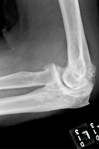
The anatomy of the knee in the context of TKA is defined by its ligamentous stabilizers, extensor mechanism, and osseous geometry. The coronal plane stability of the knee relies on the medial collateral ligament (MCL) and the lateral collateral ligament (LCL). The deep MCL is a primary restraint to valgus stress, and iatrogenic transection during TKA exposure requires immediate primary repair or the use of a constrained condylar knee (CCK) prosthesis. The extensor mechanism comprises the quadriceps tendon, the patella, and the patellar tendon. The patella acts as a fulcrum, increasing the mechanical advantage of the quadriceps. Its blood supply is derived from an anastomotic ring fed by the superior and inferior genicular arteries. Disruption of the lateral superior genicular artery during a lateral retinacular release can compromise patellar viability, leading to avascular necrosis and subsequent fragmentation or fracture.
Biomechanically, a successful TKA relies on the creation of equal and symmetric flexion and extension gaps. The extension gap is dictated by the distal femoral cut and the proximal tibial cut, while the flexion gap is determined by the posterior femoral cut and the proximal tibial cut. Complications such as instability arise when these gaps are mismatched. For example, a tight extension gap and a loose flexion gap (often due to excessive posterior femoral resection or an undersized femoral component) will result in mid-flexion and deep-flexion instability. Understanding these biomechanical principles is absolutely critical when diagnosing the etiology of a failed primary TKA and when templating for revision arthroplasty components.
Exhaustive Indications and Contraindications
The decision to proceed with operative intervention for orthopedic complications requires a meticulous risk-benefit analysis, balancing patient-specific physiological factors with the mechanical realities of the pathology. For upper extremity nerve palsies, indications for surgery depend heavily on the mechanism of injury and the time elapsed. In cases of known iatrogenic transection of the spinal accessory nerve, early primary nerve repair or nerve grafting is indicated. For closed injuries or neurapraxic/axonotmetic lesions, a period of non-operative rehabilitation focusing on the strengthening of periscapular stabilizers is indicated for 3 to 6 months. If clinical and electromyographic recovery is absent after 6 to 12 months, muscle transfer procedures, such as the Eden-Lange procedure (transfer of the levator scapulae and rhomboids), become indicated.
In the management of recurrent or persistent carpal tunnel syndrome, the indications for revision surgery are highly specific. Revision is indicated in patients with persistent symptoms who have clear evidence of incomplete release (often demonstrable on ultrasound or MRI) or those with recurrent symptoms after a symptom-free interval, suggesting perineural fibrosis. However, strict contraindications exist. As highlighted in board-relevant scenarios, a patient with persistent pain, normal EMG/NCS findings, and heavy psychosocial overlay (e.g., active workers' compensation claims, ongoing use of vibratory tools like rivet guns) is a poor candidate for revision. Normal electrodiagnostic studies in this setting strongly contraindicate revision carpal tunnel release, as the pathology is exceedingly unlikely to be median nerve compression at the wrist, and surgery will predictably fail or exacerbate symptoms.
For TKA complications, indications for revision arthroplasty are broadly categorized into septic and aseptic etiologies. Periprosthetic joint infection (PJI) meeting the Musculoskeletal Infection Society (MSIS) criteria is an absolute indication for surgical intervention, typically a two-stage exchange arthroplasty utilizing an antibiotic-eluting spacer. Aseptic loosening, characterized by progressive radiolucent lines and component migration, is an indication for single-stage revision. Instability that is recalcitrant to physical therapy and bracing also warrants revision, often requiring an upgrade in prosthetic constraint. Extensor mechanism disruptions (e.g., complete patellar tendon ruptures) are absolute indications for surgical reconstruction, as patients are rendered unable to ambulate without severe compensatory mechanisms.
Contraindications to revision TKA must be respected to prevent catastrophic patient harm. Absolute contraindications include active systemic bacteremia, profound immunocompromise precluding wound healing, and a medically unstable patient unable to tolerate major anesthesia. Relative contraindications include severe, uncorrectable peripheral vascular disease, which may necessitate amputation rather than revision, and profound neurological deficits (e.g., severe Parkinson's disease or Charcot arthropathy) that guarantee early mechanical failure of the revision construct. Lack of a functional extensor mechanism was historically a contraindication to revision, though modern reconstructive techniques have made this a relative, rather than absolute, barrier.
| Pathology / Procedure | Primary Indications | Absolute/Relative Contraindications |
|---|---|---|
| Spinal Accessory Nerve Palsy / Eden-Lange | Failure of 6-12 months conservative management; lateral winging; severe pain/weakness. | Spontaneous EMG recovery; stiff/frozen shoulder (must restore passive ROM first). |
| Revision Carpal Tunnel Release | Incomplete prior release; recurrent CTS with positive EMG/NCS; dense perineural fibrosis. | Normal EMG/NCS; proximal compression (cervical spine); primary CRPS diagnosis. |
| Revision TKA for Aseptic Loosening | Progressive radiolucent lines >2mm; component migration; intractable mechanical pain. | Active local/systemic infection; medically unfit for anesthesia; lack of functional soft tissue envelope. |
| Two-Stage Exchange for TKA PJI | Chronic PJI meeting MSIS criteria; failed DAIR procedure; presence of sinus tract. | Medically unstable patient; uncorrectable vascular compromise (consider amputation). |
Pre-Operative Planning, Templating, and Patient Positioning
Pre-operative planning is the crucible in which surgical success is forged, particularly in the complex revision setting. For periscapular tendon transfers such as the Eden-Lange procedure, planning begins with a thorough clinical examination to ensure full passive range of motion of the glenohumeral joint. Any capsular contracture must be addressed with aggressive physical therapy prior to tendon transfer, as a stiff shoulder will negate the biomechanical advantages of the transfer. Electromyography must confirm the irreversible denervation of the trapezius and the robust, healthy innervation of the donor muscles (levator scapulae, rhomboid major, and rhomboid minor). The patient is typically positioned in the lateral decubitus or prone position, with the operative arm draped free to allow for intraoperative assessment of scapular tracking and tensioning of the transfers.
When planning a revision carpal tunnel release, the surgeon must obtain and meticulously review the previous operative notes to determine the original surgical approach (endoscopic versus open) and any intraoperative complications encountered. Advanced imaging, such as high-resolution ultrasound or magnetic resonance neurography, is increasingly utilized to assess the integrity of the transverse carpal ligament, the cross-sectional area of the median nerve, and the presence of space-occupying lesions or dense scar tissue. The patient is positioned supine with the arm extended on a hand table. A pneumatic tourniquet is applied to the proximal arm, but the surgeon must be prepared to deflate the tourniquet if prolonged microsurgical neurolysis or the harvesting of a hypothenar fat pad flap is required to ensure adequate perfusion of the nerve and the flap.
Revision TKA demands an exhaustive pre-operative radiographic and clinical workup. The surgeon must first definitively rule out infection via joint aspiration, sending fluid for cell count, differential, and extended cultures. Once aseptic failure is confirmed, full-length standing lower extremity radiographs are obtained to assess the mechanical axis. Computed tomography (CT) is invaluable for quantifying osteolysis and classifying bone loss according to the Anderson Orthopaedic Research Institute (AORI) classification system. This dictates the need for structural allografts, highly porous metaphyseal cones, or sleeves. Templating involves selecting the appropriate degree of constraint—ranging from posterior stabilized (PS) to constrained condylar knee (CCK) to rotating hinge—based on the integrity of the collateral ligaments.
Patient positioning for revision TKA is typically supine on a standard radiolucent operating table. A bump is placed under the ipsilateral hip to internally rotate the leg to a neutral position. A sterile tourniquet is often utilized, though many surgeons prefer to perform the exposure and implant removal without a tourniquet to minimize ischemic time, inflating it only during cementation. The previous surgical incisions must be carefully mapped. If multiple parallel incisions exist, the most lateral viable incision is generally utilized to preserve the blood supply to the anterior skin flap, as the cutaneous blood supply to the knee arises predominantly from the medial side.
Step-by-Step Surgical Approach and Fixation Technique
The surgical execution of the Eden-Lange procedure for trapezius palsy requires meticulous soft tissue handling and secure osseous fixation. A longitudinal incision is made midway between the spinous processes and the medial border of the scapula. The levator scapulae, rhomboid minor, and rhomboid major are sequentially identified and detached from their insertions on the medial border of the scapula. Care is taken to protect the dorsal scapular nerve and artery, which run deep to the rhomboids. The muscles are then mobilized laterally. The levator scapulae is transferred to the spine of the scapula, while the rhomboids are transferred to the infraspinatus fossa. Fixation is achieved using heavy non-absorbable sutures passed through transosseous drill holes or via robust suture anchors. The transfers are tensioned with the scapula held in an anatomically reduced position, effectively substituting for the stabilizing vectors of the paralyzed trapezius.
In revision carpal tunnel release, the surgical approach must prioritize the identification of the median nerve in virgin, unscarred tissue to prevent iatrogenic transection. An extended palmar incision is utilized, extending proximally across the wrist crease in a zigzag fashion if necessary. The median nerve is identified proximally in the distal forearm, deep to the antebrachial fascia, and distally in the mid-palm. Once identified in healthy tissue, the nerve is carefully traced into the zone of prior surgery. Dense perineural fibrosis is addressed with an epineurotomy under loupe or microscopic magnification. To prevent recurrent scarring, a barrier flap is often interposed. The hypothenar fat pad flap is a workhorse for this indication; it is mobilized on its vascular pedicle from the ulnar artery branches and rotated over the median nerve. Alternatively, synthetic nerve wraps or pronator quadratus muscle flaps may be employed.
The surgical approach for revision TKA begins with the management of the extensor mechanism. A standard median parapatellar arthrotomy is utilized, but if patellar eversion is difficult due to arthrofibrosis or patella baja, the surgeon must immediately employ an extensile exposure to prevent catastrophic avulsion of the patellar tendon. A quadriceps snip (an oblique incision directed superolaterally through the rectus femoris tendon) is the first-line extensile measure. If this is insufficient, a tibial tubercle osteotomy (TTO) is performed, creating a bone block of at least 6-8 centimeters in length to ensure adequate surface area for subsequent healing. The implants are then systematically removed using flexible osteotomes, Gigli saws, and specialized extraction tools, prioritizing the preservation of host bone.
Following implant extraction, the joint line must be meticulously re-established. The femoral joint line is typically 25-30 millimeters distal to the medial epicondyle. Metaphyseal bone defects are addressed with titanium sleeves or tantalum cones, which provide immediate rigid fixation and allow for biologic ingrowth. The trial components are inserted, and the flexion and extension gaps are balanced. If the collateral ligaments are incompetent, a rotating hinge prosthesis is selected. The final components are implanted with antibiotic-loaded polymethylmethacrylate (PMMA) cement applied only to the articular surfaces (hybrid fixation) if metaphyseal sleeves are utilized. The extensor mechanism is repaired, and if a TTO was performed, it is rigidly fixed with cerclage wires or cortical screws.
Complications, Incidence Rates, and Salvage Management
Despite meticulous surgical technique, complications in both upper extremity reconstruction and TKA revision are inevitable and require rapid, decisive management. Following periscapular tendon transfers like the Eden-Lange procedure, the most common complication is stretching out or attenuation of the transfer, occurring in up to 15-20% of cases. This leads to recurrent lateral winging and weakness. Failure of the osseous fixation at the scapula is less common but catastrophic. Salvage for a failed Eden-Lange procedure is severely limited; the definitive end-stage salvage is a scapulothoracic fusion. This procedure involves decorticating the medial border of the scapula and the underlying ribs, utilizing robust wire or plate fixation and autogenous bone graft to permanently fuse the scapula to the thorax, thereby providing a stable base for glenohumeral motion at the expense of scapulothoracic rotation.
Complications following revision carpal tunnel release are notoriously difficult to manage. Persistent pain and recurrent numbness occur in 10-30% of revision cases, particularly in the demographic highlighted earlier (workers' compensation, normal preoperative EMG). Complex Regional Pain Syndrome (CRPS) Type II (causalgia) can develop secondary to iatrogenic injury to the palmar cutaneous branch of the median nerve or the main trunk itself. Management of CRPS involves aggressive multimodal pain management, stellate ganglion blocks, and intensive hand therapy focusing on stress loading. If a definitive iatrogenic neuroma is identified, salvage involves neuroma excision and burying the proximal nerve stump deep into the pronator quadratus muscle. For end-stage recalcitrant neuropathic pain without an identifiable structural cause, spinal cord stimulation may be the only viable salvage.
In the realm of TKA, Periprosthetic Joint Infection (PJI) is the most dreaded complication, occurring in 1-2% of primary TKAs and up to 5-10% of revision TKAs. Failure to eradicate infection after a two-stage exchange occurs in approximately 15-20% of cases. Salvage options for recalcitrant PJI include a repeat two-stage exchange, chronic suppressive oral antibiotic therapy (if the organism is susceptible and the implant is stable), resection arthroplasty, arthrodesis (using an intramedullary nail or external fixator), or ultimately, above-knee amputation. Amputation is reserved for cases of intractable infection with severe bone loss and an inadequate soft tissue envelope, prioritizing the patient's systemic survival over limb salvage.
Extensor mechanism disruption post-TKA is another catastrophic complication, with an incidence of 0.1% to 2.5%. Primary repair of a ruptured patellar tendon or quadriceps tendon in the setting of a TKA is universally doomed to fail due to the poor vascularity and the mechanical forces across the joint. Salvage management requires complex reconstruction. Historically, extensor mechanism allografts (incorporating the tibial tubercle, patellar tendon, patella, and quadriceps tendon) were the gold standard, but they carry high rates of delayed failure, graft stretching, and disease transmission. Currently, the use of synthetic mesh (e.g., Marlex mesh) has gained significant traction. The mesh is woven through the remaining host tissue and cemented into the proximal tibia, acting as a permanent, non-biologic scaffold that allows for immediate, aggressive fibrous ingrowth.
| Complication | Estimated Incidence | Primary Salvage / Management Strategy |
|---|---|---|
| Failed Eden-Lange Transfer | 15 - 20% | Scapulothoracic arthrodesis. |
| CRPS post-Revision CTR | 5 - 10% | Multimodal analgesia, stellate ganglion blocks, spinal cord stimulator. |
| Recalcitrant TKA PJI | 15 - 20% (of revisions) | Suppressive antibiotics, Knee Arthrodesis, or Above-Knee Amputation. |
| TKA Extensor Mechanism Disruption | 0.1 - 2.5% | Synthetic mesh reconstruction (Marlex) or complete Extensor Mechanism Allograft. |
Phased Post-Operative Rehabilitation Protocols
Rehabilitation is the final, critical phase of surgical intervention, dictating the ultimate functional outcome. For patients undergoing tendon transfers for scapular winging, the post-operative protocol must balance the need for rigid immobilization to allow osseous integration with the need to prevent glenohumeral stiffness. Phase I (0-6 weeks) consists of strict immobilization in an abduction sling or orthosis, positioning the shoulder in 30-45 degrees of abduction and neutral rotation to remove all tension from the transferred levator and rhomboids. Only passive range of motion of the elbow, wrist, and hand is permitted. Phase II (6-12 weeks) initiates passive and active-assisted range of motion of the shoulder in the supine position to minimize gravitational forces. Phase III (12+ weeks) introduces active range of motion and progressive resistance training, focusing heavily on biofeedback to train the transferred muscles to fire in their new biomechanical roles.
Following revision carpal tunnel release, particularly when a hypothenar fat pad flap is utilized, early motion is encouraged to prevent recurrent adhesion formation. Phase I (0-2 weeks) involves bulky soft dressings with immediate active tendon gliding exercises (straight, hook, fist, and tabletop positions) to maintain independent excursion of the flexor digitorum superficialis and profundus tendons. Phase II (2-6 weeks) focuses on scar desensitization techniques, utilizing varying textures, fluidotherapy, and silicone gel sheeting. Gentle nerve gliding exercises are introduced to mobilize the median nerve within its newly reconstructed bed. Phase III (6+ weeks) focuses on progressive grip and pinch strengthening, though patients must be counseled that maximal grip strength may take up to a year to return, and some permanent deficit may remain.
The rehabilitation following revision TKA is highly variable and dependent on the degree of constraint and the management of the extensor mechanism. In a standard aseptic revision utilizing metaphyseal cones and a CCK bearing, patients are typically allowed immediate weight-bearing as tolerated. Phase I (0-4 weeks) emphasizes early restoration of full extension to prevent flexion contractures, utilizing prone hangs and extension splinting. Continuous passive motion (CPM) machines may be used to facilitate early flexion. If a tibial tubercle osteotomy was performed, weight-bearing may be restricted, and active terminal knee extension is strictly prohibited for 6 weeks to prevent avulsion of the osteotomy.
Rehabilitation following an extensor mechanism reconstruction with synthetic mesh is notoriously rigid. Phase I (0-6 weeks) requires absolute immobilization of the knee in full extension using a cylinder cast or a locked hinged knee brace. Weight-bearing is permitted in full extension only. This prolonged immobilization is mandatory to allow for dense fibrous tissue ingrowth into the synthetic mesh. Phase II (6-12 weeks) introduces heavily controlled, progressive flexion, typically advancing the brace by 10 to 15 degrees per week. Active extension is still strictly avoided. Phase III (12+ weeks) allows for the gradual initiation of active extension and isometric quadriceps strengthening. Patients must be counseled that an extensor lag of 10 to 15 degrees is common and often an acceptable trade-off for a stable, functional limb.
Summary of Landmark Literature and Clinical Guidelines
The evidence base governing the management of these complex complications is built upon several landmark studies and consensus guidelines. In the management of spinal accessory nerve palsy, the seminal work by Bigliani et al. (1996) defined the modern modifications of the Eden-Lange procedure. Their long-term follow-up demonstrated that transferring the levator scapulae and rhomboids reliably restored the force couples necessary for shoulder elevation and eliminated the pain associated with lateral scapular winging. Furthermore, guidelines from the American Academy of Orthopaedic Surgeons (AAOS) emphasize the critical role of electromyography at the 3-to-6-month mark post-injury to differentiate between neurapraxia (warranting continued observation) and axonotmesis/neurotmesis (warranting surgical intervention).
In the realm of compression neuropathies, the literature strongly supports the prognostic indicators for revision carpal tunnel release. Stutz et al. and subsequent meta-analyses have definitively shown that patients presenting with normal electrodiagnostic studies, active workers' compensation claims, and a history of heavy vibratory tool use have unacceptably high failure rates following revision surgery. These studies serve as the foundation for the clinical guideline that a normal EMG/NCS in the setting of recurrent symptoms should prompt an exhaustive search for alternative diagnoses (e.g., cervical radiculopathy) rather than repeat surgical decompression at the wrist.
For TKA complications, the Musculoskeletal Infection Society (MSIS) criteria, updated in 2018, serve as the international gold standard for diagnosing periprosthetic joint infection. These criteria utilize major criteria (e.g., sinus tract communicating with the joint, two positive cultures with the same organism) and minor criteria (e.g., elevated synovial leukocyte count, elevated serum CRP/ESR, positive alpha-defensin) to guide surgical decision-making. The literature surrounding the two-stage exchange arthroplasty, originally popularized by Insall, remains the benchmark for PJI eradication in North America, boasting success rates of 80-85%.
Finally, the literature regarding extensor mechanism reconstruction in TKA has undergone a paradigm shift. Historically, the work by Booth and later Browne established the use of complete extensor mechanism allografts. However, due to the high rates of late graft attenuation and failure (up to 30-40% at 5 years), recent landmark studies by Abdel, Hanssen, and the Mayo Clinic group have championed the use of synthetic Mar
