Deep Surgical Site Infection & Osteomyelitis After Ankle ORIF: A Case Study
Key Takeaway
Diagnosing post-ORIF ankle infection requires observing clinical signs: pain, erythema, swelling, and purulent discharge. Key lab indicators include elevated WBC, ESR, and CRP. MRI with contrast is critical, revealing soft tissue edema, fluid collections, and marrow changes consistent with osteomyelitis. Prompt diagnosis ensures effective management and improved outcomes.
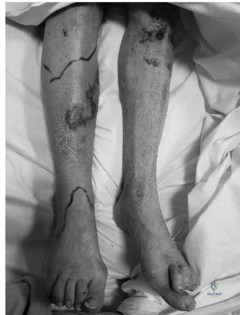
A 48-year-old male presents 6 weeks post-ORIF of a Gustilo II pilon fracture with purulent discharge from the medial incision. He is systemically unwell, diabetic, and a smoker. You suspect a deep fracture-related infection (FRI). What is your structured approach to assessment and diagnostic confirmation in the acute clinic setting?
Candidate: I would perform a thorough clinical exam looking for a sinus tract, systemic signs of sepsis, and hardware stability. I would order inflammatory markers (CRP, ESR, WBC). Radiographically, I’d check for osteolysis or hardware loosening. If I suspect FRI, I would avoid superficial swabs and plan for formal surgical debridement and deep tissue sampling.
Candidates often suggest taking superficial wound swabs in the clinic. These yield skin commensals and are clinically useless for deep infections. They also frequently fail to mention the 2018 International Consensus Meeting (ICM) criteria for FRI, instead relying on vague "clinical suspicion."
I would apply the 2018 ICM criteria for FRI. Confirmation is met if there is a sinus tract communicating with the implant or if there is frank purulence. I would assess systemic host factors (Cierny-Mader Type B) including HbA1c and smoking status. Diagnostics include serial CRP/ESR trends and plain radiographs, followed by a CT-MARS to evaluate for sequestra or occult loosening. I would explicitly state that diagnostic aspiration is contraindicated to avoid introducing infection into a joint, and that definitive diagnosis requires deep intraoperative tissue sampling.
Radiographs show periosteal reaction and lucency around the medial malleolar screws. Given the 6-week post-op timeline, what is your surgical strategy for this patient, and why?
Candidate: At 6 weeks, a biofilm is mature and the hardware is loose. Therefore, hardware retention is no longer an option. I would plan a two-stage approach: Stage 1 involves hardware removal, aggressive debridement, and placement of an antibiotic-loaded PMMA spacer, stabilized with an external fixator. Stage 2 would be definitive reconstruction once infection is cleared.
Suggesting "washout and hardware retention." This is only indicated for *very early* acute post-operative infections (usually < 2-3 weeks). By 6 weeks, the biofilm is established; retention leads to persistent infection, chronic osteomyelitis, and treatment failure.
The strategy is "eradication before reconstruction." I would perform a radical oncologic-style debridement until reaching bleeding, healthy bone (the "paprika sign"). I would send the hardware for sonication to maximize sensitivity for biofilm pathogens. The dead space must be obliterated with antibiotic-loaded PMMA (using heat-stable antibiotics like Vancomycin/Tobramycin), and the limb stabilized with a multiplanar circular fixator to allow soft tissue management and access for serial debridement or eventual definitive arthrodesis.
