

A 28-year-old rugby player presents with recurrent anterior shoulder instability. You have obtained the initial radiographs. Based on the provided image, describe your assessment of this shoulder and your immediate management priorities.
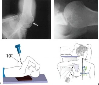
Candidate: The radiograph shows an anterior-inferior glenohumeral dislocation. The humeral head is sitting subcoracoid. My priority is to perform a neurovascular exam before attempting a gentle closed reduction under sedation. Post-reduction, I would order a CT scan to evaluate for bony Bankart or Hill-Sachs lesions.
Candidates often jump straight to "reduction." They fail to explicitly mention the neurovascular examination (specifically the axillary nerve) *before* manipulation, which is a medicolegal absolute. They also often forget to mention post-reduction imaging to ensure concentric reduction and screen for occult fractures.
The candidate should state: "This is a classic anterior glenohumeral dislocation. My management involves: 1. Assessment: Focused neurovascular exam, documenting axillary nerve function. 2. Reduction: Closed reduction under appropriate sedation. 3. Verification: Immediate post-reduction radiographs to confirm concentric reduction and identify associated fractures (e.g., Hill-Sachs). 4. Advanced Imaging: Because this is a recurrent instability case in a young athlete, I would mandate a 3D CT scan to quantify glenoid bone loss and assess for a 'bipolar' injury, which will dictate my future surgical management."
The patient's CT scan is provided below. How do you interpret this, and how does it influence your surgical decision-making regarding the 'Glenoid Track' paradigm?
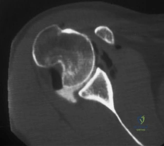
Candidate: This CT shows an osseous defect on the anterior-inferior glenoid. I would use the Pico method to quantify the bone loss. If the loss is greater than 15-20%, a standard Bankart repair will likely fail. I need to check if the Hill-Sachs lesion is 'on-track' or 'off-track' relative to the remaining glenoid track width.
Candidates describe the defect as "large" or "small" without mentioning the objective tools (Pico method for glenoid, Glenoid Track concept for Hill-Sachs). Failing to link the bone loss to the surgical choice—specifically neglecting to mention when to transition from soft-tissue repair to bony augmentation (Latarjet)—is a major oversight.
A sophisticated answer defines the "Glenoid Track" as 83% of the glenoid diameter. "I would calculate the percentage of glenoid bone loss. If <13.5% and the Hill-Sachs is 'on-track', I would proceed with arthroscopic Bankart. If 'off-track' but sub-critical glenoid loss, I would perform a Bankart with Remplissage. If glenoid bone loss is 'critical' (>15-20%), the biomechanics are compromised, and I would recommend an open Latarjet procedure to restore the osseous block and the sling effect of the conjoint tendon."
You have decided to proceed with an open Latarjet procedure. What are the critical anatomic structures at risk during this surgery, and how do you protect them?
Candidate: The main risks are the musculocutaneous nerve and the axillary nerve. I protect the musculocutaneous nerve by keeping my dissection close to the coracoid and not retracting the conjoint tendon too aggressively. For the axillary nerve, I stay superior during the subscapularis split and avoid deep, uncontrolled dissection inferiorly.
Candidates often forget the specific distance of the musculocutaneous nerve from the coracoid tip (3-5 cm) or provide vague "be careful" advice. They also often fail to mention the cephalic vein, which is the key superficial landmark in the deltopectoral approach.
The candidate demonstrates anatomical mastery: "1. The Cephalic Vein: Identified and retracted laterally to enter the deltopectoral interval. 2. Musculocutaneous Nerve: Located within the conjoint tendon, 3-5 cm distal to the coracoid tip. I avoid medial retraction of the conjoint tendon to protect it. 3. Axillary Nerve: At risk during inferior capsular release and subscapularis splitting. I perform a split of the subscapularis at the junction of the superior 2/3 and inferior 1/3, which is a safe zone to protect the nerve, and avoid excessive inferior dissection."
Detailed Chapters & Topics
Dive deeper into specialized chapters regarding shoulder-and-elbow-cases-hillsachs-lesion