Flexor Digitorum Longus Tendon Debridement: Etiology, Anatomy, & Surgical Indications
Key Takeaway
Flexor Digitorum Longus (FDL) tendon debridement is a surgical procedure for chronic FDL tenosynovitis, tendinopathy, or mechanical impingement, unresponsive to 3-6 months of conservative care. It addresses inflammation or degenerative changes within the FDL tendon and its sheath, often presenting as posteromedial ankle/midfoot pain. A precise understanding of FDL anatomy is crucial for successful outcomes.
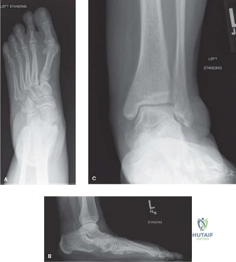
A 28-year-old professional ballet dancer presents with a 6-month history of refractory pain along the posteromedial ankle. She describes a "catching" sensation during pointe work. On examination, there is tenderness along the course of the flexor digitorum longus (FDL) and painful resisted toe flexion. An MRI scan has been performed.
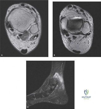
Candidate: Given her history and the MRI findings, this looks like FDL tenosynovitis or possibly a longitudinal split tear. I would exhaust conservative measures like physiotherapy and eccentric exercises. If she fails that, I would consider surgical debridement or synovectomy. I must be careful of the neurovascular bundle during the approach.
The candidate focuses solely on the FDL. A failing response ignores the "big picture" of the medial ankle. Specifically, failing to mention the exclusion of Posterior Tibial Tendon Dysfunction (PTTD) or the risk of missing the Master Knot of Henry pathology is a critical oversight. Examiners look for the ability to prioritize differential diagnoses in the medial column.
I would structure my management by first confirming the diagnosis, ensuring the MRI has ruled out concomitant PTTD or Tarsal Tunnel Syndrome, as these are common differentials. Given she is a ballet dancer, I would assess for mechanical impingement at the Master Knot of Henry. My response would detail: 1) Clinical assessment (resisted lesser toe flexion vs hallux flexion); 2) Imaging (confirming integrity of the PTT and spring ligament); 3) Conservative protocol (6 months of eccentric loading/orthotics); 4) Surgical plan (medial approach, neurovascular protection, and decompression/debridement of the FDL, potentially addressing the Master Knot of Henry).
During the surgical debridement of the FDL, you are deep in the tarsal tunnel. How do you identify and protect the critical neurovascular structures, and what is your plan if you identify a longitudinal tear?
Candidate: I would identify the FDL as the tendon posterior to the tibialis posterior. I would carefully retract the neurovascular bundle, which lies posterior and lateral to the FDL. If there is a longitudinal tear, I would debride it and then suture it closed.
Failing to define the 'threshold' for repair versus excision is a major red flag. Candidates often forget to mention the specific suture technique or the danger of iatrogenic tarsal tunnel syndrome by closing the retinaculum too tightly. They also fail to mention checking for a communicating slip from the FHL.
Safety: Identify the posterior tibial nerve and artery posterior to the FDL; use blunt retractors (e.g., Senn or Hohmann) to maintain a plane of safety. Tear management: Evaluate the cross-sectional involvement. If <50%, I would perform a marginal excision and tubularization using a running locking stitch (e.g., 4-0 PDS). If >50%, I must consider tenodesis. Critical technical step: Ensure the flexor retinaculum is reconstructed loosely to allow for gliding while preventing bowstringing, effectively preventing post-operative tarsal tunnel syndrome.
