Pediatric Gartland Type III Supracondylar Humerus Fracture: Case Study on Closed Reduction & Pinning
Key Takeaway
For a Gartland Type III displaced pediatric supracondylar humerus fracture, the recommended treatment is closed reduction and percutaneous pinning. This involves meticulous anatomical reduction followed by crossed-pin fixation (medial and lateral pins) to stabilize the fracture, with critical attention to avoiding iatrogenic neurovascular injury, ensuring optimal patient outcomes.
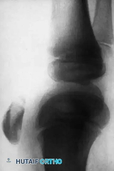
A 6-year-old boy presents to the ED following a fall from monkey bars. He is holding his left elbow in pain. On examination, there is significant swelling, an S-shaped deformity, and the hand is pink but pulses are subjectively diminished. What is your immediate management priority, and how do you classify the following radiographic appearance?
Candidate: I would immediately assess the neurovascular status. The X-ray shows a supracondylar humerus fracture. Since it's displaced with no cortical contact, I'd classify this as a Gartland Type III. My priority is to keep the limb elevated, avoid excessive manipulation, and plan for urgent closed reduction and percutaneous pinning under general anesthesia.
Candidates often forget to categorize the vascular status into "pink/pulseless" vs "white/pulseless." They also frequently fail to mention the specific maneuvers (pronation/supination based on displacement) or neglect to mention the specific risk of iatrogenic nerve injury during pin placement.
A perfect response follows a strict structure: 1. Assessment: Confirm the neurovascular status (differentiate pink/pulseless from white/pulseless). 2. Classification: Identify as Gartland Type III. 3. Immediate care: Splint in neutral, elevate, and NPO for urgent surgery. 4. Surgical Strategy: Closed reduction and percutaneous pinning. Crucially, mention that the reduction maneuver is dictated by the medial/lateral periosteal hinge (pro-nation for posteromedial, supination for posterolateral) and the importance of using a mini-open technique for the medial pin to protect the ulnar nerve.
You have performed a successful closed reduction. You are now preparing to place your pins. What are the biomechanical arguments for crossed-pinning versus lateral-only pinning, and how do you specifically protect the ulnar nerve during the medial pin insertion?
Candidate: Crossed pins provide better torsional stability compared to lateral-only pins, which is why I prefer them for Type III fractures. To protect the ulnar nerve, I make a small medial incision, use blunt dissection to find the epicondyle, and extend the elbow slightly to let the nerve subluxate posteriorly away from my drill trajectory.
Failing to emphasize that the medial pin should NEVER be placed percutaneously. Borderline candidates often suggest "feeling" for the epicondyle, which is a major red flag for examiners.
Acknowledge that lateral-only pins are acceptable IF they are widely divergent and engage the medial column, but note the biomechanical superiority of crossed pins for stability. Highlight the mandatory "mini-open" technique: 1cm incision, blunt dissection to bone, use of a soft tissue protector/sleeve, and extending the elbow to 60-70 degrees to allow the ulnar nerve to move posteriorly out of the hazard zone.
Post-operatively, you notice the child has a new inability to flex the DIP joint of the index finger. What is the likely pathology, and what is your management plan?
Candidate: This indicates an Anterior Interosseous Nerve (AIN) palsy. I would perform a thorough clinical exam to document the deficit, reassure the parents that it is a known complication of these fractures, and observe it over time, as the vast majority resolve spontaneously.
Panicking and suggesting immediate surgical exploration or revision. Also, failing to distinguish between an AIN injury (traction/stretch) and an Ulnar nerve injury (potential iatrogenic pin injury).
Correctly identify this as an AIN nerve palsy (branch of the median nerve). Explain that in the setting of a closed reduction, this is typically a neurapraxia due to stretch. The management is observation. The prognosis is excellent with spontaneous recovery expected within 3–6 months. Differentiate this from an iatrogenic ulnar nerve injury, which would present with sensory changes in the little finger and requires prompt re-evaluation of the medial pin placement.
