Pediatric Supracondylar Humerus Fracture: A Comprehensive Clinical & Radiographic Diagnostic Case Study

Key Takeaway
Pediatric supracondylar humerus fractures are diagnosed via clinical exam (pain, swelling, deformity), thorough neurovascular assessment, and critical X-ray findings. Look for posterior displacement, disrupted anterior humeral line, abnormal Baumann's angle, and fat pad signs. Advanced imaging is usually reserved for complex cases.
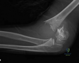
A 7-year-old child presents after a fall onto an outstretched hand. You are presented with the following AP and lateral radiographs. Describe your findings and state your immediate management plan.
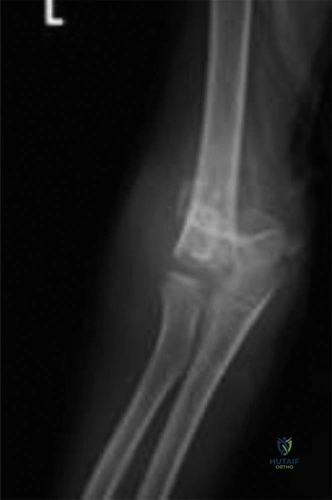
Candidate: The X-rays show a displaced supracondylar humerus fracture. There is posterior displacement of the distal fragment. I would assess the neurovascular status, classify the fracture as a Gartland Type III, and take the patient to theatre for closed reduction and percutaneous pinning (CRPP).
Failing to mention the "pucker sign" or the neurovascular assessment. Simply saying "take to theatre" without differentiating between an emergency (pulseless hand) and urgent (intact neurovascular status) case. Omitting critical radiographic metrics like the anterior humeral line or Baumann's angle.
Start with the ABCs and neurovascular status. Radiographic description: "AP view shows a complete fracture of the supracondylar humerus with medial displacement and an abnormal Baumann's angle (<64°). Lateral view shows posterior displacement of the distal fragment and the anterior humeral line failing to intersect the capitellum." Classify as a Gartland Type III. Management: If neurovascularly intact, the patient is urgent (within 12-24h). Perform CRPP using a lateral entry construct with a low threshold for a mini-open medial pin if stability is not achieved, while protecting the ulnar nerve.
During your attempted closed reduction, you find the fracture is unstable. You decide to utilize a crossed-pin construct. How do you technically perform this safely, and what is your primary concern regarding the medial pin?
Candidate: I would place the lateral pins first. For the medial pin, I am worried about the ulnar nerve. I would perform a small incision, expose the medial epicondyle, and place the pin under direct visualization to ensure I don't hit the nerve.
Suggesting a "blind" medial pin placement. Ignoring that the ulnar nerve can subluxate anteriorly when the elbow is hyperflexed for the reduction maneuver, making it even more vulnerable.
Acknowledge the high risk of iatrogenic ulnar nerve injury (up to 4%). The gold standard is a mini-open technique for the medial pin: use a 1-2cm medial incision to palpate/visualize the nerve and the medial epicondyle, then place the K-wire in a plane anterior to the nerve. If the lateral pins provide sufficient biomechanical stability, consider abandoning the medial pin altogether to eliminate the risk.
Post-operatively, the parents ask about physical therapy. What is your advice regarding the rehabilitation of this child's elbow?
Candidate: I would tell them that active motion is best. I'd advise against aggressive passive stretching because it can cause myositis ossificans. It will take time to get full extension back.
Suggesting that the child should see a physical therapist for aggressive range-of-motion exercises, or implying that immediate, full recovery is expected within weeks.
The advice should be "Active, unassisted range of motion." Explicitly state that passive stretching is strictly contraindicated as it increases the risk of heterotopic ossification (myositis ossificans). Counsel the parents that return of function is gradual, terminal extension is often the last to recover (may take months), and the child should avoid high-impact activities for 8-12 weeks.
