Pathological Femur Fracture in Fibrous Dysplasia: Clinical & Imaging Case Study
Key Takeaway
Fibrous dysplasia presenting as a pathological fracture often involves an expansile, lytic bone lesion with characteristic 'ground-glass' matrix on X-ray and CT. Clinical features include acute pain and functional limitation following low-energy trauma, often preceded by a vague ache. MRI helps assess marrow involvement and rule out malignancy or secondary changes, confirming the diagnosis.
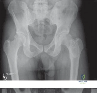
A 25-year-old male presents with a sudden "pop" and inability to bear weight after a minor slip. He reports a 6-month history of vague thigh pain. Radiographs reveal a displaced mid-femoral fracture through an expansile lesion with a ground-glass appearance. How do you approach the initial assessment and emergency management?
Candidate: I would immediately treat the fracture with analgesia and immobilization, such as a traction splint. I'd perform a neurovascular exam and order a CT and MRI to characterize the lesion, along with blood work including calcium, phosphate, and ALP. Given the ground-glass appearance and the fracture, this is likely a pathological fracture through fibrous dysplasia.
Failing to mention the "oncological emergency" aspect. A common error is focusing only on the fracture and forgetting to rule out malignancy or metabolic causes (like hyperparathyroidism) before proceeding to definitive fixation. Candidates often fail to mention the need for a multidisciplinary team or specific imaging requirements like a bone scan to differentiate mono- vs polyostotic disease.
A structured approach is required: 1. Stabilization: Assess neurovascular status, splinting, and analgesia. 2. Diagnosis: Radiographic features (ground-glass, expansile) suggestive of fibrous dysplasia, but maintain a high index of suspicion for malignancy. 3. Workup: Metabolic profile (r/o hyperparathyroidism), cross-sectional imaging (CT for matrix, MRI for soft tissue/fluid-fluid levels), and whole-body bone scan to assess for polyostotic involvement. 4. Planning: Use the Mirels' score to justify aggressive stabilization, acknowledging that these lesions require biopsy during stabilization to confirm the diagnosis and rule out secondary transformation.
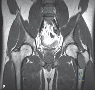
The imaging (CT and MRI) shows a lesion with significant cortical thinning and endosteal scalloping. There are no fluid-fluid levels, but the appearance is quite aggressive. Why is definitive histological diagnosis essential here, and how would you obtain it?
Candidate: I need a biopsy because, although it looks like fibrous dysplasia, there is a 0.4–4% risk of malignant transformation to osteosarcoma. I would perform an open biopsy or, in the case of a fracture, incorporate it into the surgical approach by performing a thorough intralesional curettage of multiple areas within the lesion to ensure representative sampling.
Candidates often suggest an image-guided core needle biopsy in a patient who already requires surgery for a pathological fracture. This is inefficient. Another pitfall is ignoring the risk of "seeding" or misplacing the biopsy track, which could complicate a future wide excision if malignancy were discovered.
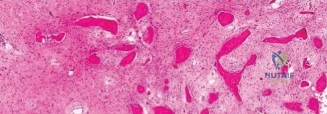
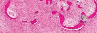
The biopsy is mandatory because imaging alone cannot definitively exclude low-grade chondrosarcoma. In the context of a pathological fracture, the biopsy should be an intraoperative incisional biopsy. One must strictly adhere to oncological principles: the biopsy track must be planned in line with a potential future extensile approach, and the curettage must be meticulous to provide enough tissue for the pathologist to distinguish the "Chinese character" pattern of fibrous dysplasia from malignant mimics.
You have decided on internal fixation with an intramedullary nail and intralesional curettage. Explain the rationale for augmenting the cavity with PMMA (cement) and the expected outcomes for the patient.
Candidate: PMMA is used to fill the void after curettage. It provides immediate structural stability, allowing earlier mobilization. It also serves as an internal cast and the exothermic reaction may help destroy any residual dysplastic cells, potentially reducing recurrence rates.
Simply stating "it fills the hole." Failing to discuss the mechanical advantage of the "load-sharing" construct combined with an IM nail. Also, failing to mention the risk of recurrence if curettage is not aggressive enough or if the augmentation doesn't cover the full extent of the lesion.
The decision is based on: 1. Biomechanical Advantage: Creating a composite construct (nail + cement) increases stability in bone with significant cortical defects. 2. Recurrence Control: The exothermic reaction acts as a thermal adjuvant to help local control. 3. Functional Recovery: It allows earlier protected weight-bearing. The candidate must also highlight the necessity of long-term surveillance, noting that while the fix is stable, the patient remains at risk for future complications like refracture or, very rarely, malignant transformation.
