Is Your Femoral Head Affected? Understanding Hip Osteoarthritis
Key Takeaway
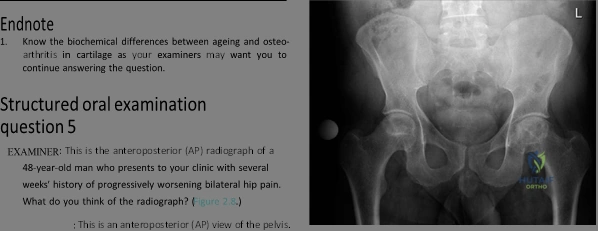
This topic focuses on Is Your Femoral Head Affected? Understanding Hip Osteoarthritis, In hip osteoarthritis, the femoral head is affected by degenerative changes. Radiographs reveal loss of joint space, osteophytes, sclerosis, and bone cysts. This reflects significant cartilage degradation. Secondary OA, from conditions like avascular necrosis or Perthes' disease, also directly impacts the femoral head, leading to similar features of osteoarthritis.
These are the radiographs of a 78-year-old lady with right hip pain. Would you care to comment on these radiographs?
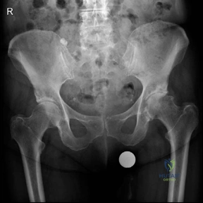
Candidate: "This is an AP radiograph of the pelvis including the lumbar spine and proximal femurs. The right hip demonstrates marked loss of joint space, subchondral sclerosis, osteophyte formation, and subchondral cysts. These findings are diagnostic of primary osteoarthritis of the hip."
Failing to describe the radiograph systematically. A poor candidate simply says "It's arthritis." They often miss the opportunity to comment on the pelvic alignment or the contralateral side, which is critical for surgical planning.
A systematic "ABC" approach: 1. Adequacy/Views: AP view of the pelvis. 2. Joint Space: Superior/axial loss. 3. Bone/Surroundings: Osteophytes (lateral/medial), subchondral sclerosis, cysts (geodes). 4. Synthesis: "Radiographic features consistent with advanced Kellgren-Lawrence Grade 4 osteoarthritis. I would now look for secondary causes like DDH or protrusio before finalizing the diagnosis."
The patient has failed all conservative management and is scheduled for a Total Hip Arthroplasty (THA). You have chosen to use a cemented Exeter stem. What are the biomechanical design principles of this implant?
Candidate: "The Exeter stem is a polished, double-tapered, collarless stem. It relies on the 'taper-slip' principle. As the stem is loaded, it subsides within the cement mantle, converting axial loads into compressive hoop stresses against the cement and surrounding bone."
Missing the term "subsidence." Candidates often think the stem should be perfectly fixed; however, the Exeter stem is designed to *subsidence* within the cement to maintain compression at the interface.
Use the buzzwords: "Polished, tapered, collarless, and subsidence." Explain that the polished surface prevents mechanical interlock (adhesion), allowing for controlled subsidence which creates compressive radial stresses, maintaining the integrity of the cement-implant interface over time.
The patient later returns with pain and a new radiograph. How would you interpret the findings in Figure 2.9?
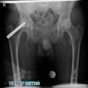
Candidate: "This is an AP radiograph showing a porous tantalum rod inserted into the femoral head for the treatment of early-stage avascular necrosis (AVN). It provides structural support to the subchondral bone."
Not recognizing the material as a "Trabecular Metal" or "Tantalum" rod. Candidates failing to recognize the specific implant often struggle to discuss the surgical implications, such as the difficulty of conversion to THA.
Identify it as a Tantalum rod used for core decompression and structural support. Mention the surgical challenge of conversion (needing specific removal kits) and be aware of the literature regarding potential "poorer outcomes" or tantalum residue leading to joint wear if it fails and proceeds to conversion.
