Orthopedic Hand Cases: A Patient's Journey, Is a Year Old a Factor?
Key Takeaway
For anyone wondering about Orthopedic Hand Cases: A Patient's Journey, Is a Year Old a Factor?, For multiple displaced metacarpal fractures, open reduction and internal fixation (ORIF) is the most appropriate management, particularly in high-energy trauma. When the patient is a 28-year-old male, ORIF restores anatomy, including longitudinal and transverse arches, decompresses interosseous muscles, and stabilizes the hand. This enables earlier rehabilitation, significantly optimizing the patient's functional outcome.
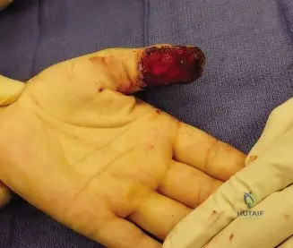
A 28-year-old snowboarder presents 4 days post-injury with a grossly swollen hand following a high-energy fall. Examination reveals a shortened, widened metacarpal profile, loss of dorsal skin wrinkles, and rotational malalignment (scissoring). Looking at this clinical appearance, what are your immediate clinical concerns, and how do you prioritize the assessment?
Candidate: I am concerned about multiple metacarpal fractures. I would check neurovascular status, look for compartment syndrome, and obtain X-rays to assess the displacement and rotation.
Candidates often jump straight to "I would order a CT scan." The examiner is testing your clinical acumen in a patient with a delayed presentation (4 days). Missing the assessment of compartment syndrome or neglecting to document the specific clinical hallmarks (rotational malalignment, intrinsic status) is a critical oversight.
A structured response is essential: "My primary priority is ruling out acute compartment syndrome, given the 4-day delay and severe swelling. I will assess the '5 Ps,' specifically looking for pain out of proportion and pain on passive stretch of the intrinsic muscles. Second, I will perform a precise neurovascular exam. Third, I will assess the mechanical integrity of the hand, focusing on the loss of the digital cascade, rotational malalignment (scissoring), and the loss of longitudinal/transverse arches. Finally, I will order orthogonal radiographs to confirm the fracture pattern and exclude CMC joint involvement."
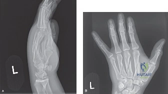
Radiographs confirm multiple shaft fractures of the 2nd, 3rd, and 4th metacarpals. The 2nd metacarpal shows a spiral pattern, while the 3rd and 4th show transverse patterns with comminution. How does this fracture personality influence your surgical strategy and implant selection?
Candidate: I would fix all of them with plates. For the spiral one, maybe a plate or screws, and for the transverse ones, definitely plates to keep them straight.
Vague terminology. Failing to distinguish between absolute stability (interfragmentary compression) and relative stability (splinting/locking plates). Failing to mention why the 2nd/3rd metacarpals require more rigid fixation due to their role as the 'fixed unit' of the hand.
Use biomechanical principles: "The spiral 2nd metacarpal fracture is amenable to absolute stability using interfragmentary lag screws, provided the fracture length is >2x the bone diameter. For the transverse/comminuted 3rd and 4th fractures, I require relative stability using low-profile locking plates acting as an internal fixator to neutralize bending and rotational forces. I must restore the longitudinal length and the natural dorsal concavity of the shafts. I will also use two separate dorsal incisions to preserve the venous and lymphatic drainage and decompress the interosseous compartments."
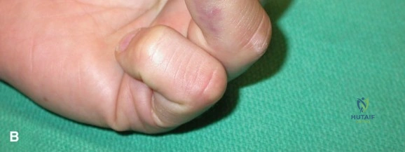
During the procedure, you have fixed the fractures. How do you ensure you have corrected the rotational deformity, and what is your immediate post-operative plan regarding rehabilitation?
Candidate: I would check if the fingers look straight. Then I'd put them in a splint for 6 weeks so the bone heals.
Suggesting long-term immobilization (6 weeks) is a major red flag in hand surgery. This will lead to stiff joints and tendon adhesions, essentially ensuring a poor outcome despite perfect radiographic alignment.
The expert response: "I confirm rotational alignment by performing the tenodesis test: flexing the wrist passively to ensure all fingertips point toward the scaphoid tubercle without scissoring. Post-operatively, I initiate an Early Active Motion (EAM) protocol within 3-5 days. The patient will be placed in an 'intrinsic-plus' splint. By 2 weeks, we begin specific tendon gliding exercises. The goal is to move the hand early to prevent adhesion of the extensor tendons over the hardware while the bone undergoes secondary healing, with protected loading until 6-12 weeks."
