High-Pressure Injection Hand Injuries: Pathophysiology, Anatomy, and Preventing DIP Joint Contracture
Key Takeaway
DIP joint contracture after high-pressure hand injuries (HPII) results from chemical irritation, tissue necrosis, and aggressive fibroproliferative responses. Preventing this severe complication requires a thorough understanding of DIP joint surgical anatomy and biomechanics, guiding early intervention to maintain range of motion and preserve critical hand function.
A 38-year-old male industrial worker presents to your emergency department with a small puncture wound on the volar aspect of his non-dominant index finger. He states he accidentally triggered a high-pressure grease gun while cleaning machinery. The finger is slightly swollen, but he has good capillary refill and sensation. How do you approach this case, and what is your immediate clinical concern?
Candidate: I would perform a thorough history and physical exam, check for systemic toxicity, and obtain radiographs to look for radiopaque material. I would admit the patient, start broad-spectrum antibiotics, and perform urgent surgical debridement of the digit to prevent tissue necrosis and secondary contracture.
The candidate focuses too much on the "good" current status (good capillary refill/sensation). A poor candidate often downplays the injury because the patient looks "fine" now, failing to convey the urgency. They might suggest simple observation or antibiotics alone, which is a dangerous error for high-pressure injection injuries (HPII).
The candidate must emphasize that this is a surgical emergency. The "deceptive innocence" of the wound is a trap. The response should be: "I treat this as a limb-threatening emergency. I will obtain baseline X-rays to assess for radiopaque material and bony involvement, but I will not wait for further imaging to proceed to theatre. The goal is urgent, wide surgical debridement, decompression, and irrigation to mitigate the chemical inflammatory cascade, which is the primary driver of future DIP joint contracture and potential amputation."
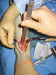
You have taken the patient to the operating theatre. You have made a mid-axial incision. Given the nature of this injury, describe your approach to the DIP joint specifically to prevent long-term functional loss.
Candidate: I would use mid-axial incisions to avoid volar scarring. I would debride the flexor sheath thoroughly, inspect the DIP joint capsule, and washout the area extensively with pulsatile lavage.
The candidate fails to explain why they are doing these steps. A poor candidate forgets to mention the exploration of the neurovascular bundles, which are often the first structures compressed by the injectate, or they fail to mention that the wound should be left open (delayed primary closure).
Structure the answer: 1) **Exposure:** Mid-axial incision preserves the volar skin (preventing secondary flexion contracture). 2) **Decompression:** Release the entire flexor tendon sheath to prevent FDP tethering. 3) **Joint Care:** Inspect the volar plate and collateral ligaments. If injectate is intra-articular, perform a formal capsulotomy and synovectomy to remove inflammatory material. 4) **Wound Management:** Do not perform primary closure; leave wounds open to allow for serial debridement and management of ongoing edema/necrosis.
Six months later, the patient returns with a stable, healed wound but a fixed 45-degree flexion contracture of the DIP joint. He is unable to participate in pinch tasks. How do you manage this?
Candidate: I would suggest surgical release, perhaps a tenolysis of the FDP tendon or a capsulotomy of the DIP joint, followed by intense physical therapy.
The candidate jumps straight to surgery. They ignore the necessity of confirming that all conservative management has been exhausted. Examiners want to see a structured approach to "salvage" that includes patient optimization and therapy verification.
State a stepwise approach: 1) **Assessment:** Verify this is a stable, non-inflammatory state. 2) **Conservative Trial:** Confirm a minimum 3-6 month period of intensive hand therapy, including dynamic/static progressive splinting and serial casting. 3) **Surgical Planning:** Only if failed, perform targeted release: FDP tenolysis, volar plate/capsular release, and potentially collateral ligament release. 4) **Rehabilitation:** Emphasize that surgery is only half the battle; the success depends entirely on the post-operative regimen of immediate active, protected motion to prevent recurrent adhesion.
