Duchenne Muscular Dystrophy: Orthopedic Management, Early Signs & Surgical Anatomy
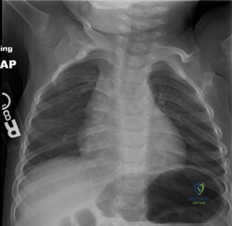
Key Takeaway
Early signs of Duchenne Muscular Dystrophy (DMD) include delayed motor milestones, waddling gait, Gower's sign, calf pseudohypertrophy, and frequent falls. Orthopedic surgeons are vital for proactive recognition, addressing musculoskeletal complications like contractures and scoliosis, and understanding the evolving surgical anatomy to optimize patient quality of life.
You are evaluating a 12-year-old male with Duchenne Muscular Dystrophy (DMD). He is currently non-ambulatory and presents to your clinic with a progressive thoracolumbar deformity. He is being considered for a posterior spinal fusion. What are your key clinical considerations in the pre-operative planning phase for this specific patient?
Candidate: I would organize a multidisciplinary team. Key areas are cardiac function—specifically checking his ejection fraction with an echo—and pulmonary function tests. I need to be aware of the risk of malignant hyperthermia-like reactions to anesthetic agents. I would also order full-length spinal radiographs and CT scans to plan for pedicle screw placement due to potential osteopenia from his chronic steroid use.
The candidate fails to address the specific "DMD-unique" nuances. Simply saying "check lungs and heart" is generic. A weak candidate misses: (1) The specific avoidance of succinylcholine/volatile agents (the rhabdomyolysis risk), (2) The specific pelvic obliquity and its implication for seating, and (3) The need to optimize bone health (DEXA/Vitamin D) pre-operatively due to steroid-induced osteopenia.
A structured, multidisciplinary approach is required: 1. Systemic Optimization: Cardiology (EF, ACE inhibitors) and Pulmonology (FVC, baseline CO2, need for post-op NIV). 2. Anesthetic Safety: Emphasize the absolute contraindication of succinylcholine/volatile gases due to the risk of rhabdomyolysis/cardiac arrest; advocate for TIVA. 3. Bone Health: Address steroid-induced osteopenia; assess DEXA/Vit D status. 4. Surgical Planning: Pedicle screw instrumentation is mandatory for superior pull-out strength in fragile bone. 5. Functional Goal: The primary indication is the preservation of sitting balance and pulmonary function, not just cosmetic correction.
Consider the radiographs below. This patient has significant lower limb contractures. Describe your approach to managing these deformities, and interpret the significance of the image provided.
Candidate: The image shows severe contractures. I would offer surgical releases for the Achilles, hamstrings, and possibly the IT band to help the patient stand or sit better. I would use percutaneous techniques where possible.
The candidate treats "the procedure" rather than the "patient goals." Failing to mention the specific goal (e.g., "facilitating brace wear" vs. "restoring normal gait") is a major oversight, as true gait restoration is rarely possible in late-stage DMD. Also, failing to identify the risk of over-lengthening (calcaneal gait) is a frequent oversight.
Start by identifying the goal: functional optimization for sitting, hygiene, or bracing (AFO/KAFO). 1. Achilles: Percutaneous lengthening is preferred to minimize wound complications, but avoid overcorrection into calcaneus. 2. Hamstrings: Medial and lateral lengthening must protect the neurovascular bundle (sciatic/popliteal). 3. IT Band: Ober-Yount procedure for hip abduction/pelvic obliquity. 4. Strategy: Emphasize that these procedures are palliative to support quality of life and mobility, not to restore normal biomechanics. Post-operative splinting and aggressive PT are mandatory to prevent recurrence.
Regarding spinal surgery in DMD, look at this image. What does it represent, and why is this construct preferred?
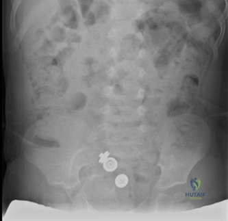
Candidate: This shows posterior spinal fusion with pedicle screws. It is preferred because it's strong and helps correct the curve.
Vague explanations. Failing to link "osteopenia" to the choice of "pedicle screws" is the key. A candidate must explain the biomechanical advantage (3-column purchase) over hooks or wires in the context of the poor bone quality prevalent in DMD patients.
The image depicts posterior spinal instrumentation utilizing pedicle screws. 1. Why Pedicle Screws: They offer superior three-column purchase and biomechanical stability, which is essential given the reduced bone mineral density (osteopenia) from steroid therapy. 2. Correction: They allow for superior 3D deformity correction, including derotation. 3. Technical Note: I would mention the need for careful preoperative CT planning for screw trajectory and the potential for prophylactic cement augmentation if bone quality is severely compromised.
